180 Kaposi’s sarcoma
Salient features
Advanced-level questions
What are the varieties of Kaposi’s sarcoma?
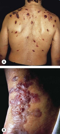
• Classic Kaposi’s sarcoma or epidemic type (Fig. 108.1B): initially described in Jews; indolent; found on the legs of elderly men; confined to the skin and may be present for decades; not fatal.
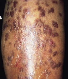
• African variety or endemic type (Fig. 180.2): violaceous skin plaques, black under Negroid skin; this is an aggressive invasive tumour that is ultimately fatal. It occurs in children and younger men. In the former it is often associated with generalized lymphadenopathy.
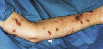
• AIDS-associated Kaposi’s sarcoma (Fig. 180.3): found in approximately one-third of patients with AIDS and more common in homosexuals. The cutaneous lesions respond to interferon-alfa and cytotoxic chemotherapy. About one-third develop a second malignancy such as lymphoma, myeloma or leukaemia. Patients eventually die from a secondary infection seen in AIDS rather than as a direct consequence of Kaposi’s sarcoma.
• Transplantation-associated Kaposi’s sarcoma, particularly in patients on high-dose immunosuppressive therapy; these often regress when therapy is stopped. Transmission of human herpesvirus 8 infection occurs from renal transplant donors to recipients and is a risk factor for Kaposi’s sarcoma (N Engl J Med 1998;339:1358–63).
What is the histology of Kaposi’s sarcoma?
Histologically all varieties are similar:
• Early or ‘patch’ stage: similar to granulation tissue and characterized by jagged, thin-walled, dilated vascular spaces in the epidermis with interstitial inflammatory cells and extravasated red cells (with haemosiderin deposition)
• Later or ‘nodular’ lesions: features are more characteristic at this stage and comprise spindle-shaped stromal cells with irregular slit-like spaces filled with red blood cells and lined by recognizable endothelium, intertwined with normal vascular channels.
Is there any difference in the prognosis of AIDS depending on whether the patient has oral or cutaneous Kaposi’s sarcoma?
How would you manage Kaposi’s sarcoma in patients with HIV infection?
• Baseline treatment includes a retroviral agent such as zidovudine, which has no direct effect on Kaposi’s sarcoma but diminishes the degree of immunosuppression (note: eczema, xerosis, warts and Kaposi’s sarcoma are associated with HIV infection but do not improve with antiretroviral therapy). Retinoic acid is a promising alternative.
• Localized mucocutaneous lesions respond well to liquid nitrogen, radiotherapy, cryotherapy, surgical excision, intralesional vinblastine or sclerosing solutions, bleomycin or interferon-alfa.
• Interferon-alfa as systemic treatment is effective in 40–50% of patients with indolent Kaposi’s sarcoma, a CD4 cell count >400 × 106 cells/l, few systemic symptoms and no opportunistic infections.
• Combination therapy of surgery, chemotherapy and radiation or with chemotherapy alone.
• Relatively marrow-sparing chemotherapy (bleomycin, vincristine or low-dose liposomal doxorubicin) is used when other treatment fails in patients with aggressive Kaposi’s sarcoma (Lancet 1997;350:1363). Complete or partial remission may be obtained in >75% of those with Kaposi’s sarcoma; shorter survival is associated with low CD4 cell count.


