146
Kaposi sarcoma
Kaposi sarcoma lesions can have varying morphologies, including multiple purple papules and plaques, and can be widespread, depending on the clinical variant.
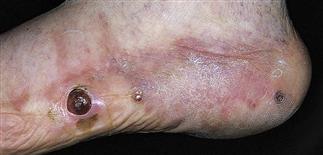
Classic Kaposi sarcoma occurs on the lower extremities; in this patient, lesions are located on the foot. Early lesions can look like malignant melanoma.
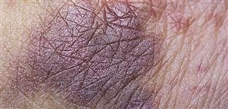
AIDs-related Kaposi sarcoma. Early lesions are flat plaques.
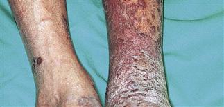
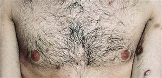
Kaposi sarcoma related to immunodeficiency syndrome is more widespread. Lesions on the lower extremities can appear eczematous. This patient has violaceous papules coalescing to plaques.
DESCRIPTION
A malignancy of lymphatic endothelial cells associated with a gamma herpesvirus, human herpesvirus 8 (Kaposi sarcoma-associated herpesvirus). Four clinical and epidemiologic subsets: classic, endemic, immunosuppression- or transplant-associated, and epidemic- or AIDS-associated.
HISTORY
Four clinical variants. • Classic. Sporadic, slowly progressive; occurs predominantly in 50- to 70-year-old men of eastern European or Mediterranean descent. • Endemic. Regions are eastern and southern Africa. Up to 50% of all childhood soft tissue tumors due to Kaposi sarcoma. Children develop an aggressive lymphadenopathic form. • Immunosuppression- or transplant-associated. Organ transplant recipients at risk. Patients of Mediterranean and eastern European descent are at increased risk for immunosuppression-associated Kaposi sarcoma, supporting the theory of genetic predisposition. • Epidemic-associated. The most common AIDS-associated cancer and is 20 times more common in homosexual men than in those who acquired HIV by another means (e.g. through hemophilia). Patients with AIDS-related Kaposi sarcoma often have systemic involvement, particularly of the gastrointestinal tract (stomach and duodenum). Fever, night sweats, and weight loss may be present.
PHYSICAL FINDINGS
Various morphologies (macules or patches, papules or plaques, nodules). • Classic. Starts with purple patches on the distal lower extremities that progress proximally and become multifocal. Individual lesions darken and thicken, eventually becoming brown and verrucous. Lesions on the lower legs can look eczematous and ulcerate. Early Kaposi sarcoma nodules can feel soft; older nodules can feel firm. • Endemic (within Africa). Involves lymph nodes in people with localized nodular lesions. Occurs most commonly in men and children. • Immunosuppression-associated. Morphologically similar to classic Kaposi sarcoma. Lesions typically improve and sometimes resolve with cessation of immunosuppressive therapy. • AIDS-associated. Lesions have a predilection for the face (especially nose, eyelids, ears), torso, and oral mucosa (especially hard palate).
TREATMENT
• Classic Kaposi sarcoma. Single lesions: surgical excision. Multiple lesions localized to one area: radiation. Extensive or recurrent lesions: combination therapy with surgery, radiation, and chemotherapy. • Endemic. Radiation and chemotherapy. • Immunosuppression-associated. Modification or discontinuation of the immunosuppressive therapy produces regression of Kaposi sarcoma. Radiation and chemotherapy useful when immunosuppressive therapy modification fails. • AIDS-associated. Radiation therapy: large, localized, and/or ulcerative lesions. Cryosurgery (3-week intervals): superficial Kaposi sarcoma. Intralesional vincristine. Antiretroviral therapy: alone or in combination with radiation or systemic therapy, such as cytotoxic drugs (Paclitaxel) and interferon alpha, directed against Kaposi sarcoma.


