15 Jugular venous pulse
Salient features
Examination
• The JVP is measured ….. cm above the angle of Louis (manubriosternal angle). Remember that the distance between the angle of Louis and the mid-right atrium can be varied and the JVP may be measured as high as at the level of the ear lobes.
• Comment on the waveform (timing it with the carotid pulse):
• Check the hepatojugular reflex.
• Tell the examiner that you would like to look for other signs of heart failure:
Diagnosis
This patient has raised jugular venous pulse with ‘v’ waves (lesion) caused by tricuspid regurgitation (see Case 19) and is in heart failure (functional status).
Questions
Advanced-level questions
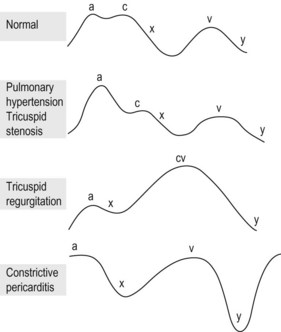
What do you know about the waveforms in the jugular pulse?
There are two outward moving waves (‘a’ and ‘v’ wave) and two inward moving waves (the ‘x’ and ‘y’ descent) (Fig 15.1):
• The ‘a’ wave is caused by atrial contraction and is presystolic. It can be identified by simultaneous auscultation of the heart and the examination of the jugular venous pulse. The ‘a’ wave occurs at about the first heart sound.
• The ‘c’ wave is caused by closure of the tricuspid valve and is not readily visible.
• The ‘v’ wave is caused by venous return to the right heart (not from ventricular contraction) and occurs nearer to the second heart sound.
• The ‘x’ descent is caused by atrial relaxation (sometimes referred to as systolic collapse).
• The ‘y’ descent is produced by opening of the tricuspid valve and rapid inflow of blood into the RV.