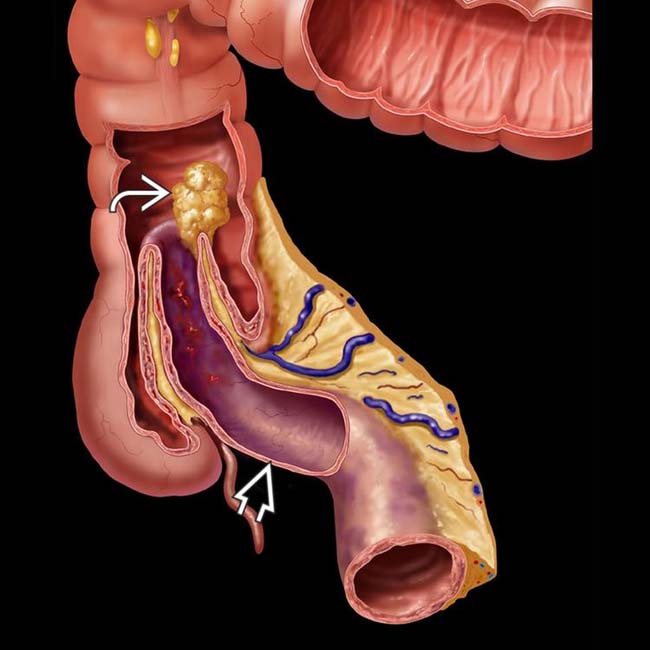
Outer layer = intussuscipiens; inner layer = intussusceptum


 as the lead mass. Note the vascular compromise and ischemia of the intussusceptum
  .
.
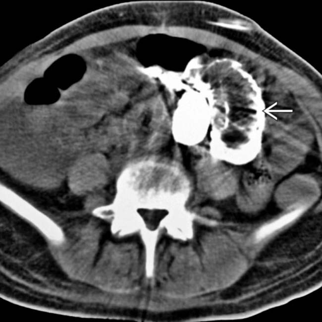
 and vessels
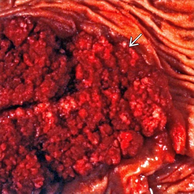
  from an ileocolonic intussusception. The lead mass proved to be carcinoma.
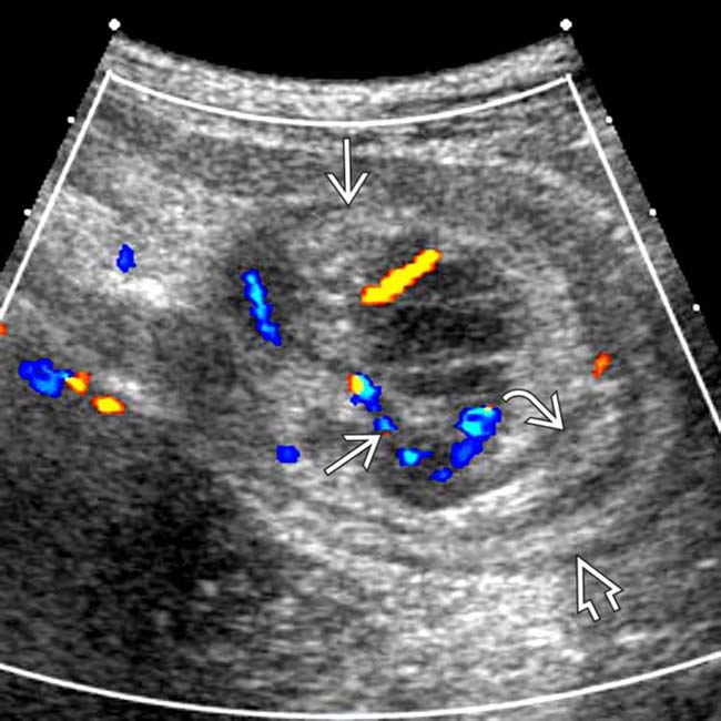
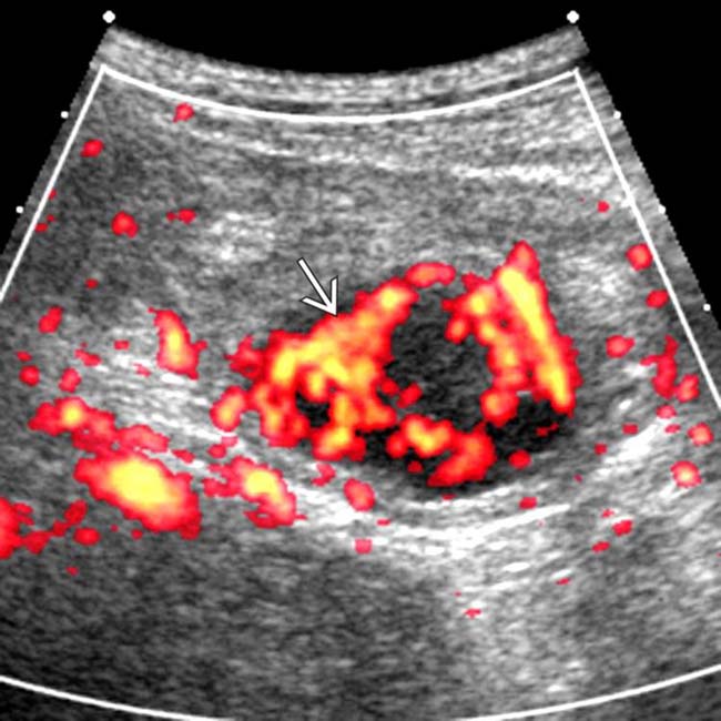
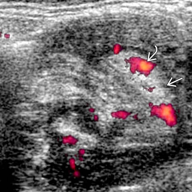
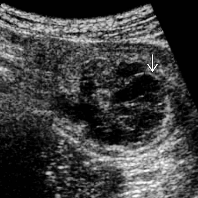
 and 2 echogenic submucosal rings representing intussusceptum
  and intussuscipiens
  .
.
 within the mass, which proved to be a metastatic melanoma.


 of the colon wall, while the intussusceptum is a small intestinal segment
  . Mesenteric fat and vessels
  accompany the intussuscepted small bowel (SB) segment.
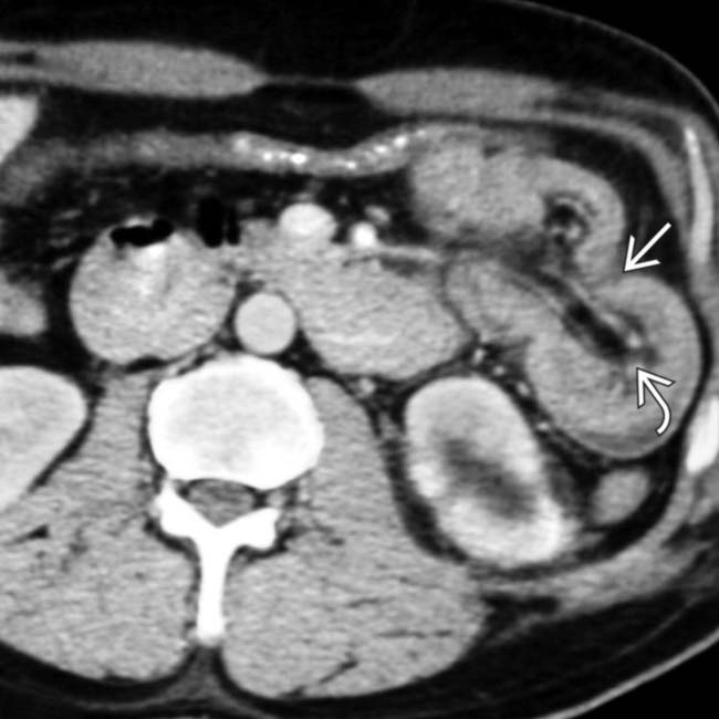
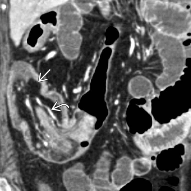
 due to jejunal intussusception. Note the intussuscepted mesenteric fat and vessels
  .
.
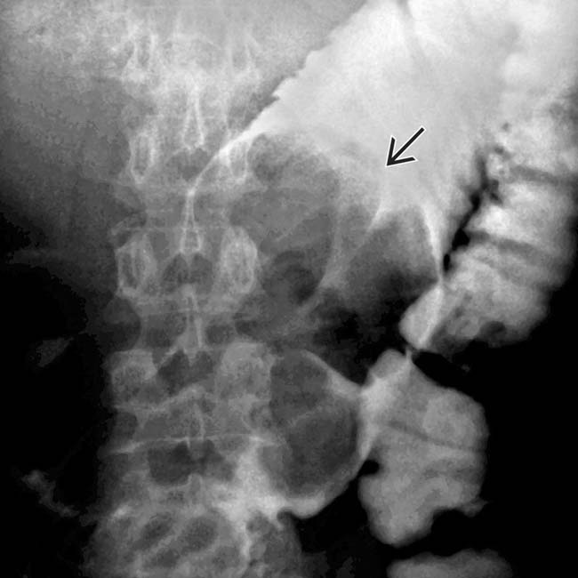
 with its mesenteric fat
  within the contrast opacified lumen of the ascending colon, the intussuscipiens in this case.
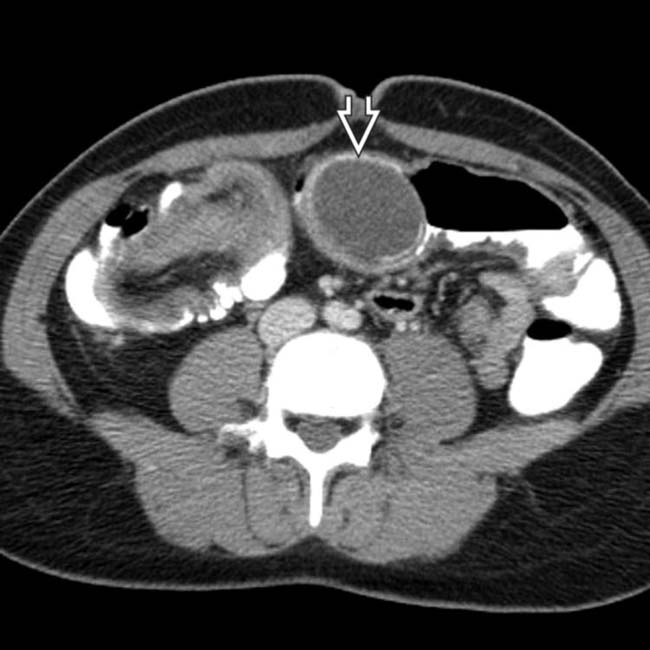
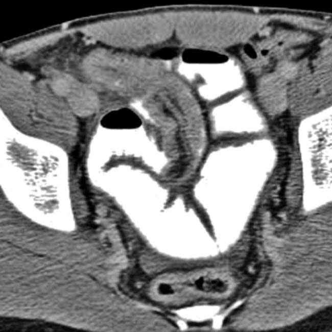
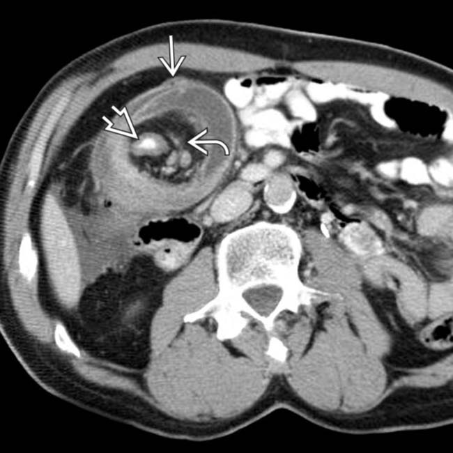
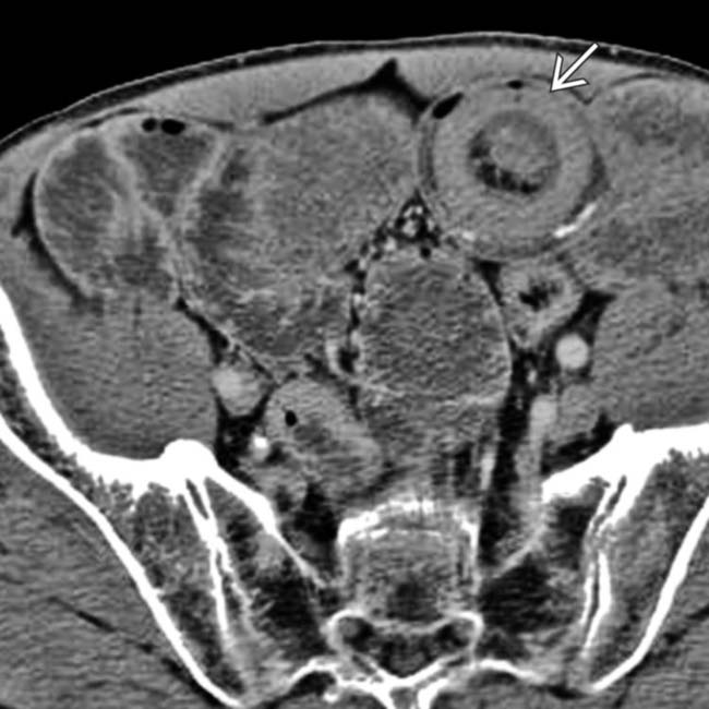
 with a calcified rim, a characteristic appearance of an appendiceal mucocele. Long-segment, obstructing intussusceptions such as this often have a lead mass when seen in adults.
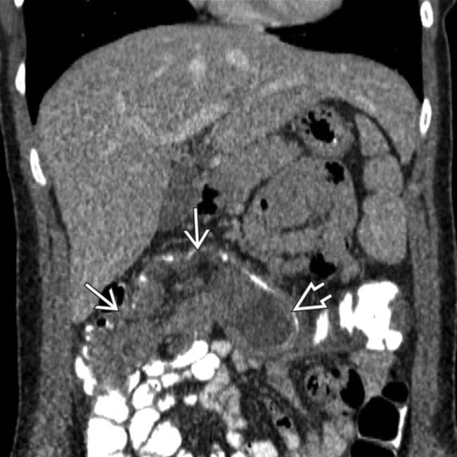
 and the mucocele
  as the lead mass.
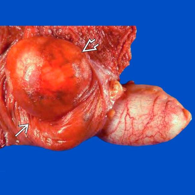
 and the intussusception
  .
.
 with accompanying mesenteric fat and vessels
  .
.
 .
.
 .
.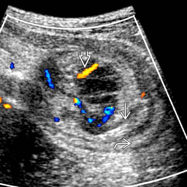
 and intussuscipiens
  . Note the invaginated mesenteric vessels
  .
.
 .
.
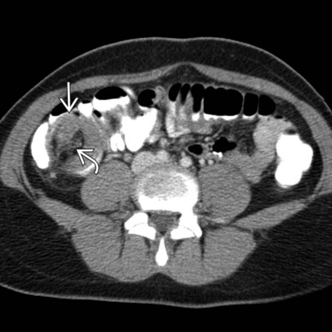
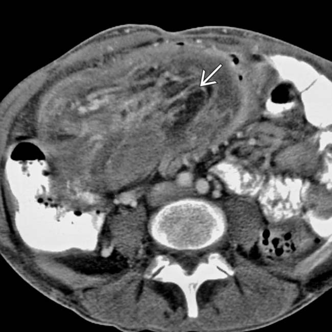
 . At surgery, the lead mass was due to inspissated mucoid material within the terminal ileum.
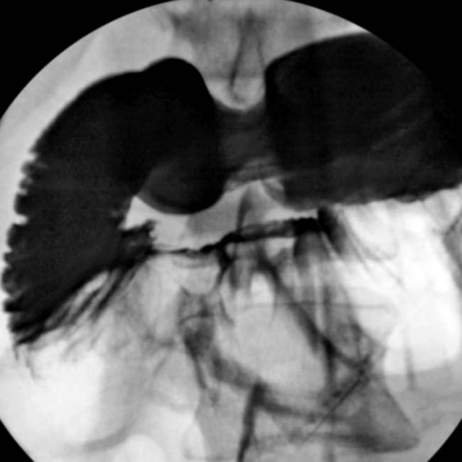
 of the intussusception.
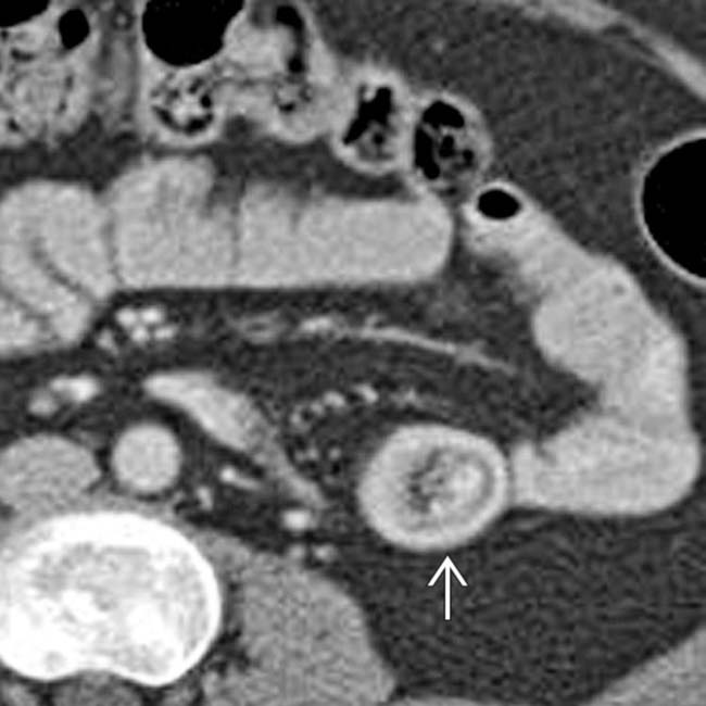
 .
.
 with an accompanying crescent of mesenteric fat. The bowel lumen is dilated due to sprue, not obstruction.