1 Introduction to the Nervous System
The brain seems bewilderingly complex the first few times you look at it. One way to ease the bewilderment is to have an overview of some vocabulary and organizing principles, which the first three chapters of this book attempt to provide. Chapter 1 is a quick introduction to the parts of the nervous system and the cells that make it up, Chapter 2 is an overview of how the parts get arranged that way during development, and Chapter 3 is a closer look at major parts and the wiring principles underlying their interconnections.
The Nervous System Has Central and Peripheral Parts
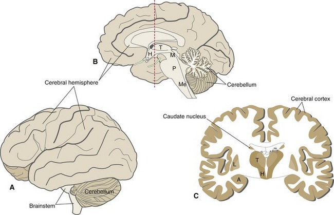
The nervous system has both central and peripheral parts, roughly corresponding to the parts inside and outside the skull and vertebral column. The peripheral nervous system (PNS) is approximately the same thing as the collection of nerves that reach pretty much every part of the head and body, collecting sensory information and delivering messages to body parts or to PNS neurons. The central nervous system (CNS) is made up of the brain and the spinal cord. The brain in turn is composed of the cerebrum (forebrain), cerebellum, and brainstem (Fig. 1-1, Table 1-1). The cerebrum, by far the largest component, is itself composed of two cerebral hemispheres and the diencephalon (from a Greek word meaning “in-between-brain,” because it’s interposed between the cerebral hemispheres and the brainstem).
| Major Division | Subdivision | Principal Function |
|---|---|---|
| Cerebral hemisphere | Cerebral cortex | Perception, cognition, memory, voluntary movement |
| Lenticular nucleus | Part of the basal ganglia: movement control | |
| Caudate nucleus | Part of the basal ganglia: movement control | |
| Amygdala | Part of the limbic system: drives and emotions | |
| Diencephalon | Thalamus | Relays information to the cerebral cortex |
| Hypothalamus | Controls the autonomic nervous system | |
| Brainstem | Midbrain Pons Medulla |
Cranial nerve nuclei, long tracts Cranial nerve nuclei, long tracts Cranial nerve nuclei, long tracts |
| Cerebellum | Coordination of movement |
Each cerebral hemisphere has a covering of cerebral cortex and encloses a series of large nuclei. Some of the enclosed nuclei (lenticular and caudate nuclei) are parts of the basal ganglia, which help control movement; another (the amygdala) is part of the limbic system, which deals with drives and emotions. The cerebral cortex is a critical structure for perception, for the initiation of voluntary movement, and for the functions we think of as distinctively human—things like language and reasoning. Corresponding to these several functions, there are cortical areas primarily concerned with sensation, others with movement, and still others with more complex activities. Because of this parceling of functions, it is possible for cortical damage to impair some abilities while leaving others more or less unaffected.
The Principal Cellular Elements of the Nervous System Are Neurons and Glial Cells
Except for some extrinsic elements such as blood and blood vessels (see Chapter 6) and meninges (see Chapter 4), the whole nervous system is made up of just two general categories of cells: neurons and glial cells (or glia). Each can be divided into a few subcategories, some characteristic of the CNS and others of the PNS (Table 1-2; see THB6 Fig. 1-27, p. 27).
| Location | Major Neurons | Major Glia |
|---|---|---|
| CNS | Motor neurons (→ skeletal muscle via PNS) Preganglionic autonomic neurons (→ autonomic ganglia via PNS) Interneurons local interneurons projection neurons |
Astrocytes (metabolic support, response to injury) Oligodendrocytes (myelin) Ependymal cells (line ventricles, secrete CSF) Microglia (response to injury) |
| PNS | Primary sensory neurons (spinal, cranial nerve, and enteric ganglia) Postganglionic autonomic neurons (sympathetic, parasympathetic, and enteric ganglia) |
Schwann cells (myelin, satellite cells) |
Neurons Come in a Variety of Sizes and Shapes, but All Are Variations on the Same Theme
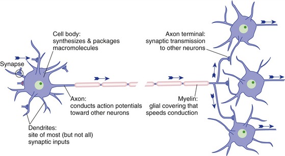
Although there are lots of variations, a typical neuron (Fig. 1-2) has a collection of tapering dendrites and a single cylindrical axon, all emerging from the cell body. The cell body is the synthetic center of the whole neuron, the dendrites receive most of the inputs (synapses) from other neurons, the axon conducts electrical impulses (action potentials) away from the cell body and toward other neurons, and axon terminals release neurotransmitter onto other neurons. So the anatomical polarization (dendrites → cell body → axon) corresponds to a functional polarization in terms of the direction in which electrical signals move.
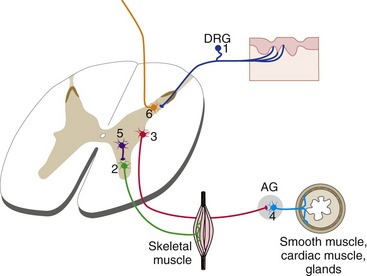
Nearly all neurons fall into one of six categories (Fig. 1-3):
Neuronal Organelles Are Distributed in a Pattern That Supports Neuronal Function
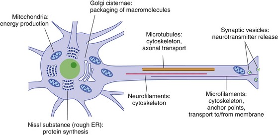
Neurons deal with the same issues as other cells, using the same organelles (Fig. 1-4). However, some of these issues are accentuated because of the specialized structure and function of neurons:
Schwann Cells Are the Principal PNS Glial Cells
Schwann cells envelop PNS neurons and their axons in three different ways. Some Schwann cells are flattened out as satellite cells that surround PNS ganglion cells (THB6 Figure 1-22, p. 23). Others have multiple indentations, each encasing part of a small (unmyelinated) axon (THB6 Figure 1-26, p. 26). Finally, many spiral around individual, larger axons (THB6 Figures 1-23 to 1-25, pp. 24 and 25), forming myelin sheaths that allow axons to conduct action potentials more rapidly (see Chapter 7). Each myelinated axon looks like a string of sausages, each sausage being a length of axon covered by the myelin formed by a single Schwann cell; the constrictions between sausages correspond to nodes of Ranvier, gaps in the myelin where nerve impulses are regenerated.
CNS Glial Cells Include Oligodendrocytes, Astrocytes, Ependymal Cells, and Microglial Cells
Astrocytes play multiple roles, generally less well understood than that of oligodendrocytes. Their cytoskeletons provide mechanical support to neighboring neurons. Astrocyte processes cover the parts of neurons not occupied by synaptic contacts and help regulate the ionic composition of extracellular fluids. They also contact CNS capillaries and help regulate local blood flow (see Chapter 6), and they assist in neuronal metabolism in multiple ways. Finally, they hypertrophy in response to CNS injury and form a kind of scar tissue (see Chapter 24).
Ependymal cells form the single-cell-thick lining of the ventricles (the fluid-filled cavities inside the CNS; see Chapter 5). At some locations they are specialized as a secretory epithelium that produces the cerebrospinal fluid that fills the ventricles.

