Introduction to excitable tissue
Basic anatomy
Neurons
Neurons or nerve cells are the main components making up the nervous system and are termed ‘an electrically excitable tissue’ by virtue of their ability to conduct and transmit electrochemical signals throughout the body. There are many different specialized neurons, however a typical neuron (Fig. 6.1) would consist of:
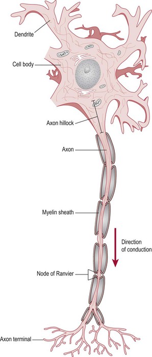
Figure 6.1 The basic structure of a myelinated neuron.
Cell body, which houses the nucleus and organelles
Dendrites, which receive signals from other neurons and carry these signals towards the cell body
Axon, which conducts signals away from the cell body and onto other neurons. The axons of some neurons are myelinated and as a result are able to conduct signals faster due to its insulation properties. Myelination occurs as a result of non-neural cells wrapping themselves around the nerve axon at intervals along its length, interspersed by gaps called the nodes of Ranvier. In the peripheral nervous system, it is Schwann cells which are responsible for this function and in the central nervous system, it is oligodendrites (S2.7).
Function of a neuron
Action potential
When the cell membrane is depolarized to a threshold level (typically −50 mV) at the axon hillock (Fig. 6.1), an ‘action potential’ is initiated. The action potential occurs as a consequence of the high concentration of voltage gated sodium and potassium ion channels at the axon hillock. When depolarized to threshold level, these channels open on mass and initiate conduction along the axon. An action potential once initiated does not fade and therefore the signal may travel over long distances.
Summation
Conduction along a neuron
Continuous conduction: This occurs in an unmyelinated axon and each ion channel must be opened in turn for conduction to continue. This results in a slow conduction of the signal.
Saltatory conduction: This occurs in a myelinated axon. In this case, the sodium and potassium channels are concentrated within the nodes of Ranvier (Fig. 6.1) and the signal is able to jump from one node to the next making conduction much quicker. Between the nodes of Ranvier, the current travels both inside the cell (insulated by the myelin) and outside the cell (in the extracellular fluid).
Synaptic transmission
Depolarization: This is termed ‘an excitatory post-synaptic potential’ or EPSP and occurs as a result of sodium (Na+) entering the cell. The membrane potential therefore becomes less negative and hence nearer to the threshold for initiating an action potential. These are often referred to as ‘excitatory synapses’.
Hyperpolarization: This is referred to as an ‘inhibitory post-synaptic potential’ or IPSP and occurs when either chloride ions (Cl−) enter or potassium (K+) ions leave the cell. In either case, the membrane potential becomes more negative and hence further from threshold potential. Therefore an action potential is less likely to occur. These are often referred to as ‘inhibitory synapses’.