Introduction and classification

Published on 03/04/2015 by admin

Filed under Hematology, Oncology and Palliative Medicine

Last modified 22/04/2025

Print this page

rate 1 star rate 2 star rate 3 star rate 4 star rate 5 star
Your rating: none, Average: 0 (0 votes)

This article have been viewed 1002 times

11

Introduction and classification

Definition

The term ‘anaemia’ refers to a reduction of haemoglobin or red cell concentration in the blood. With the widespread introduction of automated equipment into haematology laboratories the haemoglobin concentration has replaced the haematocrit (or ‘packed cell volume’) as the key measurement. Haemoglobin concentration can be determined accurately and reproducibly and is probably the laboratory value most closely correlated with the pathophysiological consequences of anaemia. Thus, anaemia is simply defined as a haemoglobin concentration below the accepted normal range.

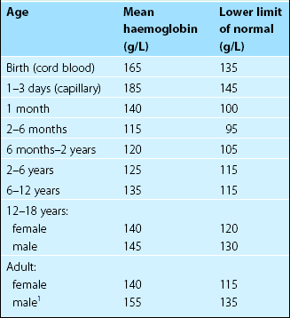
The normal range for haemoglobin concentration varies in men and women and in different age groups (Table 11.1). The definition of normality requires accurate haemoglobin estimation in a carefully selected reference population. Subjects with iron deficiency (up to 30% in some unselected populations) and pregnant women must be excluded or the lower level of normality will be misleadingly low. Normal haemoglobin ranges may vary between ethnic groups and between populations living at different altitudes.

General features

In anaemia the blood’s reduced oxygen-carrying capacity can lead to tissue hypoxia. The clinical manifestations of significant anaemia (see also p. 14) are to a large extent due to the compensatory mechanisms mobilised to counteract this hypoxia. Cardiac overactivity causes palpitations, tachycardia and heart murmurs. The dyspnoea of severe anaemia may be a sign of incipient cardiorespiratory failure. Pallor is due primarily to skin vasoconstriction with redistribution of blood flow to tissues with higher oxygen dependency such as the brain and myocardium.

Anaemia is one of the most common clinical problems presenting in general practice, in hospitals and in medical examinations. Usually characteristic symptoms and signs prompt a blood count to confirm the diagnosis but on occasion an unexpectedly low haemoglobin estimation in a ‘routine’ blood count precedes the clinical consultation. Whatever the sequence of events, anaemia is not in itself an adequate diagnosis; further enquiry to establish the underlying cause is essential.

A logical approach to anaemia demands a clear understanding of both its possible causes and its clinical and laboratory features. There are two major classifications – both have advantages and they are best used together.

Classification

Morphological classification

As already discussed (p. 18), modern electronic laboratory equipment can provide estimations of red cell indices in addition to haemoglobin concentration. Abnormal red cell indices should be confirmed by microscopic examination of blood films. The ‘morphological’ classification is based on a correlation between red cell indices and the underlying cause of anaemia. The most important measurements are of red cell size (mean cell volume or MCV) and red cell haemoglobin concentration (mean cell haemoglobin (MCH) or mean cell haemoglobin concentration (MCHC)).

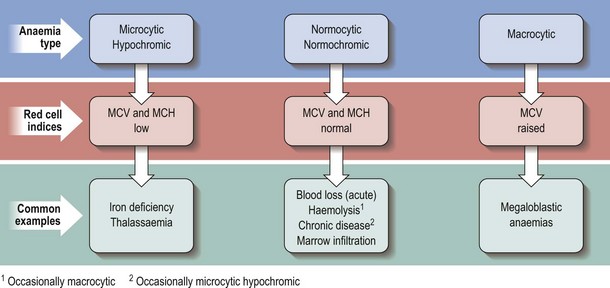
Anaemias with raised, normal and reduced red cell size (MCV) are termed macrocytic, normocytic and microcytic, respectively. Anaemias associated with a reduced haemoglobin concentration within red cells are termed hypochromic and those with a normal MCH are termed normochromic. Characteristic combinations are of microcytosis and hypochromia, and normocytosis and normochromia. As can be seen in Figure 11.1, this terminology is helpful in narrowing the differential diagnosis of anaemia. It is perhaps least helpful in normocytic anaemia as the possible causes are numerous and diverse.

The value of the blood film in diagnosis should not be underestimated. For instance, combined iron deficiency (a cause of microcytosis) and folate deficiency (a cause of macrocytosis) may cause an anaemia with a normal MCV. However, inspection of the film will reveal a dual population of microcytic hypochromic red cells and macrocytic red cells.

Aetiological classification

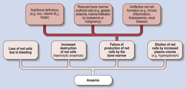
Figure 11.2 illustrates a classification of anaemia based on cause. It is less immediately helpful than the morphological classification in forming a differential diagnosis but it does illuminate the pathogenesis of anaemia. The fundamental division is between excessive loss or destruction of mature red cells, and inadequate production of red cells by the marrow.

Loss of red cells occurs in haemorrhage and excessive destruction in haemolysis. A normal bone marrow will respond by increasing red cell production with accelerated discharge of young red cells (reticulocytes) into the blood. Inadequate red cell production may result from insufficient erythropoiesis (i.e. a quantitative lack of red cell precursors) or ineffective erythropoiesis (i.e. defective erythrocytes destroyed in the marrow). Examples of insufficient erythropoiesis include bone marrow hypoplasia, as in aplastic anaemia, and infiltration of the marrow by a leukaemia or other malignancy. Inefficient erythropoiesis is seen in disorders such as megaloblastic anaemia, thalassaemia and myelodysplastic syndromes.

The above provides a useful framework for thinking about anaemia. In reality different mechanisms can operate simultaneously. The anaemia of thalassaemia is caused by both ineffective erythropoiesis and haemolysis.

Management

The treatment of specific types of anaemia is discussed in subsequent sections. However, some general statements can be made. Whenever possible, the cause of anaemia should be determined before treatment is instituted. Blood transfusion should only be used where the haemoglobin is dangerously low, where there is risk of a further dangerous fall in haemoglobin (e.g. rapid bleeding), or where no other effective treatment of anaemia is available. Prompt blood transfusion can be life-saving in a profoundly anaemic patient but it should be undertaken with great caution as heart failure can be exacerbated. Mild anaemia in the elderly should not be overlooked as it is a frequent cause of debility and has been linked with increased mortality.