Intraoperative 3-D Echocardiography
Gian Paparcuri
Transesophageal echocardiography is probably the most frequently used imaging technique in cardiac surgery. This powerful technology provides timely information, about cardiac structures (anatomy) and function (hemodynamics) without disrupting the surgical workflow.
 Enhances the illusion of depth perception.
 Offers a better appreciation of individual patient anatomy.
 Facilitates the understanding of complex cardiac pathology.


With 3-D echocardiography, people who aren’t echocardiographers can appreciate valve anatomy and physiology in three dimensions. Surgeons tend to very much appreciate 3-D images because the echo image looks exactly like what they see when the heart is open. Finally, for those of you taking the well-known echocardiography course in San Diego, expect to see a lot of 3-D. It seems like the speakers are not allowed to talk about echocardiography if they don’t complement their presentation by adding some 3-D images.


TEE Probes
Non-3-D echocardiography probes use a limited number (64–128) of piezoelectric elements (crystals) to scan tissue. Sequential (phased) activation of individual crystals generates an ultrasound beam that is steered back and forth over a 90° angle to sweep a flat, “pie-shaped” scanning plane or sector. 3-D echocardiographic probes have on their tips more than 2500 active elements, incorporated onto the probe, conforming a rectangular grid of 50 rows and 50 columns for a total of 2500 independent piezoelectric crystals, generating a matrix array.
 Conversion: raw data are placed into a Cartesian volume with each point assigned x-y-z coordinates and an echo intensity value. The product of this step is a group of points with distinctive echogenic characteristics and a known position in space, called voxels or volume of pixels, encrypting the physical characteristics and location of the smallest cube in the dataset, similar to pixel size in 2-D image resolution. Large voxels are generated when raw data are available for fewer points in the space and interpolation has to fill wider gaps. Still not a clear 3-D image.

Finally, step 4 or 3-D image display: makes 3-D dataset visible.
 1st step is segmentation: separates the object to be rendered from surrounding structures (differentiates between cardiac tissue, blood, pericardial fluid, blood, air; given their diverse physical properties and different ability to reflect US; this requires setting a threshold of echo intensity). The program excludes from further processing any point with echo intensity equal to or lower than blood, and delineates the 3-D surfaces of cardiac tissue.

Wireframe rendering: The simplest technique. Defines and connects equidistant points on the surface of a 3-D object with lines (wires) to create a mesh of small polygonal tiles. Smoothing algorithms refine the narrow angles making rudimental object appear more real. This technique is used for relatively flat surfaces such as the LV and the atrial cavities. It cannot display objects/structures with complex shapes, such as valves (requires greater anatomic detail for meaningful analysis). Processes small amount of data (fast and efficiently performed on basic computers).
Surface rendering: similar to wireframe but defines more points on the surface of a 3-D object making the lines joining them visible. Displays details of a 3-D surface and makes morphologic assessment of the corresponding anatomic structure feasible. Generates 3-D objects with rendered surfaces and a hollow core.
Volume rendering: displays 3-D objects with a rendered surface and details of its inner structure. Enables the potential display of every voxel of the 3-D object permitting a “virtual dissection”.
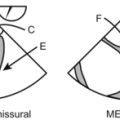
Mitral Valve
 Spatial manipulation, assessment in anatomical and surgical orientation for discussion with the surgeon.

The MV can be easily imaged using all the 3-D imaging modes described:


Stored MV 3-D images can be cropped on any plane to further delineate leaflet morphology.
The role of 3-D transesophageal echocardiography is expanding to become a powerful tool guiding surgical repair (treatment of choice for MR). In mitral stenosis, 3-D echo can consistently identify MV commissural fusion and predict the success of MV balloon valvuloplasty. Some authors have proposed planimetry by real-time 3-D echocardiography as a “gold standard” in the assessment of MS.
LV Assessment


LV Volume
 3-D-guided biplanes: by simultaneously displaying 2 perpendicular 2-D planes (ME 4C and ME 2C combination minimizes LV foreshortening) cutting the LV along its long axis at the true apex. Allows calculation (by applying modified Simpson biplane methods of disks to ES and ED frames) of LV volume, EF, mass. Limitations: still relies on geometric assumptions.

RV Assessment
RV 2-D assessment is difficult because of its crescent shape and complex geometry (a lot of assumptions). 3-D overcomes 2-D limitations allowing full-volume 3-D dataset acquisition for RV size, volume and function with good correlation with MRI and better reproducibility than 2-D.