Intestinal Surgery
Anatomy of the Small and Large Intestine
The intestines constitute the largest organ system within the abdominal cavity. Pelvic surgery always translates into some contact with the intestines. Although some individual variation may be noted, upon entering the peritoneal cavity of a person who has not previously been operated on, the greater omentum can be seen to cover the intestines (Fig. 93–1A). The omentum takes its origin from the greater curvature of the stomach and also attaches to the transverse colon (Fig. 93–1B). Beneath the omentum lies the small intestine (Fig. 93–2A). The small bowel measures 22½ feet in length, and for the most part is completely covered with peritoneum and is suspended by a wide mesentery (Fig. 93–2B). The latter extends from the upper left abdomen to the lower right portion of the posterior wall of the abdomen (Fig. 93–3). The small intestine is divided into three portions: (1) duodenum, which is uncommonly related to gynecologic surgery; (2) jejunum; and (3) ileum; all three are frequently encountered (Fig. 93–4). The duodenal–jejunal junction is secured by a fibromuscular band on the upper left side of the abdomen. This band is called the ligament of Treitz. This is a convenient initial landmark for systematic examination of the entire small intestine for a suspected injury (Fig. 93–5A through C). Another important anatomic reference point is the ileocecal junction, where a valve connects the small to the large intestine (Fig. 93–6A, B). The small intestine receives its blood supply from the superior mesenteric artery via its mesentery (Fig. 93–7). The vessel branches into a series of arches, which terminate in a small straight artery that surrounds the segment of intestinal wall. The nerve supply emanates from the superior mesenteric plexus of nerves, which is in direct continuity with the celiac plexus.
The large bowel measures approximately 5 feet in length and can be distinguished from the small intestine by the presence of appendices epiploicae (three longitudinal bands of muscle fibers) and by its larger diameter (Fig. 93–8A). The large intestine forms a three-sided framelike structure (see Figs. 93–2B, 93–8B). On the right side, the cecum frequently dips into the pelvis and importantly terminates in the vermiform appendix. The ascending colon at its hepatic flexure imperceptively joins the transverse colon. The latter is located just beneath (inferior to) the stomach and connects to the left, or descending, colon at the splenic flexure.
The left (descending) colon joins the S-shaped sigmoid portion in the left iliac fossa. The sigmoid colon enters the pelvis by passing anterior to the sacrum and crossing it from the left to right side of the pelvis (Fig. 93–9). The sigmoid colon then curves back upon itself to the midline, descending just posterior to the uterus, where it joins the straight terminal portion of the colon (i.e., the rectum) (Fig. 93–9). The sigmoid colon is completely surrounded by peritoneum and moves freely via its mesentery (sigmoid mesocolon). It is important to note that the sigmoid and the left colon are attached to the peritoneum along the lateral aspect of the abdominal wall. These attachments are not adhesions (Fig. 93–10A). The entire descending colon may be mobilized by cutting the peritoneum overlying the left gutter (Fig. 93–10B, C). At the lower pole of the aforesaid incision, the ovarian vessels and the psoas major muscle are encountered beneath the peritoneum (Fig. 93–10D). The inferior mesenteric artery supplies the left colon via three branches: the left colic, sigmoid, and superior hemorrhoidal arteries. This is an important area for collateral circulation between the middle and inferior hemorrhoidal branches of the hypogastric artery and the superior hemorrhoidal branch of the inferior mesenteric artery. The right side of the colon receives its blood supply from the ileocolic branch of the superior mesenteric artery (see Fig. 93–7).
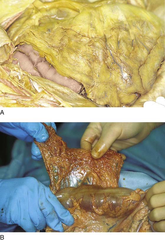
FIGURE 93–1 A. The abdomen has been opened and the peritoneal cavity entered. The greater omentum is draped over and, for the most part, covers the intestines. B. The stomach has been pulled out of the abdomen to demonstrate the omentum originating from the greater curvature and the transverse colon.
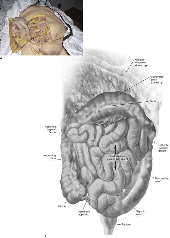
FIGURE 93–2 A. The underlying small intestine fills the abdomen and covers the pelvis. It comes into view when the omentum is retracted. B. The small and large intestines are demonstrated by retracting the omentum, which is attached to the transverse colon. Note how the large bowel frames the small intestine.
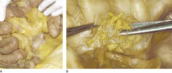
FIGURE 93–3 A. The small intestine is supported by a mesentery, which sweeps from the upper left to the lower right quadrant. B. The mesentery contains the blood supply of the small bowel. The blood vessels are buried in the fat, which is sandwiched between the two layers of peritoneum.

FIGURE 93–4 The small bowel is divided into three portions; however, the duodenum is rarely encountered during gynecologic surgery. As can be seen here, the jejunum and ileum are commonly within the operative field.
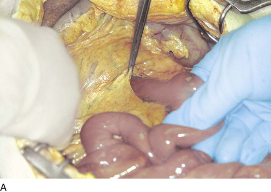
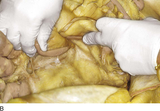
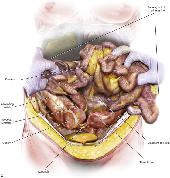
FIGURE 93–5 A. At the junction of the jejunum and duodenum is a fibromuscular ligament attached to the intestine. This is the ligament of Treitz. B. Close-up view of the ligament of Treitz. The surgeon’s finger points to the ligament. C. The small intestine is fanned out and anchored by its mesentery. The entire bowel is seen from the ileocecal junction to the ligament of Treitz.
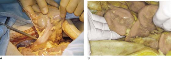
FIGURE 93–6 A. The surgeon is holding the cecum in his right hand and elevating the mobilized right colon. The ileum is tented up by a tonsil clamp. The ileocecal junction is clearly seen. B. The ileum joins the cecum at the ileocecal junction. The dissector’s hand is resting under the cecum. The other hand is pulling on the terminal ileum.
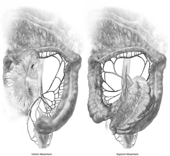
FIGURE 93–7 The small intestine is supplied by the superior mesenteric artery via a series of arcades. This major vessel also supplies the right colon and the right side of the transverse colon. The inferior mesenteric artery supplies the left transverse, left colon, sigmoid colon, and rectum.
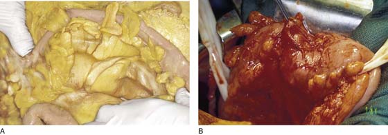
FIGURE 93–8 A. The transverse colon with its prominent appendices epiploicae and taeniae is shown here. B. The transverse colon is held by two rubber retractors. Note that the combined ascending, transverse, and descending colon frames the abdominal contents.
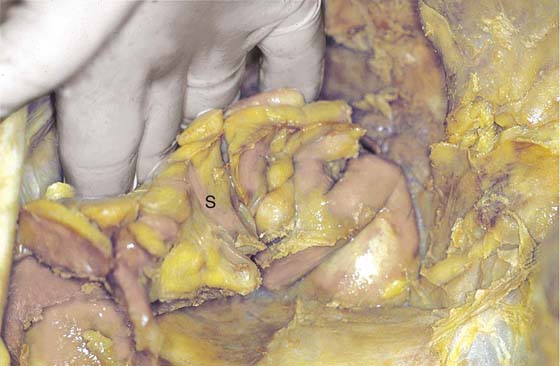
FIGURE 93–9 The sigmoid colon vectors from left to right over the sacrum, then turns upon itself back to the midline. The left colon is seen on the lower left side of the picture (l). The left colon joins the sigmoid (s), which veers to the right and descends into the pelvis.
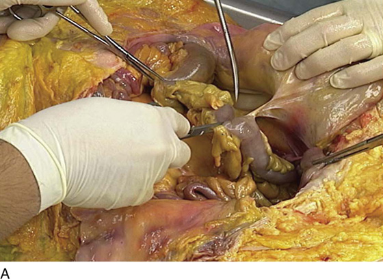
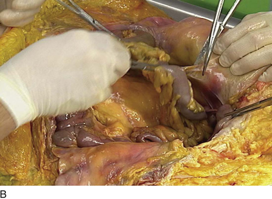
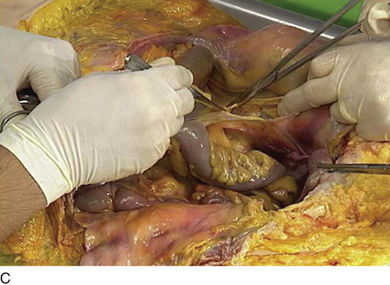
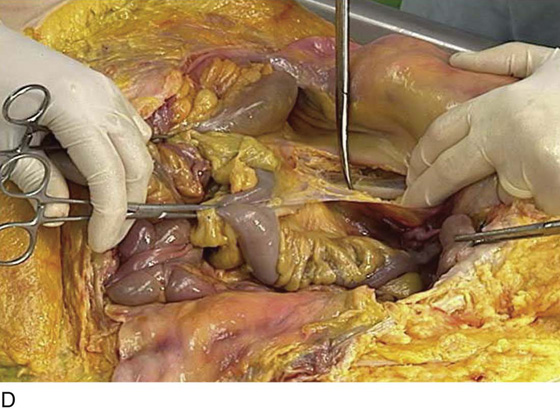
FIGURE 93–10 A. The left colon and the upper sigmoid colon attach to the lateral abdominal wall. These are normal peritoneal attachments. B. The peritoneum at the left colonic gutter may be opened. C. The avascular plane may be opened along the entire length of the left colon. D. The colon can be completely mobilized. Note the relationship of the sigmoid colon to the psoas major muscle, which is visible at the cut edge of the peritoneum.


