Limited to bowel ± mesenteric nodes


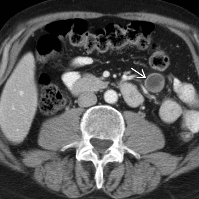
 in the mesentery. The metastases subsequently resulted in an intussusception.
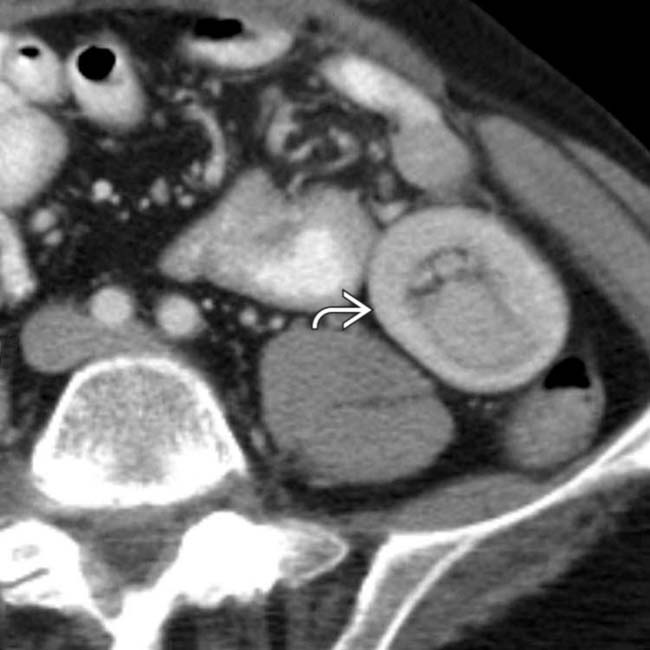
 . One of the bowel wall metastases served as the lead point of the intussusception.
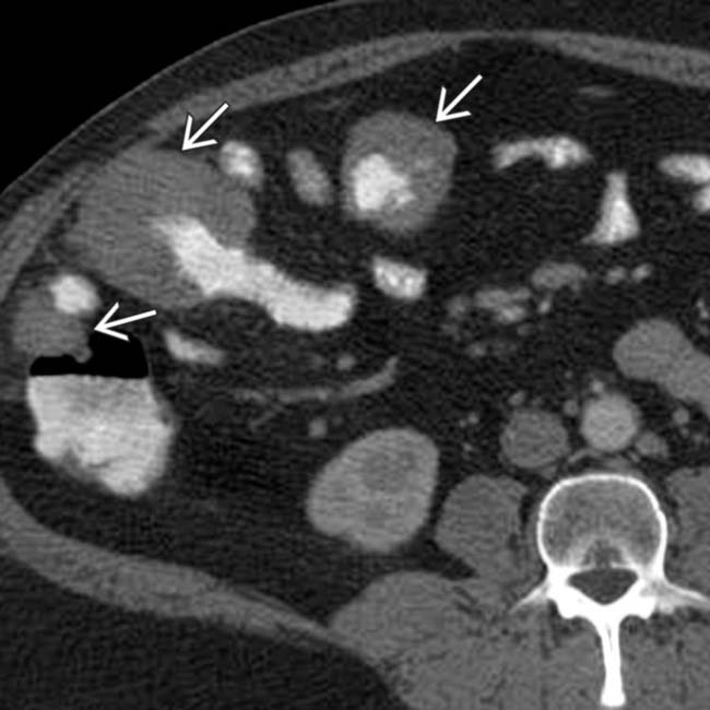
 .
.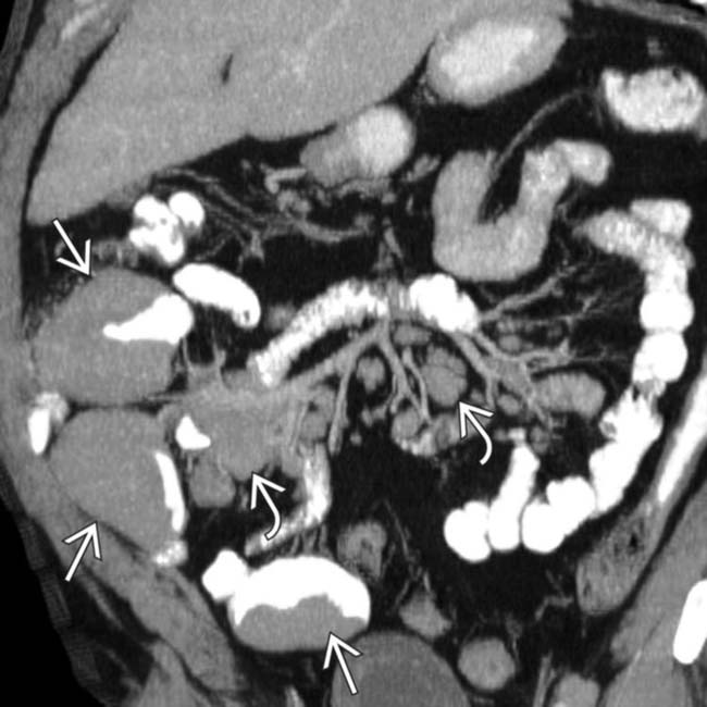
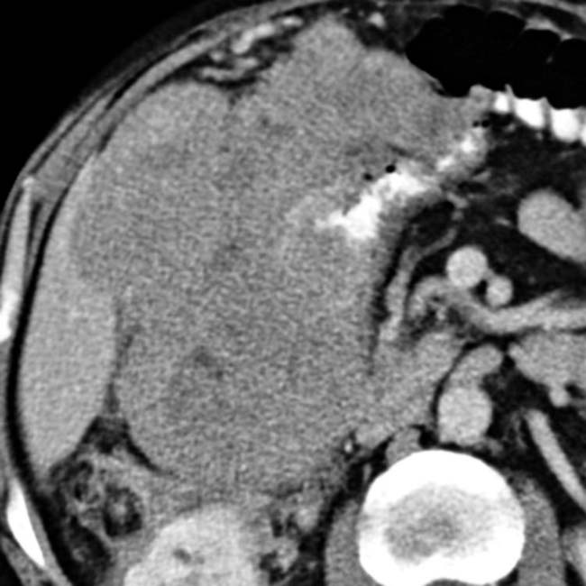
 and encasement of the mesenteric vessels, but no bowel or vascular obstruction. Multifocal masses of lymphoma
  are also seen.

IMAGING


Radiographic Findings
• Barium-enhanced fluoroscopic studies (upper GI, SB follow-through, barium enema)


CLINICAL ISSUES
Presentation


Natural History & Prognosis
• Intestinal metastases


 and lymph nodes
  . The gastric wall metastases may ulcerate, leading to the classic “target” or bull’s-eye appearance.
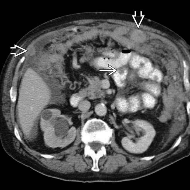
 , lymph nodes, and omentum
  , with both nodular and diffuse metastases seen. The right ureter was obstructed due to a ureteral metastasis.
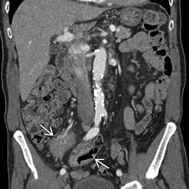
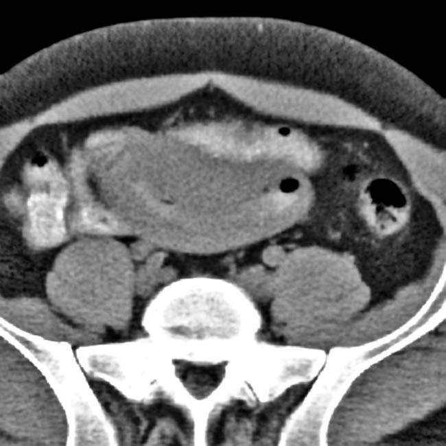
 enveloping, but not obstructing, small bowel and mesenteric vessels.
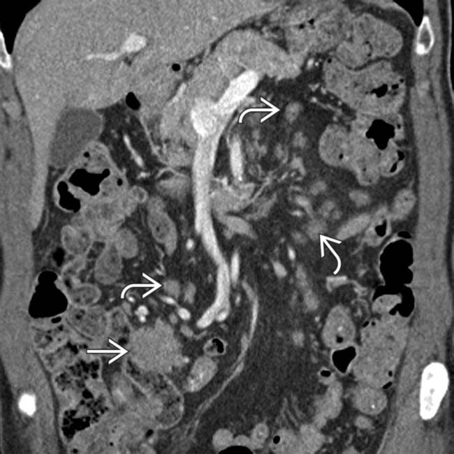
 along with extensive mesenteric lymphadenopathy
  . Non-Hodgkin lymphoma was confirmed.
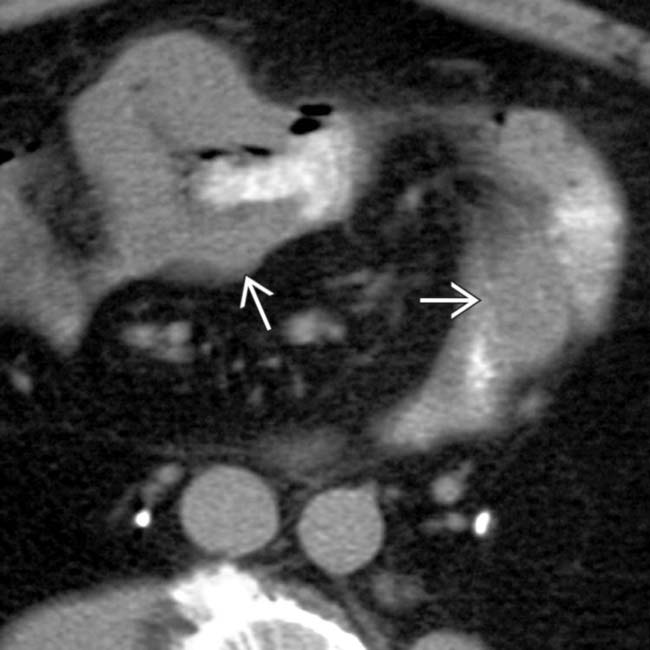
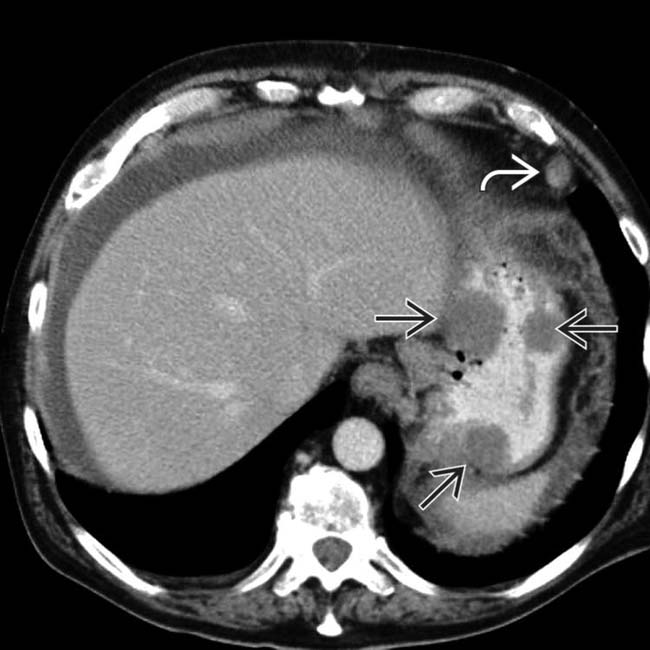
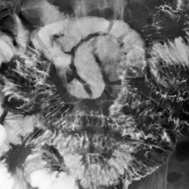
 with aneurysmal dilation of the small intestine lumen; however, no intestinal obstruction is seen.
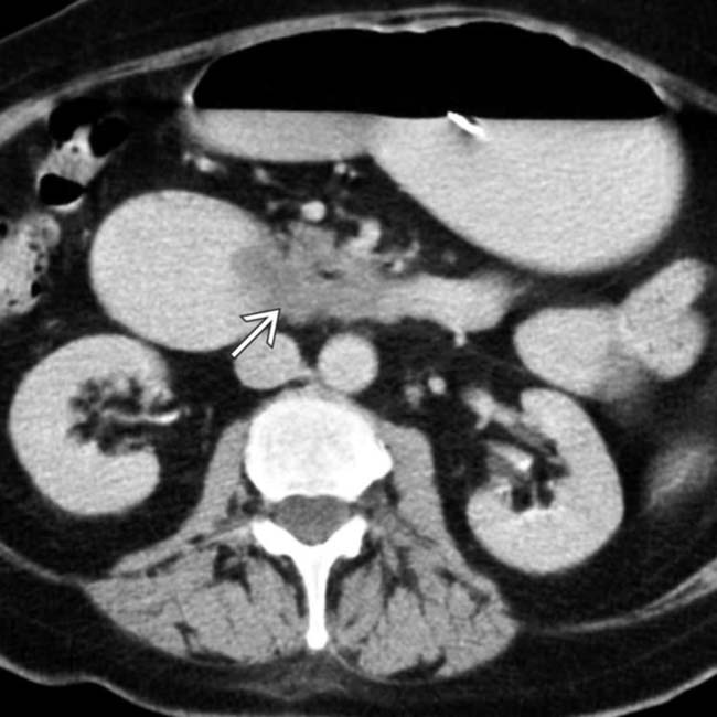
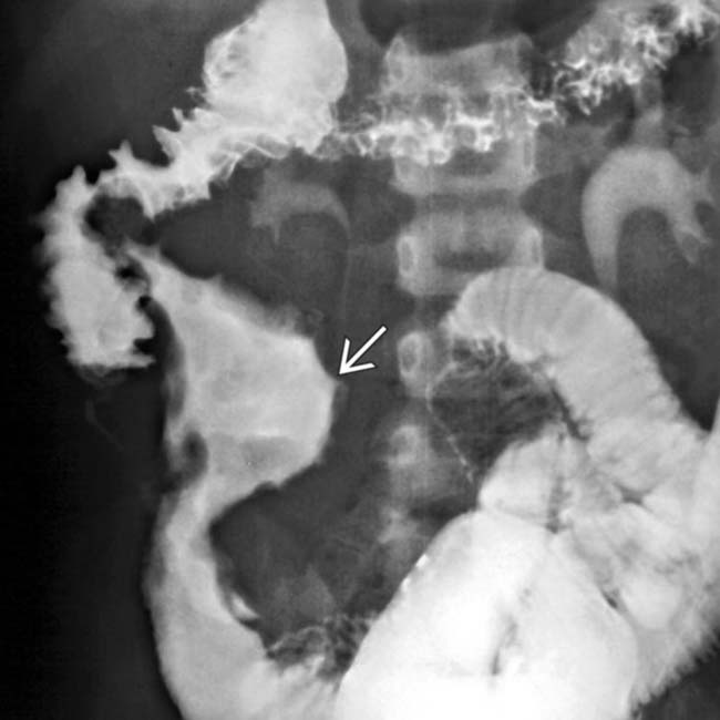
 . An intramural mass is constricting the lumen of the duodenum. At surgery, metastatic breast cancer was confirmed.
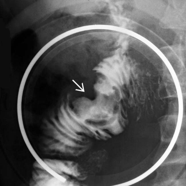
 with distorted mucosa due to melanoma.
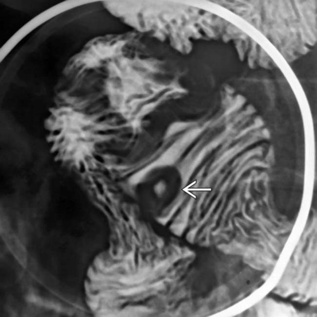
 due to metastatic melanoma.
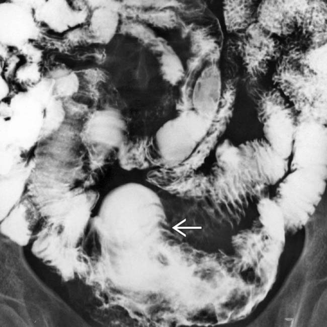
 and mucosal destruction of the distal SB segment in this patient with melanoma.
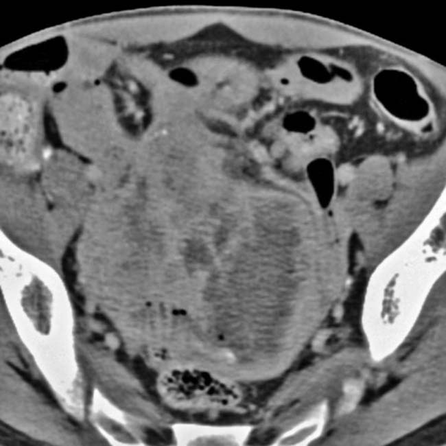
 of the lumen of the terminal ileum along with mesenteric mass effect in this patient with lymphoma.