Intestinal Metastases and Lymphoma

Published on 19/07/2015 by admin

Filed under Radiology

Last modified 22/04/2025

Print this page

rate 1 star rate 2 star rate 3 star rate 4 star rate 5 star
Your rating: none, Average: 0 (0 votes)

This article have been viewed 1933 times

 Limited to bowel ± mesenteric nodes
• Secondary lymphoma

image Involvement of spleen, liver, or thoracic nodes

IMAGING

• Intestinal metastases
• Malignant melanoma is most common primary site

image Enhancing masses within SB mesentery and bowel wall
image Bull’s-eye or “target” lesions; intussusception
• Lung and breast carcinoma metastases

image Are scirrhous tumors
image Likely to cause luminal obstruction
• Intraperitoneal metastatic spread (e.g., from ovarian and GI primary tumors)

image Serosal metastases cause clustered adhesion and fixation of SB loops and functional obstruction
• Direct invasion (e.g., from pancreatic or GYN tumor)

image Lumen of affected SB is often narrowed or obstructed
• Intestinal lymphoma
• Circumferential type: Sausage-shaped mass(es)

image Rarely obstructs; may cause aneurysmal dilation
• Polypoid form: Bull’s-eye or “target” lesions
• Mesenteric form: SB masses and nodes
• CT enterography is best protocol, with multiplanar reformation

TOP DIFFERENTIAL DIAGNOSES

• Primary small bowel carcinoma

image Solitary mass causing luminal obstruction
• Infectious and inflammatory etiologies 

image Mucosal hyperenhancement and submucosal edema

CLINICAL ISSUES

• Metastases: Most common with melanoma > lung, breast, others

image SB and mesentery are common sites of metastases from melanoma
image May arise many years after primary tumor removal
• Lymphoma accounts for 1/2 of all malignant SB tumors

image Patients with immune suppression (e.g., transplant recipients, AIDS); celiac disease
• Treatment

image Surgical resection of lesions that bleed, perforate, obstruct, or have aneurysmal dilation
image
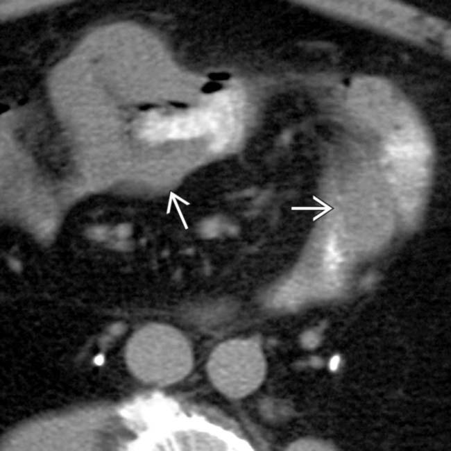
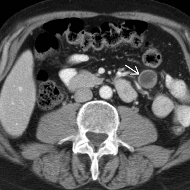
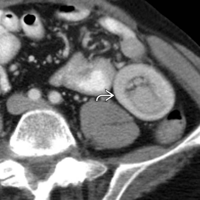
(Left) Axial CECT in a 58-year-old man who presented with a known history of malignant melanoma demonstrates 1 of several soft tissue masses image in the mesentery. The metastases subsequently resulted in an intussusception.
image
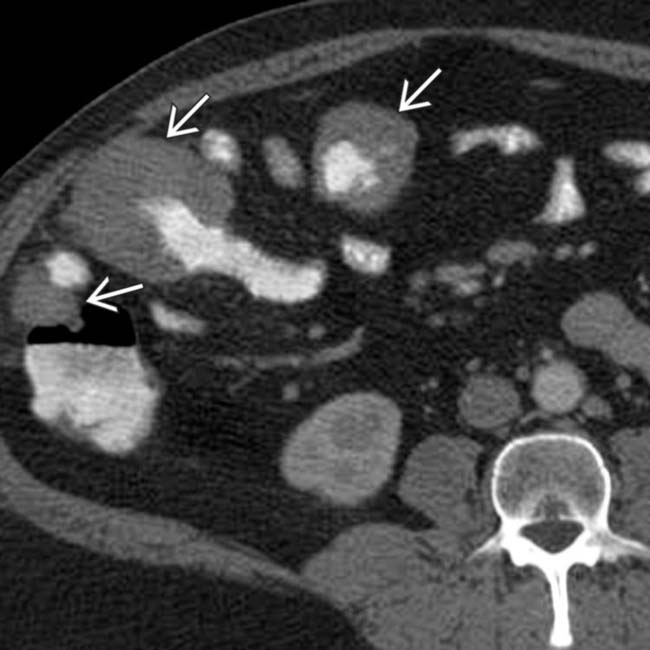
(Right) Axial CECT in the same patient 5 months later reveals the resultant long-segment intussusception image. One of the bowel wall metastases served as the lead point of the intussusception.
image
(Left) Axial CECT in a 46-year-old man who presented with a known history of non-Hodgkin lymphoma demonstrates extensive, multifocal, bowel wall thickening and aneurysmal dilatation of the lumen of the ileum image.

image
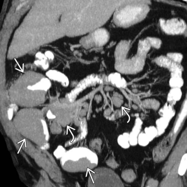
(Right) Coronal CECT reconstruction in the same patient illustrates extensive mesenteric lymphadenopathy image and encasement of the mesenteric vessels, but no bowel or vascular obstruction. Multifocal masses of lymphoma image are also seen.

TERMINOLOGY

Definitions

• Intestinal metastases from extraintestinal primary cancer
• Lymphoma: Malignant tumor of B lymphocytes

image Primary small bowel (SB) lymphoma: Limited to bowel ± mesenteric nodes
image Secondary or generalized lymphoma: Involvement of spleen, liver, or thoracic nodes

IMAGING

General Features

• Best diagnostic clue

image Bull’s-eye or “target” lesions
image Aneurysmal dilation of bowel lumen

Radiographic Findings

• Metastases to bowel
• Barium-enhanced fluoroscopic studies (upper GI, SB follow-through, barium enema)

image Most detailed study of SB is enteroclysis (tube administration of barium into SB with distention of lumen)
image Offer detailed view of mucosal and intramural extent of disease

– Less useful for extrinsic, extraluminal disease
• Malignant melanoma metastases to SB

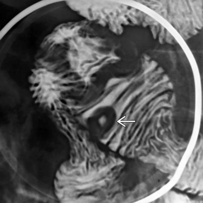
image Solitary or multiple discrete submucosal masses
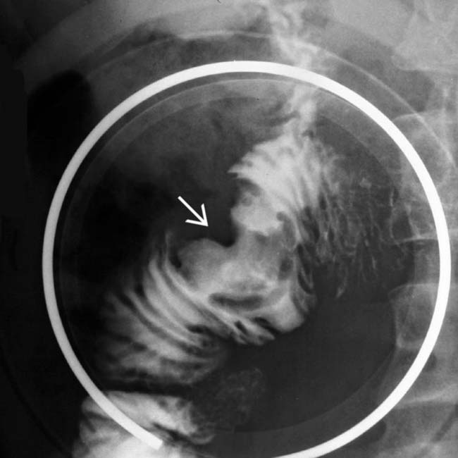
image Bull’s-eye or “target” lesions: Centrally ulcerated submucosal masses
image “Spoke-wheel” pattern: Radiating superficial fissures from central ulcer
image Giant cavitated mass (aneurysmal dilation)
image Small or large, lobulated masses

– Large collection of enteric contrast medium contiguous with lumen (melanoma, lymphoma)
• Lung and breast carcinoma metastases

image Are scirrhous tumors; likely to cause luminal obstruction
image Solitary/multiple, flat/polypoid intramural masses
• Intraperitoneal metastatic spread (e.g., from ovarian and GI primary tumors)

image Serosal metastases cause clustered adhesion and fixation of SB loops and functional obstruction
image Lack of peristalsis through affected segments
• Direct invasion (e.g., from pancreatic or GYN tumor)

image Spiculated mucosal folds, nodular mass effect, ulceration, obstruction, rarely fistula
image Lumen of affected SB is often narrowed or obstructed
• Intestinal lymphoma

image Multifocal intramural and mesenteric masses without SB obstruction
image Splenic and hepatic enlargement or focal masses
image Infiltrating lymphoma (most frequent)

– Circumferential thickening and effacement of folds
– Lumen may be compressed or dilated (aneurysmal dilation)

image Due to replacement of muscularis propria by lymphoma
image Lymphoma is not likely to cause high-grade bowel obstruction
image Polypoid lymphoma

– Single/multiple, mucosal/submucosal masses
– “Target” or Bull’s-eye lesions (if centrally ulcerated)
– Rarely lymphomatous polyposis (follicular mantle cell origin)
image Nodular lymphoma

– Multiple small submucosal nodular defects
image Endoexoenteric (cavitary form): Localized perforation into extraluminal tissue

– Barium, air, and debris fill cavity along mesenteric border of SB
– ± ulcer, fistulas, aneurysmal dilatation
image Mesenteric lymphoma

– Displaces and compresses SB loops and mesenteric vessels

CT Findings

• Demonstration of lesions is facilitated by distention of SB with water

image CT enterography is best protocol, with multiplanar reformation
• Intestinal metastases

image Malignant melanoma

– Bull’s-eye or “target” lesions (discrete mural nodules)

image Enhancing mural nodules protruding into lumen, focal thickening of intestinal wall
– Enhancing masses within SB mesentery, abdominal viscera, etc.
– Lobulated submucosal or giant cavitated lesions (aneurysmal dilation)
– Nonobstructive, large intramural mass favors melanoma or lymphoma over carcinoma
– May be lead point in intussusception
image Lung and breast carcinoma metastases

– Flat or polypoid intramural masses that compress or obstruct bowel lumen
– Mimic primary SB carcinoma
image Direct invasion

– Pancreatic head cancer: Mural thickening or circumferential narrowing of duodenal lumen
– Gynecologic cancer: Distal ileal involvement
image Intraperitoneal metastases

– Serosal metastases: Clustered SB loops adherent to each other
– Functional or mechanical SBO
– “Stellate” appearance: Mesenteric fat infiltration
– Ovarian carcinoma: Mesenteric/omental and mural intestinal masses + solid/cystic pelvic mass (± foci of calcification)
• Intestinal lymphoma

image Infiltrating form most common

– Circumferential type: Sausage-shaped mass of homogeneous density; minimal enhancement
– Aneurysmal dilation of SB lumen
image Polypoid form: Bull’s-eye or “target” lesions
image Mesenteric form

– Retroperitoneal and mesenteric adenopathy favors lymphoma as diagnosis
– Sandwich sign: Mildly enhancing, multiple discrete or confluent masses encasing mesenteric vessels

Imaging Recommendations

• Best imaging tool

image CT enterography with multiplanar reformations for total extent of disease
image Fluoroscopic-guided enteroclysis for mucosal and subtle intramural disease

DIFFERENTIAL DIAGNOSIS

Primary Small Bowel Carcinoma

• Solitary SB mass with SBO as presenting symptom

Hemorrhage

• Due to warfarin (Coumadin), trauma
• Localized bleeding may be seen as intramural mass

Vasculitis (Small Intestine)

• Henoch-Schönlein purpura

image Triad: Palpable purpura, arthritis, abdominal pain
• Systemic lupus erythematosus (SLE): Segmental bowel lesions → necrosis and perforation
• Vasculitis causes mucosal hyperenhancement and submucosal edema, not seen with neoplastic wall thickening

Crohn Disease

• Segmental mucosal hyperenhancement and submucosal edema
• Mesenteric adenopathy, fibrofatty proliferation
• Skip lesions, transmural, fistulas, fissures, aphthoid ulcerations, “cobblestoning,” “string” sign

Other Inflammatory (Whipple Disease)

• Thickened proximal SB folds, thickened mesentery, lymphadenopathy
• Micronodules in jejunum on enteroclysis

Opportunistic Infection

• Giardiasis (duodenum, jejunum)

image Thickened irregular folds with hypermotility, luminal narrowing, and increased secretions
image Ileum usually appears normal
• Mycobacterium avium-intracellulare (MAI)

image Thickened SB folds, fine nodularity
image Low-density (caseated) lymph nodes on CT
• Cytomegalovirus (CMV)

image Thickened folds, spiculation, ulcers, narrow lumen
image Often causes terminal ileitis and colitis in AIDS patients

PATHOLOGY

General Features

• Etiology

image Intestinal metastases

– Malignant melanoma more common than breast, lung, ovarian cancer
– Appendix, colon, pancreatic cancer; carcinoid are reported rarely
image Intestinal lymphoma

– Primary: Non-Hodgkin (B-cell) most common

image High grade of large cell or immunoblastic cell types; 30-50% harbor disease in mesenteric lymph nodes
– Secondary: Generalized lymphoma 

image Bowel is just 1 of multiple sites of involvement
– Enteropathy-associated lymphoma: Celiac disease
• Associated abnormalities

image Primary tumor mass in patients with intestinal metastases
image Generalized adenopathy, splenomegaly in patients with secondary lymphoma

Gross Pathologic & Surgical Features

• Solitary/multiple; polypoid, ulcerated, cavitated

Microscopic Features

• Metastases: Varies based on primary cancer
• Lymphoma: Lymphoepithelial lesions

CLINICAL ISSUES

Presentation

• Most common signs/symptoms

image Usually found while staging known primary carcinoma or lymphoma
image Pain, weight loss, palpable mass; may be asymptomatic
image Malabsorption, diarrhea
image Acute abdomen: Obstruction, perforation

Demographics

• Age

image More common in elderly patients
image Burkitt lymphoma in children involves ileocecal areas
• Ethnicity

image Mediterranean type of intestinal lymphoma: Arabs, Middle Eastern Jews
• Epidemiology

image Metastases are most common in patients with melanoma

– Lung, breast, and ovarian carcinoma are other common primary sites
image Intestinal lymphoma

– Most common malignant SB tumor, if primary and secondary forms are included

image Lymphoma accounts for 50% of all malignant SB tumors, but only 10-15% of primary SB malignancies
– More common in patients with immune suppression (e.g., transplant recipients, AIDS) or celiac disease
– SB is 2nd most frequent site of GI tract primary lymphoma

image Stomach (51%), SB (33%), colon (16%), esophagus (< 1%)

Natural History & Prognosis

• Intestinal metastases
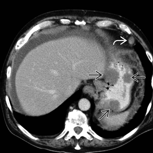
image Various forms of metastatic spread to intestine

– Hematogenous spread

image Melanoma, lung, breast cancer
image Reaches antimesenteric border of SB via small mesenteric arterial branches
image SB and mesentery are most common sites of metastases from melanoma after lung and liver
image Breast cancer: Metastasizes to stomach, duodenum, and colon more often than SB
image Breast, lung, and melanoma metastases may arise many years after primary tumor removal
– Lymphatic spread: Colon, ovarian, breast, and lung cancer, carcinoid and melanoma
– Direct invasion from contiguous neoplasms

image Pancreatic cancer: 2nd and 3rd parts of duodenum
image Cecal and gynecologic cancer: Distal ileum
– Intraperitoneal spread or seeding (carcinomatosis)

image Primary mucinous tumors of ovary, appendix, colon
image Due to natural flow and accumulation of ascitic fluid within peritoneal recesses; influences serosal implantation of cancer cells
image Common sites: Ileocecal region, SB mesentery, posterior pelvic cul-de-sac
• Complications: Bleeding, perforation, obstruction
• Prognosis: Poor

Treatment

• Surgical resection of lesions that bleed, perforate, obstruct, or have aneurysmal dilation

DIAGNOSTIC CHECKLIST

Consider

• Check for history of primary cancer or immune suppression

Image Interpretation Pearls

• Overlapping radiographic features of intestinal metastases, lymphoma, and primary carcinoma
• Imaging is important to suggest and stage malignancy

image
(Left) Axial CECT in a 69-year-old man who presented with a known history of metastatic melanoma demonstrates classic widespread metastases from melanoma, including the gastric wall image and lymph nodes image. The gastric wall metastases may ulcerate, leading to the classic “target” or bull’s-eye appearance.
image
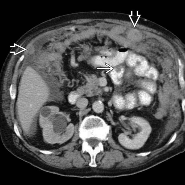
(Right) CT in the same patient shows widespread metastases, including the small bowel image, lymph nodes, and omentum image, with both nodular and diffuse metastases seen. The right ureter was obstructed due to a ureteral metastasis.
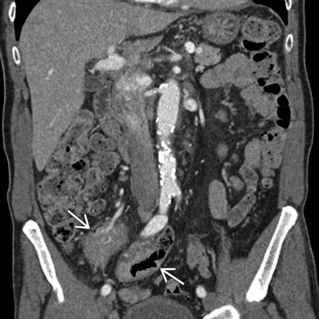
image
(Left) This elderly man presented with fever, night sweats, and weight loss. A coronal-reformatted CT shows soft tissue masses image enveloping, but not obstructing, small bowel and mesenteric vessels.
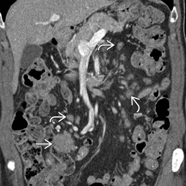
image
(Right) Another coronal section in this patient shows more of the dominant mass image along with extensive mesenteric lymphadenopathy image. Non-Hodgkin lymphoma was confirmed.
image
(Left) Axial CECT in a patient with melanoma, weight loss, and diarrhea shows large, ulcerated masses image with aneurysmal dilation of the small intestine lumen; however, no intestinal obstruction is seen.
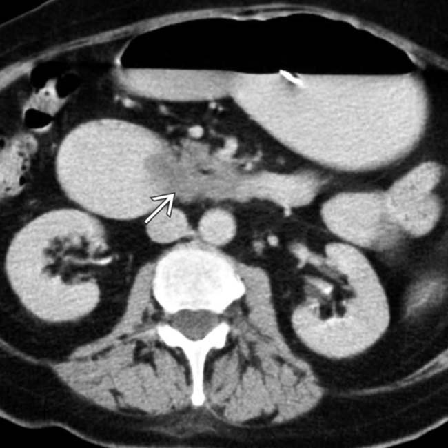
image
(Right) Axial CECT in a woman with a history of breast cancer shows dilation of the stomach and duodenum, with abrupt narrowing of the 3rd portion of the duodenum image. An intramural mass is constricting the lumen of the duodenum. At surgery, metastatic breast cancer was confirmed.
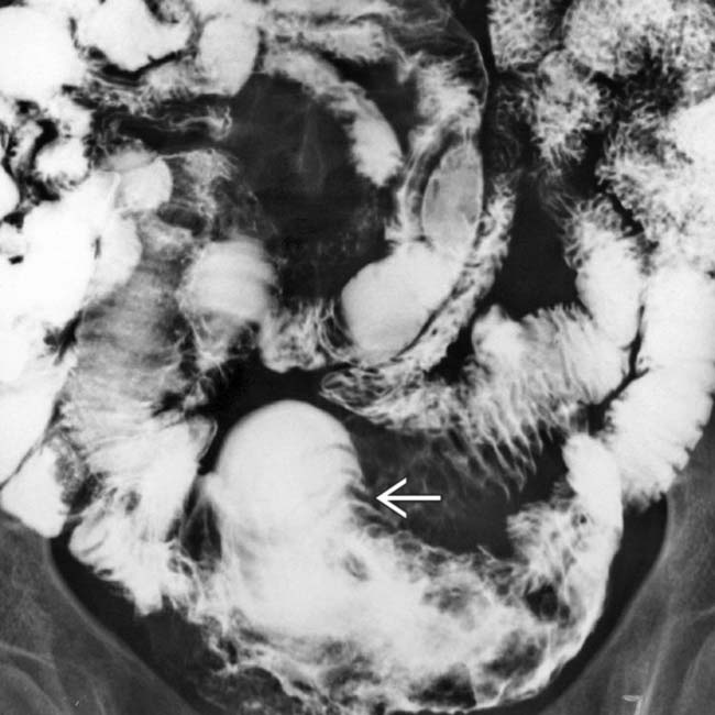
image
SBFT spot film shows an intramural mass image with distorted mucosa due to melanoma.

image
Fluoroscopic spot film from SBFT shows a bull’s-eye lesion image due to metastatic melanoma.
image
SBFT shows aneurysmal dilation image and mucosal destruction of the distal SB segment in this patient with melanoma.
image
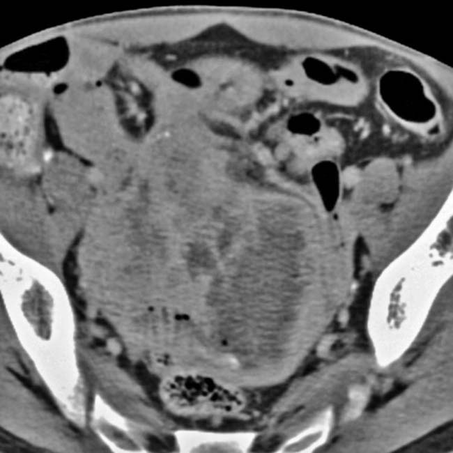
Axial CECT shows a partially necrotic mass that envelopes, but does not obstruct, multiple SB segments in this patient with melanoma.
image
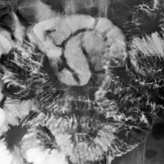
SBFT shows diffuse nodular fold thickening of most of the SB in this patient with lymphoma.
image
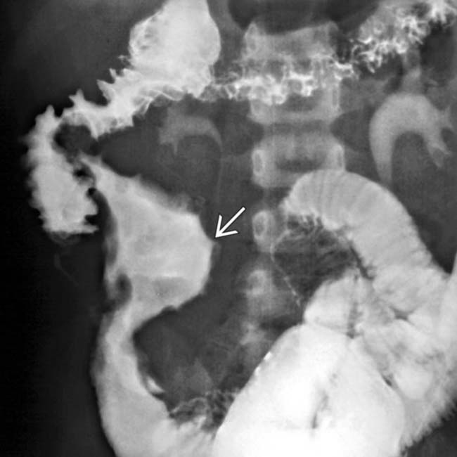
SBFT shows aneurysmal dilation image of the lumen of the terminal ileum along with mesenteric mass effect in this patient with lymphoma.
image
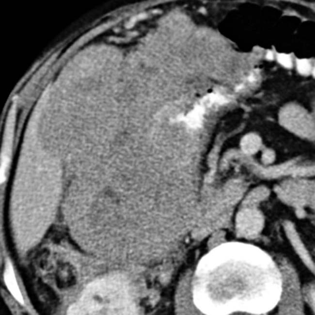
Axial CECT shows a soft tissue mass in the mesentery and wall of the colon due to lymphoma in this patient with post-transplant lymphoproliferative disorder.
image
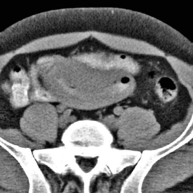
Axial CECT shows massive SB wall thickening of an ileal segment with soft tissue density due to lymphoma.

Share this: