Inflammatory bowel disease

Published on 09/04/2015 by admin

Filed under Gastroenterology and Hepatology

Last modified 22/04/2025

Print this page

rate 1 star rate 2 star rate 3 star rate 4 star rate 5 star
Your rating: none, Average: 0 (0 votes)

This article have been viewed 1121 times

Chapter 16 INFLAMMATORY BOWEL DISEASE
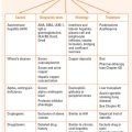
KEY POINTS

Ulcerative colitis:

Crohn’s disease:

ULCERATIVE COLITIS

The diagnosis of ulcerative colitis (UC) is relatively easy since the inflammatory process almost always involves the rectum and generally causes urgency and bleeding. People generally present promptly when rectal bleeding is noted, but the onset may be insidious and the presentation delayed. The bleeding is persistent rather than intermittent as would be expected with local anal problems, such as haemorrhoids. Proctitis may also cause discomfort, increased frequency and a change in the consistency of the stools. Rarely, the presenting symptom may be constipation. As the disease extends more proximally and more of the mucosal surface area is involved, the symptoms become more severe: diarrhoea increases (with looser or more frequent stools) and pain may become more prominent. Systemic symptoms may also develop.

In a significant minority of patients, the presentation is as a fulminant colitis with features including fevers, anorexia and weight loss; such patients are at risk of developing life-threatening complications including toxic megacolon, perforation, peritonitis and haemorrhage, and require urgent assessment and treatment in specialist centres. Given the large surface area of the colon, it is not surprising that ulcerative pancolitis results in blood loss and consequent anaemia, protein loss, hypoalbuminaemia and fluid and electrolyte losses, and the associated metabolic disturbances.

The extraintestinal manifestations of UC may affect the skin, joints, eyes and/or the liver (Table 16.2). These may occur before, during or after the onset of gut symptoms and may be associated with disease activity. These are generally more common in UC than Crohn’s disease.

TABLE 16.2 Extraintestinal manifestations of ulcerative colitis

Skin Erythema nodosum
Pyoderma gangrenosum
Aphthous stomatitis
Joints Type 1 peripheral osteopathy*
Type 2 arthritis**
Sacroileitis
Eyes Iritis
Uveitis
Episcleritis
Liver Primary sclerosing cholangitis

* Acute self-limiting inflammation affecting <5 joints, lasting <5 weeks and associated with symptom relapse and with other extraintestinal manifestations.

** Chronic arthritis affecting five or more joints with a median duration of symptoms of 3 years, and associated with uveitis but not erythema nodosum.

When assessing patients with suspected UC, particular attention should be paid to the haemodynamic status for evidence of significant intravascular losses and the abdominal examination for distension, tenderness and/or signs of peritonism. There may also be signs of anaemia or significant weight loss. Sigmoidoscopy will show an abnormal rectal mucosa: there will be erythema, loss of the normal vascular pattern, contact bleeding and mucopus or blood in the lumen. In more severe disease there will be extensive ulceration and pseudopolyp formation.

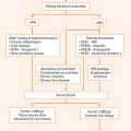
The differential diagnosis is considered in Table 16.3.

TABLE 16.3 Differential diagnosis of ulcerative colitis and Crohn’s disease

The diagnosis depends upon the exclusion of infective and other causes of colitis and the demonstration of the typical endoscopic and histological features of UC. Stool cultures and microscopy and studies for Clostridium difficile toxin should be sent. If there is a history of overseas travel, a warm stool smear should be performed to exclude amoebic dysentery. Patients suffering from UC may also have relapses secondary to gastrointestinal infections or non-steroidal antiinflammatory drug (NSAID) usage.

Ulcerative colitis can generally be differentiated from Crohn’s disease using endoscopic and pathological criteria (Table 16.1). UC affects the colon exclusively and almost always involves the rectum from which it extends proximally in continuity. Sigmoidoscopy will show evidence of proctitis although occasionally patients can have ‘rectal sparing’, particularly if there has been use of corticosteroid or 5-aminosalicylate enemas. Biopsies will show that the inflammation involves the more superficial layers of the mucosa, macrophages are less common and granulomata are not present. Sigmoidoscopic biopsies will help differentiate between UC and other causes of colitis, such as ischaemic colitis and radiation colitis.

Assessment and management of UC

Symptoms in patients suffering from UC characteristically remit and relapse. If gut or systemic symptoms do occur, investigations should be used to determine the activity of the disease, to determine how much of the bowel is involved and to decide whether or not complications have arisen.

The main parameters of disease activity in UC are the number of motions a day, the presence of blood and the associated systemic symptoms, including fevers, malaise and weight loss. Useful laboratory indices include haemoglobin, white cell count, ESR, C-reactive protein, electrolytes, creatinine and albumin. The C-reactive protein is generally a more sensitive assay for inflammation than the ESR. Sigmoidoscopy will demonstrate the changes of proctitis described above. Either endoscopy or radiology may be used to determine the extent of the disease; sigmoidoscopy and colonoscopy are regarded as more sensitive and are safe (as long as there is no evidence of toxic megacolon—see below). If the disease is restricted to the rectum or the sigmoid colon, the prognosis and clinical course is more favourable (although there is a subgroup of patients who are unresponsive to the standard treatment regimens). However 20% or more of these patients may develop disease extension over time but surgery is rarely necessary.

The aims of therapy are to induce remission in active disease and to maintain remission/prevent relapse. The severity of the disease and the site(s) of the affected colon are used to determine which agents may be used and their route of administration.

Proctitis and distal colitis

Patients with mild disease (less than four stools daily, no fever or weight loss) limited to the left side may respond to local therapy using 5-aminosalicylic acid and/or steroids as an enema, as a foam or as a suppository depending on the extent of the disease. There may be problems retaining the medication in the early stages of treatment because of urgency and diarrhoea. Side effects of topical steroids are minimal in the short term, but do occur if they are used long term. Local therapy is used once (or twice) a day until a response is achieved, and then decreased to every second or third day, generally at night before retiring. They are sometimes used in this fashion as maintenance therapy.

In moderate disease (4–8 bloody stools daily, low-grade fever, moderate abdominal pain) oral therapy is preferred. One agent is sulfasalazine, which consists of the active moiety, 5-aminosalicylic acid linked to a carrier molecule, sulfapyridine, which prevents it from being absorbed in the proximal gut. The azo bond joining the molecules is broken down by bacteria in the terminal ileum and colon. The majority of the 5-aminosalicylic acid remains in the colon and is excreted in the faeces. The sulfapyridine is absorbed and is responsible for the majority of the side effects. Since the side effects (especially nausea, anorexia and headache) are dose-related, sulfasalazine is generally introduced at a low dose and increased gradually over a week to 3–4 g/day. If these or other side-effects occur, there are alternative ways of administering the 5-aminosalicylic acid: olsalazine comprises two molecules of 5-aminosalicylic acid linked by an azo bond; various other formulations allow for the release of 5-aminosalicylic acid in the terminal ileum and colon by coating it in resins or encapsulating it in microspheres which result in pH-dependent release of the agent.

Disease proximal to the splenic flexure

If the disease extends proximally to the splenic flexure, oral therapy should be considered. If the disease is mild, the agent of choice is sulfasalazine or one of the other 5-aminosalicylates. A Cochrane meta-analysis showed that the newer 5-aminosalicylic acid preparations were superior to placebo and tended towards therapeutic benefit over sulfasalazine, which was not as well tolerated. However, considering their relative costs, a clinical advantage to using the newer 5-aminosalicylic acid preparations as first-line therapy in place of sulfasalazine appeared unlikely.

If there has been no response over 2–4 weeks, oral corticosteroids in doses of 30–60 mg/day may be commenced. Steroids are sometimes used as initial therapy because of a more rapid clinical response. After a response has been induced, the dosage can be reduced by 5 mg/week or 10 mg/fortnight. Once remission has been attained, steroids should be ceased.

Azathioprine or 6-mercaptopurine are often useful in steroid-unresponsive patients or those who require high doses of steroids to remain well. The dose of azathioprine is between 2 and 2.5 mg/kg body weight and 6-mercaptopurine 1–1.5 mg/kg. Adverse effects include bone marrow suppression, liver test abnormalities, pancreatitis and a clinical syndrome characterised by high fevers and/or myalgia. Blood counts should be performed weekly for the first month, then on a regular basis every 1–3 months. Thiopurine methyltransferase (TPMT) testing can be considered to identify patients with low activity of this enzyme; they are at higher risk of bone marrow suppression and liver toxicity from azathioprine. The dose should be reduced if patients are on allopurinol therapy. If no response is seen within 3–6 months, azathioprine therapy should be withdrawn. Other approaches, including methotrexate, should be reserved for patients who are refractory to standard regimens.

The tumour necrosis factor (TNF) neutralising antibody, infliximab, has been also shown to be effective in inducing and maintaining remission in active ulcerative colitis, including fulminant colitis (see below). The major adverse events are infusion reactions and increased susceptibility to serious and/ or opportunistic infections (particular care is recommended to exclude latent infection with tuberculosis). There is concern about an increased risk of lymphoma and cancer (especially lung cancer in smokers). Patients may develop antibodies to double-stranded DNA but clinical features of systemic lupus erythematosis, especially end-organ damage, are very rare.

Acute severe or fulminant colitis and toxic megacolon

About 10%–15% of patients will develop acute severe or fulminant colitis, which is characterised by the presence of:

Initial therapy is with bed rest, parenteral fluids, high-dose intravenous steroids (hydrocortisone 300–400 mg day or equivalent) and close monitoring (note: there is a significant risk of hypokalaemia if hydrocortisone is used). There is an increased risk of thromboembolic disease and appropriate prophylaxis should be considered for hospitalised patients. If there is no clear improvement within 3–7 days consideration should be given to surgery.

If fulminant colitis is present, there is a 10%–20% chance that there will be progression to toxic megacolon. This, in turn, carries a significant mortality. The main risk is from perforation, so patients should be monitored closely for evidence of deterioration as manifest by worsening signs of toxicity or increasing colonic dilatation on progress abdominal X-rays (diameter >6 cm in the transverse colon and/or >9 cm in caecum). Patients who have evidence of acute fulminant colitis should be managed in centres where there is expertise in the combined medical and surgical management of this problem. Cyclosporine or infliximab may avert the need for surgery in the short term and possibly the long term in such patients.

CROHN’S DISEASE

Crohn’s disease is a chronic, often debilitating disease that affects any part of the gastrointestinal tract. Patients present in many ways, but diarrhoea, abdominal pain and weight loss are common symptoms. The condition is rarely fatal, but causes significant morbidity, particularly in young adults; the peak age of onset is in the second and third decades.

The diagnosis of Crohn’s disease is often very hard to make:the symptoms may be non-specific and the disorder is rare, although it may be increasing in incidence. This difficulty in diagnosis is reflected in frequent delays—the mean time from onset of symptoms to diagnosis is in the order of three years in some studies.

When the left side of the colon is involved, the symptoms are similar to those described above for ulcerative colitis. When the right colon or terminal ileum is involved, there may be less disturbance of bowel function while pain may be more prominent. Pain may be ‘inflammatory’ due to ulceration or inflammation extending through the intestinal wall, or ‘obstructive’ from strictures (generally small intestinal colic). The strictures themselves may be either inflammatory when the disease history is of short duration or fibrotic if there has been long-standing Crohn’s disease. Occasionally the presentation mimics acute appendicitis and the diagnosis of Crohn’s disease is made at surgery.

The diarrhoea is often multifactorial, but generally reflects gut inflammation. It may be secondary to the many complications of Crohn’s disease including fistula formation, bacterial overgrowth, bile salt malabsorption or fat malabsorption. A number of medications used for the treatment of Crohn’s disease can also cause diarrhoea. Weight loss is also multifactorial: any of the factors listed above may cause significant loss of weight.

Systemic symptoms are often more prominent in Crohn’s disease; patients often learn to recognise the fevers, anorexia, lethargy and malaise which herald a period of active Crohn’s disease. A patient’s ‘well-being’ may be used as a subjective measure of disease activity.

Another common association of Crohn’s disease is the presence of perianal complications including anal fissures, abscesses and fistulae. The inflammation and ulceration in Crohn’s disease is transmural and fistulae can occur between any of the viscera and cause symptoms including pneumaturia, faecal staining of urine or vaginal secretions, recurrent urinary tract infections, bleeding and dyspareunia.

Examination may demonstrate evidence of weight loss, localised tenderness or a mass in the right iliac fossa and perianal abnormalities, including anal induration.

The differential diagnosis of Crohn’s disease is similar to that of UC, but includes mycobacterial and Yersinia infections and disorders of the appendix.

Assessment

Symptoms in patients suffering from IBD characteristically remit and relapse. If gut or systemic symptoms do occur, investigations should be used to determine the activity of the disease (which is not always easy), to determine how much of the bowel is involved (the extent of the disease will determine what treatment will be offered) and to decide whether or not complications have arisen.

Activity indices are available, but are complicated and are generally only used in clinical trials. Parameters include the number of bowel actions per day, the presence of abdominal pain, the sense of well-being, the presence of complications (e.g. fistulae, rashes, ocular manifestations, arthritis), the requirement of opiates to control diarrhoea, the haematocrit, the presence of an abdominal mass, and the degree of weight loss. An elevated ESR or C-reactive protein may also be an indicator of active disease.

Antibody testing remains of uncertain value, but patients who are antineutrophil cytoplasmic antibody (pANCA) negative but anti-Saccharomyces cerevisiae antibody (ASCA) positive or anti-OmpC antibody positive may be more likely to have Crohn’s disease.

Endoscopic investigations (colonoscopy and terminal ileoscopy) and radiology (small bowel series and barium enema) are the main modalities used to determine the activity and extent of disease. Unlike patients with UC, sigmoidoscopy is not always helpful in determining the presence of recurrent disease. Abdominal ultrasound and CT scan may help determine whether or not there is active disease (thickening of the bowel wall, absent peristalsis, increased vascularity) and identify complications such as abscess or fistula formation. MRI may have a role in defining the extent of perianal disease and its response to therapy. The role of videocapsule endoscopy in the diagnosis of Crohn’s disease and in the monitoring of its activity is still being defined. White cell scans use radiolabelled autologous leucocytes to demonstrate sites of active inflammation and may help to define the nature of strictures seen in a small bowel series (either inflammatory or fibrotic).

Management

Current therapeutic modalities include lifestyle modification, drug therapy, nutritional support and surgery.

There is evidence that relapse rates are lower in patients with Crohn’s disease who are successful in giving up smoking compared to patients who continue to smoke. Patients should be counselled accordingly.

Drugs may be used to induce or maintain remission or both.

Corticosteroids (e.g. prednisone 40–60 mg/day) are the cornerstone of remission induction therapy; they may be successful in improving symptoms in 60%–70% of patients but have no effect on the endoscopic features or pathology of the disease. In contrast to UC where the dosage reduction is relatively rapid, steroids are withdrawn stepwise over a longer period of time—3 or 4 months or more. If symptoms recur on steroid reduction, the dosage may be increased to a level at which symptoms are controlled and then reduced at a slower rate. However, steroids should not be used as maintenance therapy because of the significant side effects, which include diabetes, hypertension, osteoporosis, increased susceptibility to infection, changes in mentation including insomnia, hypomania and psychosis, and many others.

There is an increased risk of thromboembolic disease and appropriate prophylaxis should be considered for hospitalised patients.

Azathioprine (2–2.5 mg/kg body weight) or 6-mercaptopurine (1–1.5 mg/kg) are useful for both inducing remission and as maintenance agents. They are also of benefit for the treatment of fistulae and perianal disease. As experience with these agents grows, they are becoming more widely used; there is good evidence that they are safe for use in pregnancy and in children. Particular care should be taken if there is evidence of sepsis (abdominal mass, ischiorectal abscesses, etc). Side effects include bone marrow suppression, liver test abnormalities, pancreatitis, myalgias, fevers and nausea. They are well tolerated in about 80%–90% of patients treated.

A Cochrane review (1998) provided the following data for the use of azathioprine and 6-MP in inducing remission in CD: the odds ratio of a response to azathioprine or 6-MP therapy compared with placebo in active Crohn’s disease was 2.36 (95% CI: 1.57 to 3.53). This corresponded to a NNT (number needed to treat) of about 5 to observe an effect of therapy in one patient. Treatment ≥17 weeks increased the odds ratio of a response to 2.51 (95% CI: 1.63 to 3.88), suggesting that there is a minimum length of time for a trial of therapy. A steroid sparing effect was seen with an odds ratio of 3.86 (95% CI: 2.14 to 6.96), corresponding to a NNT of about 3. Adverse events requiring withdrawal from a trial, principally allergy, leucopenia, pancreatitis and nausea, were increased on therapy with an odds ratio of 3.01 (95% CI: 1.30 to 6.96). The NNH to observe one adverse event in one patient treated with azathioprine or 6-mercaptopurine was 14.

Immunosuppressive agents are also effective as maintenance therapy, and, if tolerated, are generally used for years. After at least 4 years of continuous therapy, there is a 20% risk of relapse if treatment is ceased. Recommencement of therapy is generally effective in this context.

Sulfasalazine or the other oral aminosalicylates are sometimes used, particularly for mild colonic Crohn’s disease, but are not much better than placebo. The dosage regimens are as described above for UC. It has a limited role in maintaining remission: meta-analyses indicate that the number needed to treat is in the order of 16.

Antibiotics such as metronidazole and ciprofloxacin may also have a role in the treatment of perianal disease or in the postoperative setting. They are not generally used for long-term maintenance therapy.

A number of new ‘biologicals’ have been developed. The first was infliximab, a monoclonal antibody directed against tumour necrosis factor alfa (TNFα), an important cytokine involved in inflammation. About 60%–80% of patients will have an initial response to infliximab and more than half of these will benefit from regular infusions every 8 weeks over a period of a year or longer. Other anti-TNF agents that are effective for the treatment of Crohn’s disease are adalimumab and certolizumab. Other biologicals using different strategies for the control of inflammation in Crohn’s disease are in various stages of development.

Surgery is not curative, but is reserved for the treatment of complications or when medical therapy is ineffective. In historical studies, up to 70% of patients will require surgery at some stage; of these, 45%–50% will require more than one operation. It seems likely that the newer treatments and the use of effective maintenance therapy will reduce these figures in time.

Crohn’s colitis is probably associated with an increased risk of colon cancer when longstanding, and colonoscopy surveillance is recommended (Chapter 18).

The agents that are generally regarded as being safe for use in pregnancy are the 5-aminosalicylates, corticosteroids, azathioprine and possibly infliximab. Metronidazole and methotrexate are teratogens.

Recent epidemiological studies suggest that mortality rates for IBD are similar to that of the general population for the majority of patients. However, older patients with IBD and newly diagnosed cases with severe disease are at increased risk of dying.

SUMMARY

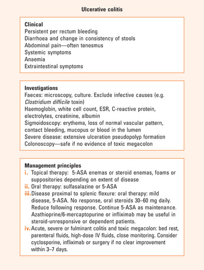
UC almost always involves the rectum and causes urgency and bleeding. It has a remitting and relapsing course. Investigations focus on the activity of the disease, the distribution of the disease and to decide whether complications are present. The main parameters of disease activity are the number of motions per day, the presence of blood and associated constitutional symptoms (Figure 16.1). Extraintestinal manifestations affect the skin, joints, eyes and/or the liver. Endoscopy or radiology may be used to determine the extent of the disease. The aims of therapy are to induce remission in active disease and to maintain remission/ prevent relapse. The mainstay of therapy are the corticosteroids and 5-aminosalicylates. These agents may be administered either orally or as enemas, foams or suppositories. In acute, severe or fulminant colitis, parenteral fluids, high dose intravenous steroids and close monitoring is essential. Consider cyclosporine/infliximab or surgery if there is no clear improvement within 3–7 days.

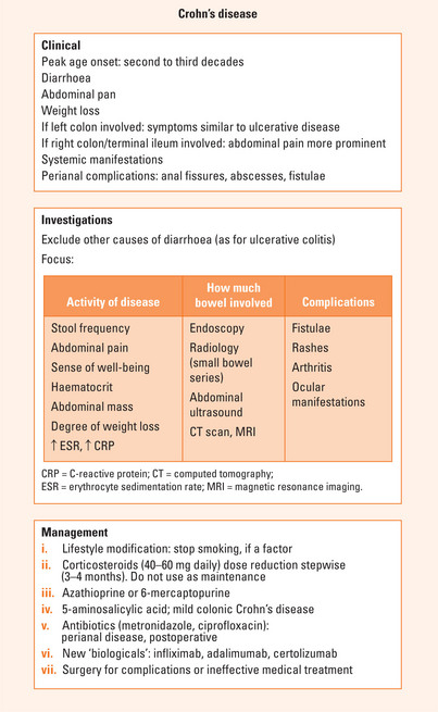
Crohn’s disease is a chronic, debilitating disease of multifactorial aetiology that affects any part of the gastrointestinal tract. There is a genetic predisposition (NOD2/CARD15) in 30%–50% of patients and other potential susceptibility genes are being assessed. Diarrhoea, abdominal pain and weight loss are common symptoms (Figure 16.2). Left-sided CD is similar in presentation to UC. Pain is a prominent symptom in right-sided CD. Systemic symptoms are often more prominent in CD than in UC. Perianal complications are prominent in CD. Activity indices are available but are generally only used in the research situation. A patient’s well-being may be used as a subjective measure of disease activity.