Infertility

Published on 09/03/2015 by admin

Filed under Obstetrics & Gynecology

Last modified 22/04/2025

Print this page

rate 1 star rate 2 star rate 3 star rate 4 star rate 5 star
Your rating: none, Average: 0 (0 votes)

This article have been viewed 1500 times

Infertility

William Ledger

Estimates of the prevalence of infertility in different parts of the world give remarkably similar results, with a 12-month prevalence rate ranging from 3.5–16.7% in more developed nations and from 6.9–9.3% in less-developed nations, with an estimated overall median prevalence of 9%. Only half of all infertile couples seek medical help, with the proportion being similar in developed and less-developed nations. Based on these estimates and on the current world population, 72.4 million women are currently infertile and of these, 40.5 million are currently seeking infertility medical care. Although firm evidence is hard to find, it seems that the prevalence of infertility in the Western world is increasing due to a number of factors, including the increase in number of young people with sexually transmitted diseases, increase in obesity and increasing numbers of women deferring plans for childbearing until later in life.

Primary infertility is defined as infertility without a previous pregnancy or live birth and secondary infertility as failure to conceive after one or more pregnancies, whether successful or ending in miscarriage, ectopic pregnancy or voluntary termination. Improved methods for investigation of infertility frequently reveal a problem in both partners, leading to the concept of relative subfertility. A highly fertile female partner will often compensate for a male with poor sperm quality and conceive without difficulty, and vice versa.

If conception does not occur after 12 months of regular sexual intercourse then the couple should be considered to be potentially infertile, as 80% of couples normally conceive within 1 year. It is therefore reasonable to proceed with investigations at this time. However this definition should be tempered by common sense. For example, a woman who has lost both Fallopian tubes because of ectopic pregnancies or a man who is known to have had testicular torsion in his youth should not be denied early investigation and treatment.

Both partners should be seen and investigated together, as infertility may result from male or female factors and is often associated with a combination of both. At the completion of all investigations, about one quarter will be given a diagnosis of ‘unexplained infertility’. Long-term follow-up studies of couples with unexplained infertility have shown that 30–40% will conceive over a 7-year period after investigation. Many ‘unexplained’ cases involve women over age 35 years who may later be shown to have a poor response to ovarian stimulation and oocyte abnormalities if in vitro fertilization (IVF) is performed. Age, particularly female age, undoubtedly affects fertility. IVF success rates fall sharply for women age 35 or older (Table 17.1), and natural fertility appears to decline slowly but irrevocably from the late 20s onwards. The effect of age on the male is less pronounced, but older men exhibit more sperm abnormalities and DNA fragmentation.

Table 17.1

Female age and IVF outcomes in the UK (2007 and 2008)

  Under 35 35–37 38–39 40–42 43–44 Over 44
2007 32.3% 27.7% 19.2% 11.9% 3.4% 3.1%
2008 33.1% 27.2% 19.3% 12.5% 4.9% 2.5%

image

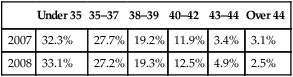
(Data sourced from www.HFEA.gov.uk.)

The relative incidence of causative factors will vary according to country and whether the problem is primary or secondary. Furthermore, in many couples there are multiple reasons for the infertility. Table 17.2 shows the pattern of causative factors of primary infertility in a Western population.

Table 17.2

Causes of infertility

Diagnosis* Primary infertile group (n = 167) N (%) Secondary infertile group (n = 151) N (%) P-value
Ovulation problems 54 (32.3) 35 (23.2) 0.069
Sperm quality problems 49 (29.3) 36 (23.8) 0.268
Blocked Fallopian tubes 20 (12) 21 (13.9) 0.607
Unexplained infertility 49 (29.3) 45 (29.8) 0.928
Endometriosis 19 (10.7) 15 (10) 0.677
Others 23 (13.8) 32 (21.2) 0.081

image

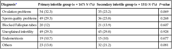
*Women have reported more than one diagnosis.

Data derived from a Scottish general practice-based survey (Bhattacharya et al. 2009). Self-reported cause of infertility amongst women who reported a diagnosis (North East Scotland). The data reflect unsuccessful attempted conception for 12 months or longer and/or had sought medical help with conception.

History and examination

The initial consultation should involve both partners. Many clinics use a pro forma questionnaire to elicit basic information, allowing better use to be made of the time available in the consultation. Basic investigations, including baseline blood tests for both partners, and semen analysis, can be organized through the General Practice with results available at the initial meeting.

The history should include the following:

• Age, occupation and educational background of both partners.

• Number of years that conception has been attempted and the previous history of contraception.

• Previous conceptions of either partner in this or previous relationships.

• Details of any complications associated with previous pregnancies, deliveries and postpartum.

• Full gynaecological history including regularity, frequency and nature of menses, cervical smears, intermenstrual bleeding and vaginal discharge.

• Coital history, including frequency of intercourse, dyspareunia, post-coital bleeding, erectile or ejaculatory dysfunction

• History of sexually transmitted diseases and their treatment.

• A general medical history to include concurrent or previous serious illness or surgery, particularly in relation to appendicitis in the female or herniorrhaphy in the male; a history of undescended testes or of orchidopexy.

Examination of both partners should be considered, although examination of the male is unlikely to reveal anything of significance in the presence of a normal semen analysis, and of the woman may well be equally unremarkable if there is a normal high quality pelvic ultrasound. Azoospermic men should be examined for congenital bilateral absence of the vas deferens (CBAVD) which is associated with cystic fibrosis mutations.

Female infertility

General factors such as age, serious systemic illness, inadequate nutrition, excessive exercise and emotional stress may all contribute to female infertility. The majority of cases of female infertility follow from disorders of tubal or uterine anatomy or function, or ovarian dysfunction leading to anovulation. Less frequently observed disorders include cervical mucus ‘hostility’, endometriosis and dyspareunia.

Disorders of ovulation

Disorders of ovulation are divided into four categories, defined by the World Health Organization (WHO):

• Type I – hypogonadal hypogonadism resulting from failure of pulsatile gonadotrophin secretion from the pituitary. This relatively rare condition can be congenital (as in Kallman’s syndrome) or acquired, for example, after surgery or radiotherapy for a pituitary tumour. Serum concentrations of luteinizng hormone (LH) and follicle-stimulating hormone (FSH) and oestradiol are abnormally low/ undetectable and menses will be absent or very infrequent.

• Type II – normogonadotropic anovulation, most commonly caused by polycystic ovary syndrome (PCOS; see Chapter 16). Serum concentrations of FSH will be normal and LH normal or raised. Serum anti-Müllerian hormone (AMH) will be elevated and there may also be elevation of serum testosterone or free androgen index.

• Type III – hypergonadotropic hypogonadism, frequently described as ‘premature ovarian failure’ describes cessation of ovulation due to depletion of the ovarian follicle pool before age 40 years. Serum gonadotrophin concentrations will be greatly raised and AMH low/undetectable, with postmenopausal (low) concentrations of oestradiol.

• Type IV – hyperprolactinaemia, with elevated serum prolactin and low/normal serum FH and LH. Frequently due to a pituitary microadenoma although it is important to rule out a space occupying macroadenoma using pituitary MRI or CT.

Anovulation is usually associated with amenorrhoea or oligomenorrhoea. Alterations in the menstrual cycle are commonly associated with periods of stress and also with excessive weight gain or obesity, worsening the impact of PCOS on ovulation, or at the other extreme, with anorexia nervosa or excessive exercise leading to hypogonadal (type I) anovulation.

Tubal factors

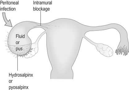
The Fallopian tube must first collect the ovum from its site of ovulation from the ruptured Graafian follicle and then transport the ovum to the ampullary segment, where fertilization occurs. The fertilized ovum must then be transported to the uterine cavity to arrive at the correct point in the menstrual cycle at which the endometrium becomes receptive to implantation (the ‘implantation window’). Tubal factors account for about 10–30% of cases of infertility: this figure varies considerably according to the population involved. Occasionally, congenital anomalies occur but the commonest cause of tubal damage is infection. Infection may cause occlusion of the fimbrial end of the tube, with the collection of fluid (hydrosalpinx) or pus (pyosalpinx) within the tubal lumen (Fig. 17.1).

The commonest cause of acute salpingitis in UK is infection with Chlamydia trachomatis, but it may also result from infection with other organisms such as Neisseria gonorrhoeae, Escherichia coli, anaerobic and haemolytic streptococci, staphylococci and Clostridium welchii. The incidence of tubal damage is approximately 8% after the first episode of pelvic infection, 16% after two and 40% after three episodes. Tubal or uterine tuberculosis has begun to be seen more frequently in the UK in the immigrant population or their relatives.

Disorders such as appendicitis associated with peritonitis or inflammatory conditions including Crohn’s disease or ulcerative colitis can result in peritubal and peri-ovarian adhesions, leaving the internal structure of the Fallopian tube relatively unaffected.

Uterine factors

Implantation is less likely to occur if there is distortion of the uterine cavity due to submucous fibroids or congenital abnormalities such as an intrauterine septum. These disorders are often amenable to surgical correction. Subserous or entirely intramural fibroids do not appear to affect implantation. The effect of adenomyosis on implantation is unclear, although the disorder has been linked to recurrent implantation failure and miscarriage. Intrauterine adhesions or synechiae following over-vigorous curettage or infection (Asherman’s syndrome) result in inadequate endometrial development, absent or light periods and recurrent implantation failure.

Investigation of infertility

Investigation of the female partner

All women presenting with infertility should have their rubella immunity checked and, if seronegative, be offered vaccination before undertaking further treatment for their infertility. They should also be advised to take folic acid supplementation from the outset of investigation and treatment of their fertility problem, to reduce chances of spina bifida in their child.

Detection of ovulation

The assessment of ovulation depends on the menstrual history. In the presence of a regular menstrual cycle ovulatory status can be investigated by changes in basal body temperature, cervical mucus or hormone levels, by endometrial biopsy or by ultrasound. However, measurement of basal body temperature (BBT) is difficult for many women to achieve and requires daily charting, increasing stress with a daily reminder of failure to conceive. Hence measurement of BBT is no longer recommended. Similarly, many women find assessment of cervical mucus changes difficult and challenging and this method is also not recommended. Ovulation can be inferred by detection of the LH surge in blood or urine, with a peak that occurs approximately 24 hours before ovulation. Modern commercially available LH surge detection kits can provide reassurance and allow timing of intercourse. Formation of the corpus luteum can be demonstrated by measurement of serum progesterone in the luteal phase of the cycle. A mid-luteal concentration above 25 nmol/L is usually accepted as evidence of ovulation, although values vary from laboratory to laboratory.

Assessment of ovarian reserve

Advancing female age is one of the strongest prognostic factors that determines the success or otherwise of IVF treatment. Ovarian reserve testing using measurement of AMH in serum and/or antral follicle count (AFC) with transvaginal ultrasound allows an individual estimate of ‘ovarian reserve’ to be made. An age-related low AMH or low AFC predicts poor oocyte yield at IVF and a lower than average chance of pregnancy, whereas higher than average values predict a better ovarian response to gonadotrophin stimulation. However, although these markers are helpful in identifying predicted oocyte quantity after stimulation, they do not identify oocyte quality with the same precision. Quality (potential for fertilization and implantation leading to healthy live birth) seems to be more closely related to female age, such that a young ‘poor responder’ to stimulation has a good chance of pregnancy, whereas an older ‘good responder’ may obtain a larger than usual number of oocytes but there is still a reduced chance of pregnancy.

Investigation of tubal patency

It is essential to establish tubal patency before beginning ovulation induction or intrauterine insemination. Tubal patency need not be established if the couple are to proceed directly to IVF if, for example, there is a severe male factor. However, uterine anatomy should then be checked with high-resolution transvaginal ultrasound or hysterosalpingography (HSG).

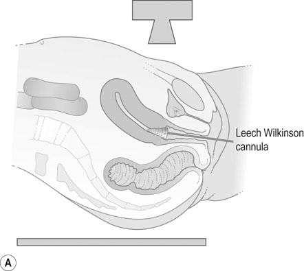
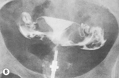
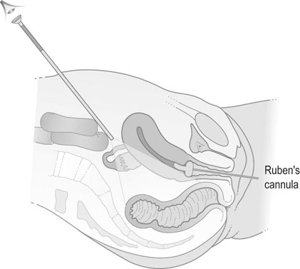
Hysterosalpingography

A radio-opaque contrast medium is injected into the uterine cavity and Fallopian tubes. General anaesthesia is unnecessary. The contrast medium outlines the uterine cavity and will demonstrate any filling defects. It will also show whether there is evidence of tubal obstruction and the site of the obstruction (Fig. 17.2). HSG should be performed within the first 10 days of the menstrual cycle to avoid inadvertent irradiation of a newly fertilized embryo. Women should be screened for C. trachomatis infection or given appropriate antibiotic prophylaxis before HSG in order to reduce the risk of reactivation of infection leading to pelvic abscess formation.

Laparoscopy and dye insufflation

Laparoscopy enables direct visualization of the pelvic organs and allows assessment of pelvic pathologies such as endometriosis or adhesions. Methylene blue is injected through the cervix in order to test tubal patency. Laparoscopy can be combined with hysteroscopy to assess the uterine cavity. A ‘see-and-treat’ policy allows for rapid surgical treatment of minor degrees of endometriosis or adhesions, although surgery that may result in damage to pelvic structures is better left to another occasion to allow full discussion of the implications of surgery to take place with the patient and her partner. Laparoscopy almost invariably requires general anaesthesia and there are small but significant risks of damage to pelvic structures including bowel, bladder and ureter at laparoscopy, so less invasive methods are preferred as first line investigations unless there is a specific indication, such as a history of pelvic inflammatory disease or appendicitis with peritonitis (Fig. 17.3).

Investigation of the male partner

The most useful investigation of the male partner is by semen analysis (Box 17.1). Semen should be collected by masturbation into a sterile container after 3 days abstinence and examined within 2 hours of collection. The sample is best collected in a private facility adjacent to the andrology laboratory to avoid cooling during transportation and allow accurate identification of the male partner.

The recently revised lower reference limits and 95% confidence intervals for sperm parameters (WHO 2010) are given in Table 17.3.

Table 17.3

Lower reference limits (5th centiles and their 95% confidence intervals) for semen characteristics

Parameter Lower reference limit
Semen volume (mL) 1.5 (1.4–1.7)
Total sperm number (106 per ejaculate) 39 (33–46)
Sperm concentration (106/mL) 15 (12–16)
Total motility (PR+NP, %) 40 (38–42)
Progressive motility (PR, %) 32 (31–34)
Vitality (live spermatozoa, %) 58 (55–63)
Sperm morphology (normal forms, %) 4 (3.0–4.0)
Other consensus threshold values
pH 7.2
Peroxidase-positive leukocytes (106/mL) <1.0
MAR test (motile spermatozoa with bound particles, %) <50
Immunobead test (motile spermatozoa with bound beads, %) <50
Seminal fructose (µmol/ejaculate) ≥13
Seminal neutral glucosidase (mU/ejaculate) ≥20

image

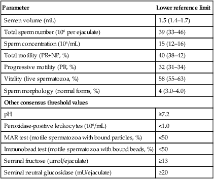
(Data from Cooper TG, Noonan E, von Eckardstein S, et al (2010) World Health Organization reference values for human semen characteristics. Human Reproduction Update 16:231–245.)

The major features of the semen analysis are:

• Volume: 80% of fertile males ejaculate between 1 mL and 4 mL of semen. Low volumes may indicate androgen deficiency and high volumes abnormal accessory gland function.

• Sperm concentration: The absence of all sperm (azoospermia) indicates sterility, although sperm may well be recoverable by percutaneous epididymal aspiration (PESA) or testicular aspiration (TESA) or testicular biopsy. The lower limit of normality is between 15 million and 20 million sperm/mL, but the findings should not be accepted on a single sample as there is significant fluctuation from day to day. Abnormally high values, in excess of 200 million sperm/mL, may be associated with subfertility.

• A normal analysis should show good motility in 60% of sperm within 1 hour of collection. The characteristic of forward progression is equally important. The World Health Organization grades sperm motility according to the following criteria:

• Sperm morphology shows great variability even in normal fertile males, and is less predictive of subfertility than count or motility. It is important to look for leukocytes as they may indicate the presence of infection. If pus cells are present, the semen should be cultured for bacteriological growth.

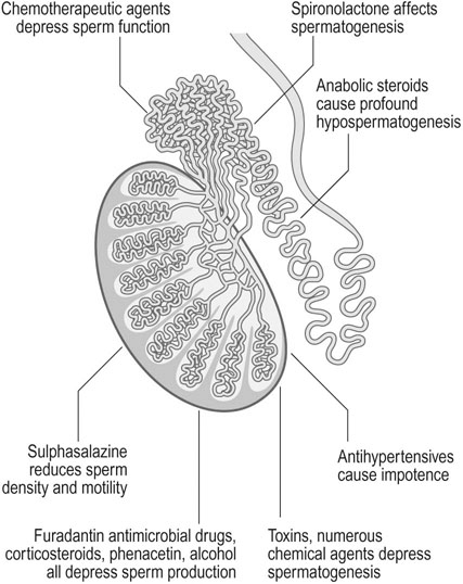
Spermatogenesis and sperm function may be affected by a wide range of toxins and therapeutic agents. Various toxins and drugs may act on the seminiferous tubules and the epididymis to inhibit spermatogenesis. Chemotherapeutic agents, particularly alkylating agents, depress sperm function and sulphasalazine, frequently used to treat Crohn’s disease, reduces sperm motility and density. Patients who are prescribed chemotherapy or pelvic radiotherapy should be offered sperm cryopreservation before treatment to allow them to start a family later in life once their disease has been successfully treated (Fig. 17.4).

Additionally, antihypertensive agents can cause erectile dysfunction and anabolic steroids used for bodybuilding may produce profound hypospermatogenesis.

Analysis of sperm DNA

Standard tests of sperm concentration, motility, and morphology are poorly predictive of the ability of a couple to conceive. The integrity of sperm chromosomal DNA is essential for normal fertilization and transmission of paternal genetic information, and tests of sperm DNA integrity generally correlate with routine semen variables, including impaired sperm concentration or motility. Sperm DNA is protected from damage while the sperm is transported through the male and female reproductive tracts, and damage to sperm DNA may lead to impaired fertility.

The most frequently used measure of sperm DNA damage is the sperm chromatin structure assay (SCSA) that measures the stability of sperm chromatin in acid media with acridine orange. The dye gives rise to green fluorescence when bound to intact DNA and red when bound to fragmented DNA; the proportion of sperm with fragmented DNA is determined by flow cytometry and expressed as the DNA fragmentation index (DFI). Other commonly used tests include the deoxynucleotidyl transferase-mediated dUTP nick end labelling (TUNEL) assay in which fluorescence-activated cells are sorted by flow cytometry, the single cell electrophoresis assay (Comet) that measures single-strand and double-strand DNA breaks using electrophoresis and the Halo (SCD) test that identifies sperm with fragmented DNA because they fail to produce the characteristic halo when mixed with aqueous agarose following acid/salt treatment. Each assay has its strengths and weaknesses and results imputing normality or abnormality do not always concur between assays.

In clinical studies, sperm DNA integrity is impaired among infertile compared with fertile men and with poor semen quality. Time-to-pregnancy studies with apparently normally fertile couples at the time of stopping contraception showed that results of the SCSA test were significantly associated with the probability of pregnancy. However, IVF and intracytoplasmic sperm injection (ICSI) studies have been less conclusive in relating sperm DNA integrity results to fertilization or pregnancy rates. At present, assessment of sperm DNA damage should remain as a research tool and routine use as a diagnostic test should await further evidence of ability to discriminate between those couples who will, or will not, conceive.

Treatment of female subfertility
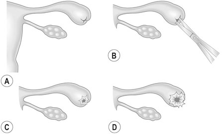
If the history, examination and systematic investigation in both partners is normal, and the duration of infertility is less than 18 months, the couple should be reassured and advised regarding coital frequency and simple lifestyle changes that may improve chances of conception. Both partners should be advised to stop smoking and limit their intake of alcohol. Women or men with a body mass index of more than 30 should be encouraged to join a supervised programme of weight loss.

However, if the woman is over 30 years of age then this ‘wait and see’ policy is unwise, since delay will have a significant adverse impact on her lifetime chance of conception using IVF. The couple should be referred rapidly to a specialist infertility clinic that has access to the full range of assisted reproductive technologies (ART) including IVF and ICSI, intrauterine insemination (IUI) and donor sperm and oocyte treatments.

Anovulation

In the presence of WHO group II anovulation with stigmata of PCOS, normal FSH and prolactin levels, the drug of choice remains clomiphene citrate. Clomiphene will produce ovulation in 80% of subjects leading to pregnancy in about one half of those who ovulate. Clomiphene is administered from day 2–6 of the cycle with an initial dosage of 50 mg/day, increased to 100 and 150 mg/day where necessary. Ovulation can be monitored by measurement of day 21 progesterone levels, although restoration of a regular menstrual cycle is frequently followed by pregnancy as ovulation resumes. Rates of twin pregnancy of 6–10% have been reported, with higher order pregnancies being reported in approximately 1 : 1000 patients. Ultrasound monitoring of follicle growth is recommended, with abstention from intercourse if there are more than two mature follicles, to reduce the incidence of multiple pregnancy. More recently, the aromatase inhibitor letrozole has been used as an oral alternative to clomiphene, with an increase in percentage of women who ovulate and possibly better pregnancy rates. However letrozole remains unlicensed for treatment of infertility.

Second-line management of anovulation may involve laparoscopic ovarian diathermy (LOD), which induces ovulation in over 70% of PCOS patients. LOD has the advantage of inducing natural mono-ovulation with a risk of multiple pregnancy no higher than background and, when successful, allowing a drug-free and more natural conception. Alternatively, ovulation may be induced with daily injection of recombinant or urinary derived FSH although this may be costly and monitoring with ultrasound and blood tests is required due to the possibility of over response and risk of multiple pregnancy. Careful management using a low-dose step up regime can produce acceptable pregnancy rates with a low multiple pregnancy rate.

Anovulation associated with hyperprolactinaemia, in the absence of macroadenoma, can be treated with a dopamine receptor agonist such as cabergoline. Cabergoline is preferred to bromocriptine due to its ease of administration and reduced incidence of side effects.

Tubal pathology

Tubal microsurgery has been almost completely supplanted by IVF in the management of tubal infertility. Laparoscopic surgery may still be necessary to perform salpingectomy or tubal clipping prior to IVF in the presence of hydrosalpynx to reduce chances of contamination of the endometrial cavity with ‘toxic’ hydrosalpyngeal secretions, or for preliminary ovarian cystectomy or myomectomy.

Salpingolysis to release peritubal adhesions still has a place if the fimbrial ends of the tubes are well preserved. However, it is important not to lose too much time if the woman is over 30 years of age. At times the blocked tubal end can be opened, i.e. salpingostomy (Fig. 17.5). There is an increased risk of ectopic pregnancy after all forms of tubal surgery.

Intrauterine insemination

Placement of a sample of sperm into the uterine cavity using a soft, flexible catheter has been performed for many years. The technique has been enhanced by preparation of the semen sample by washing and isolation of motile sperm, and by stimulation of ovulation using low dose gonadotrophins. IUI can be seen as a specific treatment for coital dysfunction and for abnormalities of cervical mucus, but is also used frequently to treat unexplained infertility and mild male factor infertility. IUI requires healthy, patent Fallopian tubes. Live birth rates in good quality centres are between 15% and 20% per cycle, although IUI remains a cost effective alternative to IVF because of the lower doses of gonadotrophins, reduced level of monitoring and simplified laboratory requirements.

In vitro fertilization and embryo transfer

In vitro fertilization and its many variants have revolutionized the management of infertility since the birth of Louise Brown in 1978. Globally, there are now more than 5 million children who have been born following IVF and generally reassuring data on safety of IVF, ICSI and embryo cryopreservation have been compiled by the European Society for Human Reproduction and Embryology and the American Society for Reproductive Medicine. Essentially, IVF involves stimulation of multiple ovarian follicle development using recombinant or urinary derived gonadotrophins, with concurrent use of a gonadotrophin-releasing hormone (GnRH) agonist or antagonist to prevent a premature LH surge and ovulation before oocytes are harvested. Oocytes are collected using transvaginal ultrasound guided needle follicle aspiration, with the oocytes being isolated from the follicular fluid and cultured in the presence of a washed sample of the partner’s sperm. Fertilized oocytes (embryos) can be cultured for up to 5 days, at which point they reach the blastocyst stage of division and it becomes possible to make a detailed assessment of their morphological quality. The ‘best’ one or two blastocysts are then transferred to the uterine cavity using a simple catheter, with remaining blastocysts being cryopreserved for use later if conception does not follow the fresh embryo transfer.

There are many variations on this theme – embryos can be transferred on day 2 or 3 of development rather than as blastocysts, all embryos may be cryopreserved without a fresh transfer if there is risk of ovarian hyperstimulation syndrome (OHSS) and embryos can be biopsied for pre-implantation genetic diagnosis (PGD) of chromosomal or genetic disorders with transfer only of those screened as normal. ICSI is widely used in cases of moderate to severe male factor infertility to inject a single spermatozoon directly into the cytoplasm of the oocyte, giving similar fertilization and pregnancy rates to IVF. Female age remains the main determinant of outcome and an increasing number of women are resorting to treatment with donated oocytes from younger women as a means of achieving pregnancy at a time of life when their own ovarian reserve is too low to allow them the opportunity of healthy pregnancy and live birth. Whilst treatment with donor oocytes has a high chance of a healthy live birth, the child does not have the DNA of his or her birth mother and the shortage of altruistic donors has led to development of an international market in oocytes from paid donors. Payment to oocyte or sperm donors remains illegal in UK, although at time of writing an Human Fertilisation and Embryology Authority (HFEA) consultation is underway to assess societal attitudes to this ethical dilemma.

In the UK, ART is regulated by the HFEA that oversees all aspects of IVF treatment. The HFEA has proven a useful interface between the public and Government on one hand, and IVF clinics on the other, allowing for ethical debate and imposition of ‘best practice’ in areas of clinical and laboratory safety. HFEA also collates treatment results from all clinics in UK, providing a snapshot of what can be achieved by ART. Fertilization rates after IVF are between 60% and 80% depending largely on the age of the woman, and most patients who undertake IVF will have an embryo transfer. However, implantation rates remain relatively low leading to a live birth in 30–40% of cycles in most centres.

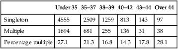
The most frequent cause of obstetric and paediatric problems in offspring from IVF is the result of multiple pregnancy leading to premature birth. Transfer of two, three or more embryos in a single IVF cycle was commonplace in the early days of ART, but many countries, led by those in Scandinavia, have adopted a policy of single embryo transfer (SET) in the majority of IVF cycles. Multiple pregnancy rates remain at approximately 20% in UK, but are falling steadily (Table 17.4). Improved success rates from transfer of frozen embryos after cryopreservation using vitrification have made single embryo transfer a more attractive option to couples, since their chances of a live birth after sequential transfer of one fresh then one frozen embryo are equivalent to those seen after transfer of two fresh embryos, but without the risk of multiple pregnancy. The higher percentage of multiple pregnancies seen in the older patient groups reflects the lower overall chances of pregnancy, leading patients and practitioners to resort to desperate measures in order to try and achieve a pregnancy.

Table 17.4

Multiple pregnancy rates after IVF/ICSI – HFEA data

  Under 35 35–37 38–39 40–42 43–44 Over 44
Singleton 4555 2509 1259 813 143 97
Multiple 1694 681 255 136 31 38
Percentage multiple 27.1 21.3 16.8 14.3 17.8 28.1

image

Ovarian hyperstimulation syndrome

OHSS is the consequence of over dosing with gonadotrophins leading to excessive follicle development and high circulating concentrations of oestrogens and vascular endothelial growth factor (VEGF). OHSS is potentially lethal and is almost completely avoidable if a conservative approach to ovarian stimulation is used. In its severe form, the condition results in marked ovarian enlargement with fluid shift from the intravascular compartment into the third space, leading to ascites, pleural effusion, sodium retention and oliguria. Patients may become hypovolaemic and hypotensive and may develop renal failure as well as thromboembolic phenomena and adult respiratory distress syndrome. The pathophysiology of this condition appears to be associated with an increase in capillary vascular permeability.

Treatment

If the haematocrit is below 45% and the signs and symptoms are mild, the patient can be managed at home, but where there is significant ascites as judged by ultrasound examination, she should be hospitalized. Baseline electrolyte values and liver and kidney function should be assessed. Volume expansion can be performed using human albumin, sometimes with crystalloid, and, if there is severe ascites or pleural effusion, fluid should be drained to reduce the fluid load. Drugs such as indomethacin and angiotensin-converting enzyme inhibitors may be useful in reducing the severity of the episode. Eventually, the cysts will undergo resorption, and the ovaries will return to their normal size. Further attempts at ovarian stimulation should take into account the dosage regimes used during the episode of ovarian hyperstimulation syndrome.

Treatment of male infertility

Specific treatment is possible in only a small proportion of infertile males. Testicular size is important and with the finding of small testes, azoospermia, high FSH and low AMH levels, it is unlikely that any therapy will help.

Where FSH levels are normal and testicular size is normal, ductal obstruction should be suspected and testicular biopsy performed. If normal spermatogenesis is demonstrated, then it is necessary to proceed to vasography and exploration of the scrotum. Surgical anastomosis may re-establish fertility. Gonadotrophins are effective in the rare cases of men with hypogonadotrophic hypogonadism, and dopamine agonists are used in men with hyperprolactinaemia. Unproven but widely practised treatments for male infertility include ligation of a varicocele and anti-oxidants and supplements to reduce sperm DNA fragmentation.

The most successful treatment for male infertility is ICSI. ICSI involves the direct injection of a single immobilized sperm into the cytoplasm of the oocyte. This technique produces pregnancy rates similar to those of in vitro fertilization. Anxieties remain concerning the slightly higher rate of abnormality in children conceived after ICSI. Abnormalities are mainly observed in the development of the genital tract (hypospadias, testicular maldescent) although cases of imprinting disorders such as Angelman and Beckwith–Widemann syndromes may also be seen more commonly after ICSI.

Donor insemination

If sperm cannot be obtained for ICSI, or if the man is a carrier of a genetic disorder that the couple wish to avoid transmitting to their child, then donor insemination should be discussed with the couple.

The implications of the procedure, from both a legal and personal point of view, should be discussed in depth, with independent counselling for both partners. Anonymity for sperm donors was removed in the UK in 2004 and the resultant shortage of donor sperm has led many couples to seek treatment overseas or to import sperm from Denmark, the US and elsewhere. Children conceived using donor oocyte or sperm collected after 2004 are legally allowed to meet their genetic parent under supervised conditions when they reach the age of 18. They can also check whether a potential partner was conceived using the same donor by referring to the HFEA database, although the name of the donor will not be released if the treatment preceded the change in the law regarding anonymity.

Waiting lists for treatment in UK are lengthy, although this does at least give adequate time for reflection.