Mucosal hyperenhancement, marked submucosal edema, ascites


 , and ascites. C. difficile colitis typically presents as a pancolitis, as in this example.
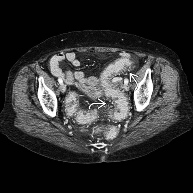
 and hyperemia of the entire colon and rectum.
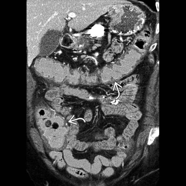
 of the entire colon. C. difficile (pseudomembranous) colitis was confirmed.
IMAGING
General Features


Fluoroscopic Findings
• Contrast enema
 Small nodules or inflammatory polyps ± diffuse mucosal granularity (may simulate ulcerative colitis)
 Possible extrinsic mass with inflammatory changes → distortion, short strictures (may simulate carcinoma)
 Tuberculosis


 Small nodules or inflammatory polyps ± diffuse mucosal granularity (may simulate ulcerative colitis) Possible extrinsic mass with inflammatory changes → distortion, short strictures (may simulate carcinoma)


 Tuberculosis
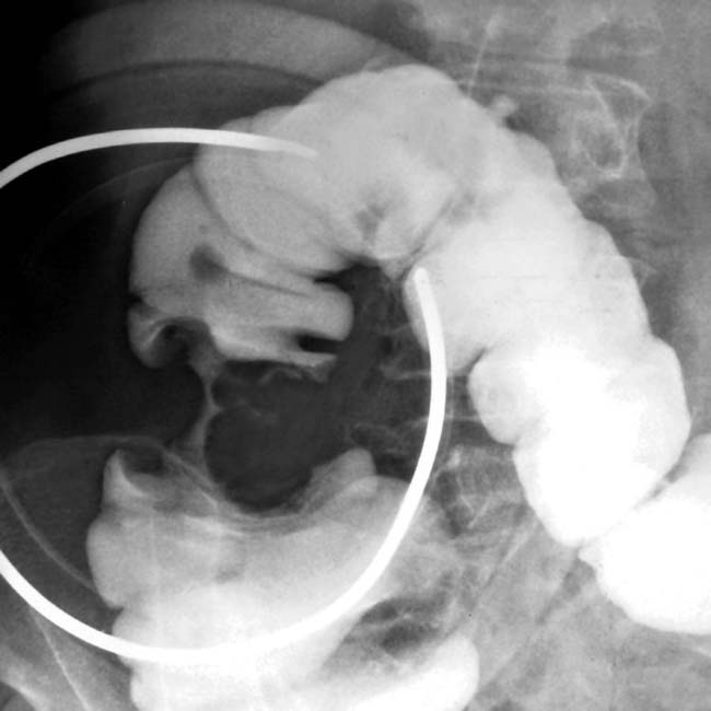
– Oval/circumferential transverse ulcers; loss of demarcation between distorted terminal ileum and ascending colon


CLINICAL ISSUES
Presentation
• Most common signs/symptoms


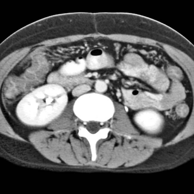
 affecting both the ascending colon and distal ileum. A small amount of ascites
  is also seen.
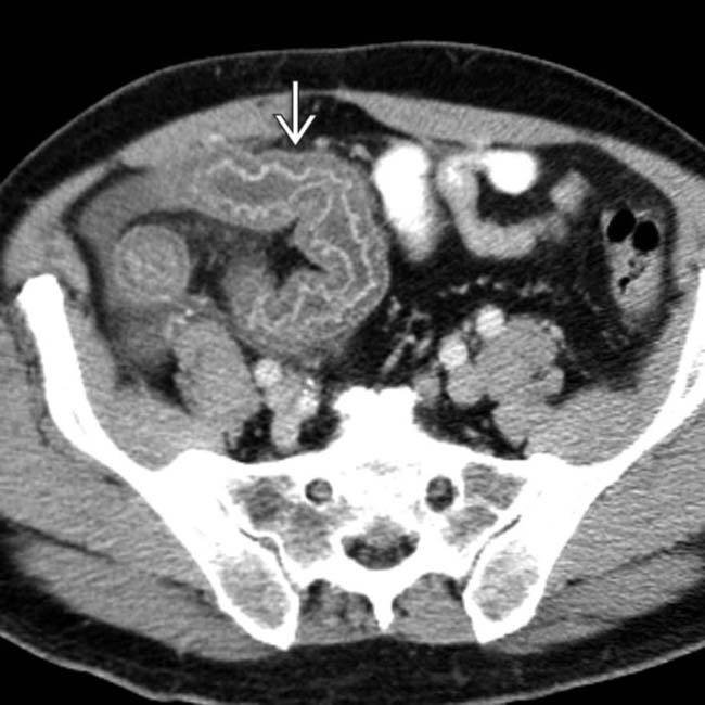
 . The etiology was C. difficile infection, an unusual cause of small bowel inflammation.
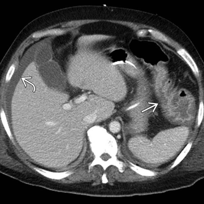
 . The splenic flexure shows mucosal hyperenhancement and submucosal edema
  .
.
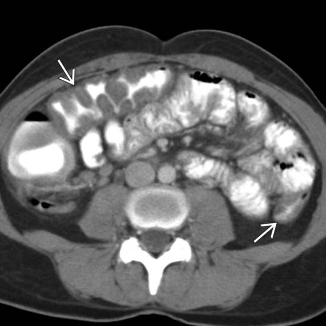
 . These colonic segments seem to have a thin wall, while others are thick walled
  .
.
 with loss of normal transverse folds.
 causing an “accordion” appearance of the colonic wall.
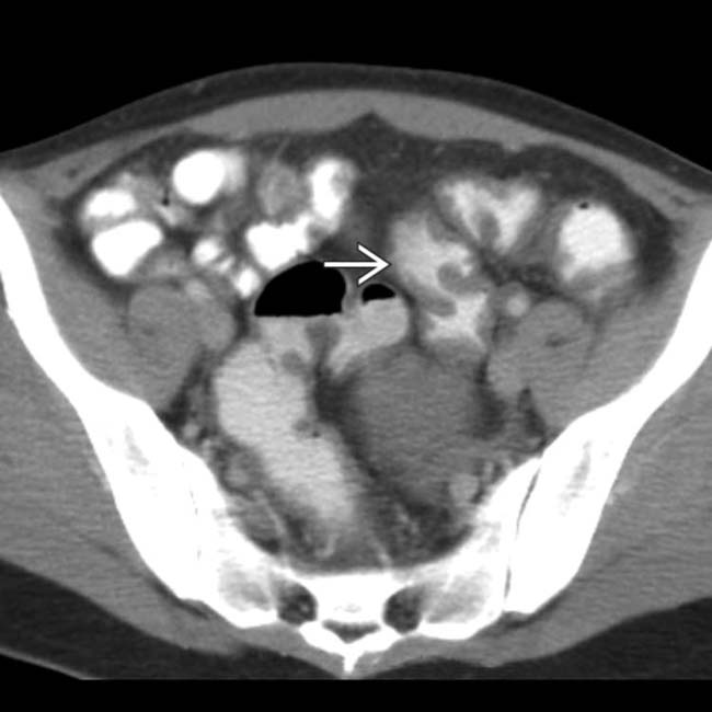
 but not the small bowel. Campylobacter colitis was the final diagnosis.
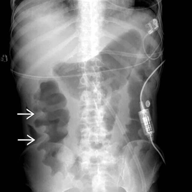
 .
.
 is massively thickened. On endoscopy (not shown) the colonic mucosa was ischemic and biopsy showed cytomegalovirus (CMV) infiltrating the colonic wall and inducing hemorrhagic necrosis.
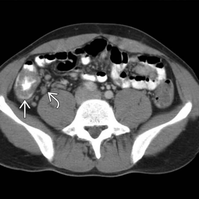
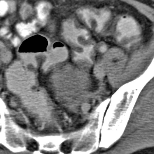
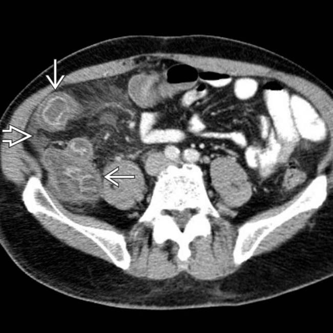
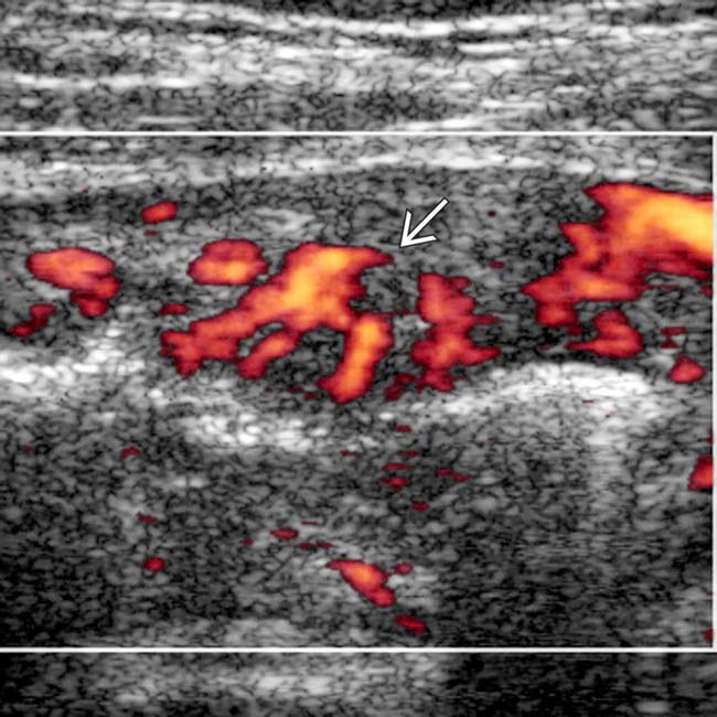
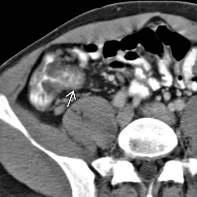
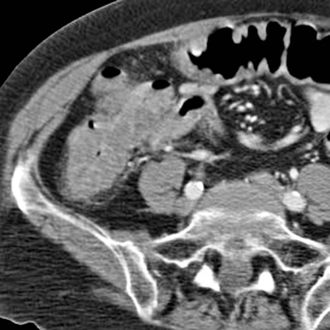
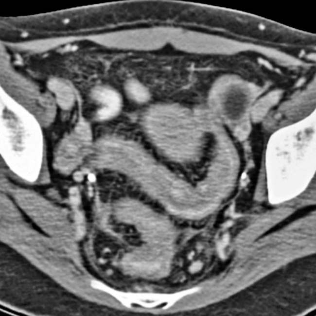
 and RLQ lymphadenopathy
  .
.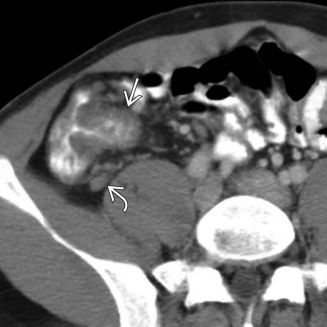
 and additional enlarged nodes
  . This is a typical example of Yersinia enterocolitis with mesenteric adenitis. Most forms of infectious colitis spare the distal small bowel.
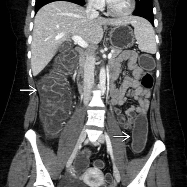
 .
.
 throughout the colon. The small bowel was spared.
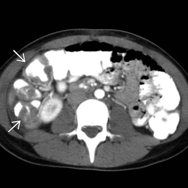
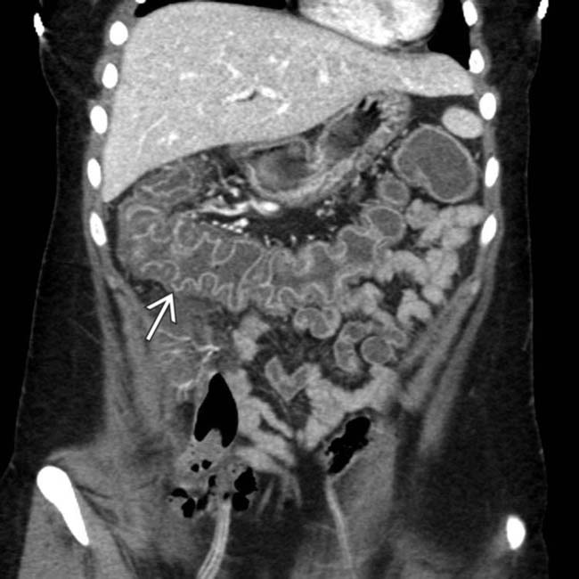
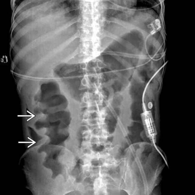
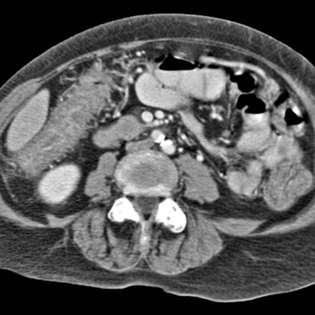
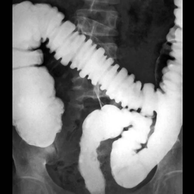
 primarily affecting the ascending colon. Note that the presence of dense contrast material within the colon impairs evaluation for mucosal inflammation.
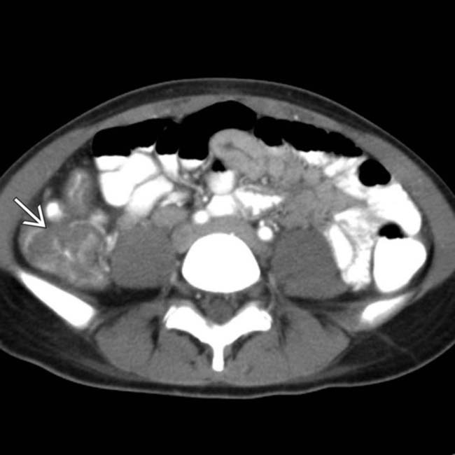
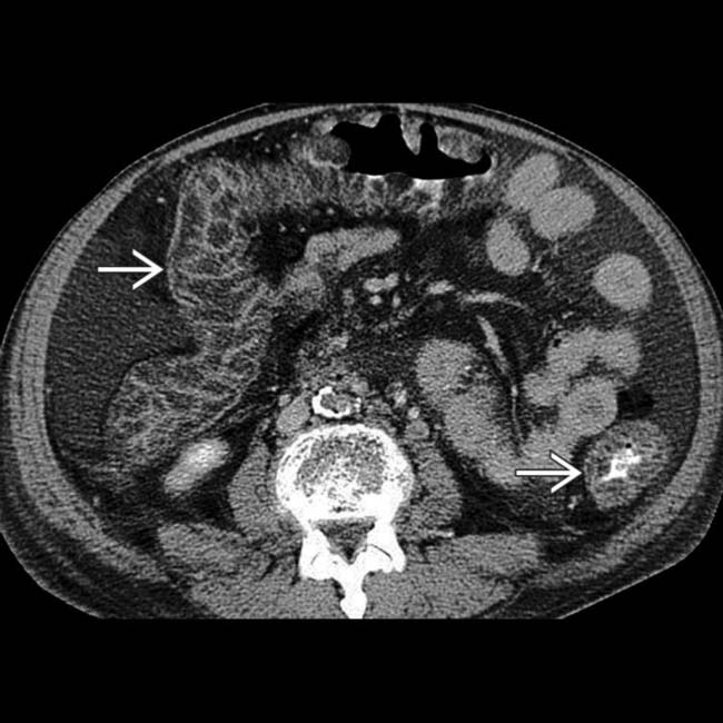
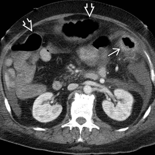
 . The left side of the colon seems uninvolved. Preferential involvement of the right colon is characteristic of Salmonella (typhus), which is endemic in some populations.
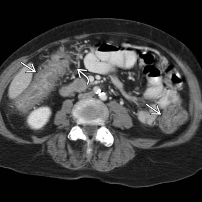
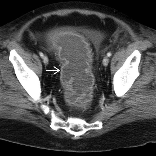
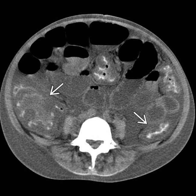
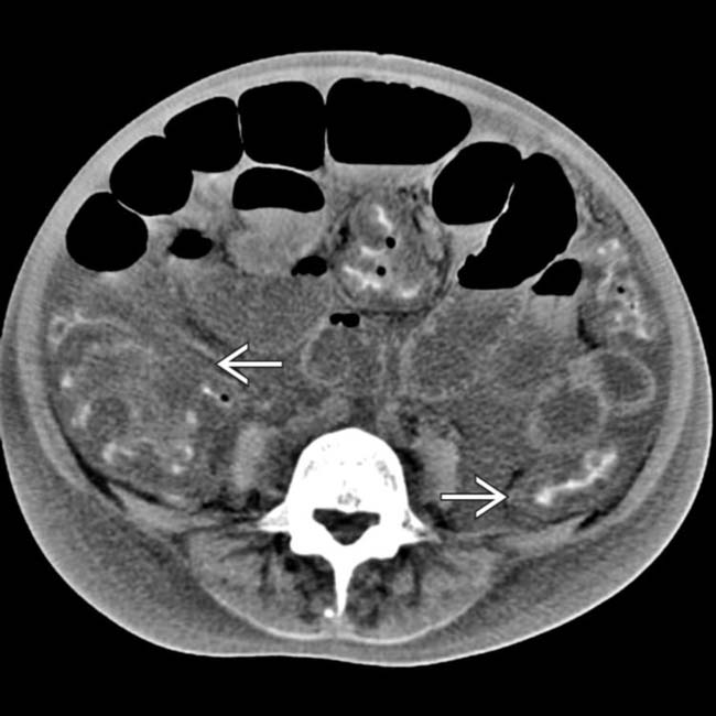
 and adjacent mesenteric hyperemia
  . The rectum and small bowel were spared. Campylobacter colitis was confirmed.
 .
.
 .
.
 .
.
 is massively thickened. The biopsy showed cytomegalovirus infiltrating the colonic wall and inducing hemorrhagic necrosis.
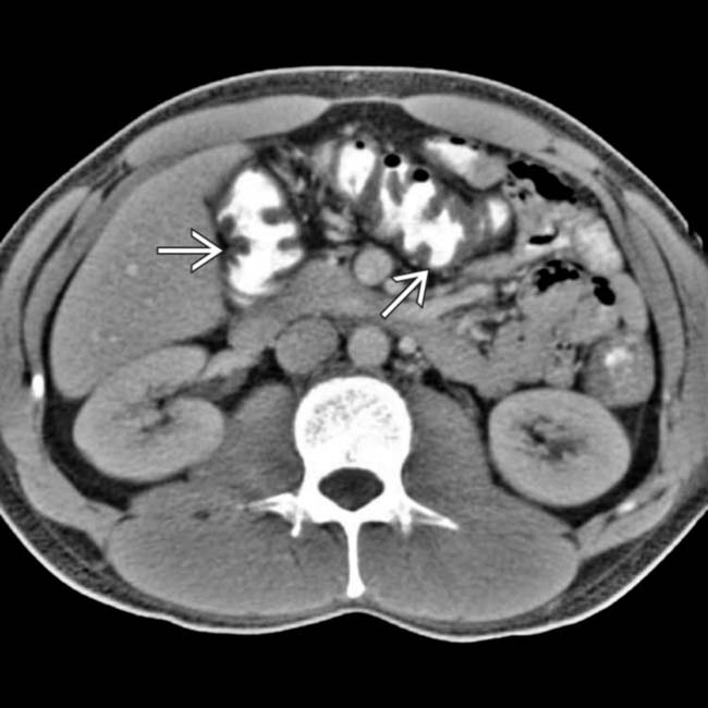
 .
.
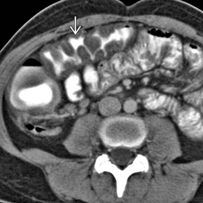
 . Yersinia tends to involve the right colon preferentially, and almost always involves the terminal ileum, unlike most causes of acute infectious colitis.
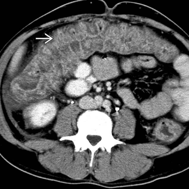
 of the transverse colon.
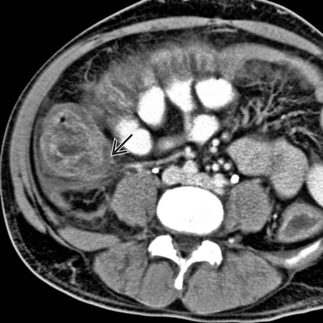
 .
.
 .
.