47. Incontinentia Pigmenti
Definition
Incontinentia pigmenti is a rare, X-linked, dominant inherited disorder involving skin pigmentation. Melanin is lost from the basal cells of the epidermis and collects in the dermis as free pigment or as aggregates of melanophages. Incontinentia pigmenti is also known as Bloch-Sulzberger syndrome.
Incidence
The incidence of incontinentia pigmenti is 1:40,000. The disorder is more common among Caucasian populations than other races. It affects females almost exclusively. Male fetuses typically expire in utero, although some do survive. The overall male to female ratio is 1:37.
Etiology
Almost all cases of incontinentia pigmenti occur as the result of a deletion in the NEMO gene; about half are spontaneous mutations.
Signs and Symptoms
Four Stages of Skin Change
1. Vesicular stage: linear vesicles; pustules; bullae with erythema along the lines of Blaschko; present at birth
2. Verrucous stage: warty; keratotic papules and plaques; occurs between 2 and 8 weeks of age
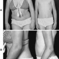
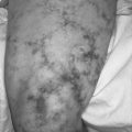
3. Hyperpigmentation stage: macular hyperpigmentation in a swirl pattern along the lines of Blaschko; changes often involve nipples, axilla, and groin; occurs between 12 and 40 weeks of age
4. Hypopigmentation stage: streaks and/or patches of hypopigmentation along with cutaneous atrophy; onset in infancy remaining throughout adulthood
Central Nervous System (10% to 40% of Patients)
• Ataxia
• Hyperactivity
• Mental retardation
• Microcephaly
• Seizures
• Spasticity
• Stroke
Ocular Changes
• Band keratopathy
• Blue sclera
• Cataracts
• Congenital glaucoma
• Exudative retinal detachment
• Foveal hypoplasia
• Leukocoria
• Microphthalmia
• Optic atrophy
• Retinal pigmentary changes
• Retrolental mass formation
• Strabismus
Skeletal/Structural (≈14% of Patients)
• Acheiria
• Ear abnormalities
• Extra ribs
• Hemivertebrae
• Scoliosis
• Skull deformities
• Somatic asymmetry
• Spina bifida
• Syndactyly
Teeth and Jaw Changes (65% to 90% of Patients)
• Delayed eruption
• Hypodontia
• Microdontia
• Micrognathia
• Prognathia
• Round, conical, or peg-shaped teeth
Medical Management
Currently there is no specific treatment available for incontinentia pigmenti. Lesions of stage 1 (vesicular) skin changes should be kept clean and as undisturbed as possible. Frequent, meticulous dental care and hygiene are very important.
Complications
• Blindness or reduced visual acuity due to ophthalmic changes
• Mental retardation (seen predominantly in patients with structural brain deformity or ischemic brain injury)
• Secondary bacterial infection during vesicular stage (rare)
• Seizures (seen predominately in patients with structural brain deformity or ischemic brain injury)
Anesthesia Implications
The predominance of dental and jaw abnormalities associated with incontinentia pigmenti constitutes a significant portion of anesthesia concerns. The dental abnormalities predispose the patient to potential dental injury during direct laryngoscopy. Extra care should be taken with airway manipulation. Micrognathia or prognathia in a patient with incontinentia pigmenti can have a significant impact on the anesthetists ability to secure the airway. A difficult airway cart with a fiberoptic bronchoscope should be immediately available in the event that attempts via direct laryngoscopy prove unsuccessful. It may be more prudent, based on the situation revealed by the preoperative physical examination combined with the anesthetists experience and skill level, to forgo direct laryngoscopy in favor of the fiberoptic approach. The level of sedation provided by the anesthetist to facilitate the fiberoptic intubation will be dictated, in large part, by the age—chronologic and/or mental development—of the patient.
Frequently the patient with incontinentia pigmenti is blind, thus requiring more detailed explanations and direction for transfer to the operating room bed. The anesthetist should tailor the depth of explanation to the patient by the patients age, both chronologic and developmental.
The patient with associated spastic paralysis should not receive succinylcholine to facilitate intubation. The patient with a high-level spinal cord lesion is more likely to develop autonomic hyperreflexia.
Choice of anesthetic technique, general versus regional, will be predicated on the patient’s age (as described above) and any associated anomalies. The very young patient or one mentally challenged may best be served by choosing general anesthesia. General anesthesia may also be the better choice for the patient with associated spinal cord lesions.