Advances in Anesthesia, Vol. 28, No. 1, 2010
ISSN: 0737-6146
doi: 10.1016/j.aan.2010.09.002
Perioperative Implications of Obstructive Sleep Apnea
Obstructive sleep apnea (OSA) was first described by novelist Charles Dickens in The Posthumous Papers of the Pickwick Club, published in 1937. The character Joe was an obese boy who was constantly hungry, red in the face, and always falling asleep in the middle of his duties. Dickens [1] writes:
OSA is a common cause of daytime sleepiness for millions of Americans. It is estimated that in adults between the ages of 30 and 60 years, 9% of women and 24% of men have OSA, whereas 2% of women and 4% of men have OSA with daytime sleepiness [2]. The prevalence of OSA increases with age [3]. OSA is associated with an increased incidence of hypertension [4], coronary artery disease [5,6], cardiovascular morbidity and mortality [7], cor pulmonale [8], stroke [9,10], neurocognitive dysfunction [11], motor vehicle accidents [12], and a decreased quality of life [13].
Definition of OSA
OSA is clinically characterized by recurrent episodes of upper-airway obstruction that result in cessation (apnea) or reduction (hypopnea) in airflow during sleep, often accompanied by hypoxia and/or hypercarbia. The diagnosis of OSA is based on the apnea-hypopnea index (AHI), which is a count of the number of apneas and hypopneas per hour of sleep. Apnea is defined as the cessation of airflow for at least 10 seconds in the presence of thoracoabdominal ventilatory efforts and hypopnea as a 50% or more reduction in breathing amplitude. The occurrence of more or less frequent oxygen desaturations or electroencephalography (EEG) arousals is taken as an additional diagnostic tool to identify these conditions [14]. Obstruction is often inferred from thoracoabdominal paradox, the shape of the airflow signal, or when snoring intensity increases during the event [15].
Anatomic Factors
Numerous studies have shown that patients with OSA have structurally narrower and more collapsible airways than patients who do not have OSA who are matched in age and body mass index (BMI, calculated as weight in kilograms divided by the square of height in meters) [16,17]. Craniofacial and upper-airway abnormalities have been shown to predispose patients with OSA to upper-airway obstruction [18]. Micrognathia with an inferior-located hyoid bone has been shown to be a major risk factor for OSA in Japanese patients. This risk factor was most significant in obese patients with large tongues [19]. An anatomic imbalance between upper-airway soft-tissue volume and bony enclosure size may result in pharyngeal airway obstruction during sleep and general anesthesia. Tsuiki and colleagues [20] measured tongue cross-sectional area and craniofacial dimensions through lateral cephalograms in 50 adult male patients with OSA and 55 adult male individuals who did not have OSA and found that when maxillomandibular dimensions were matched, patients with OSA had a significantly larger tongue for a given maxillomandibular size compared with individuals who did not have OSA. Schwab and colleagues [21] used volumetric analysis techniques with magnetic resonance imaging to study the upper-airway soft-tissue structures in sleep apnea and control individuals with a variety of ethnic backgrounds. These investigators showed that the volume of soft-tissue structure surrounding the upper airway is enlarged in patients with OSA. They also showed a greater risk for sleep apnea the larger the volume of the tongue, lateral pharyngeal walls, and soft tissue.
Effect of Lung Volumes on Pharyngeal Patency
Lung volume normally decreases during sleep [22]. Obesity is associated with further reductions in lung volumes caused by increased elastic resistance and decreased compliance of the chest wall, resulting in reduced total respiratory compliance. Decreased pulmonary compliance leads to decreased expiratory reserve volume, functional residual capacity (FRC), vital capacity, and total lung capacity [23]. Whereas the decrease in FRC is responsible for the development of severe hypoxemia during obstructive episodes in obese patients with OSA, the decrease in lung volumes has been shown to contribute to pharyngeal airway obstruction. Van de Graaff [24] studied the effect of thoracic traction on upper-airway resistance during breathing. In anesthetized dogs with denervated upper airways, thoracic traction generated during inspiration reduced upper-airway resistance significantly. These findings indicate that tonic and phasic forces generated by the thorax can improve upper-airway patency. The increased patency during inspiration cannot be attributed solely to activity of upper-airway muscles. Likewise in humans, Heinzer [25] reported a significant decrease in respiratory events during non-rapid eye movement (REM) sleep in patients with OSA when lung volume was increased by negative extrathoracic pressure during sleep. Small changes in lung volume during sleep in patients with OSA have been shown to cause large changes in the continuous positive airway pressure (CPAP) required to prevent flow limitation in the upper airway [26]. Therefore, lung volume has an important effect on the upper airway in patients with OSA and is one factor that increases the risk of OSA in obese patients. Tagaito and colleagues [27] examined the structural interaction between a passive pharyngeal airway and lung volume before and during lung inflation in 8 anesthetized and paralyzed patients with sleep-disordered breathing. Increasing lung volume by applying negative extrathoracic pressure (thereby leaving the transpharyngeal pressure unchanged) improved pharyngeal collapsibility, especially in obese patients. Increased lung volume during inspiration is believed to provide caudal tracheal traction that increases the longitudinal tension on the upper airway independent of upper-airway muscle activity, causing unfolding of the pharyngeal mucosa and stiffening the airway. Traction on the trachea could be explained as a sum of mediastinal traction and the force generated by changes in intrathoracic pressure [28].
Neuromuscular Control
Neuromuscular control of the upper airway plays an important role in OSA pathogenesis. Because upper-airway dilator muscles are important in maintaining pharyngeal patency, reductions in activity of these muscles may be important in mediating REM-related obstructive apnea. Sauerland and Mitchell [29] showed that the electromyographic activity of the largest upper-airway dilator muscle, the genioglossus (GG), was increased during inspiration and markedly reduced during expiration. Mezzanotte and colleagues [30] reported that patients with OSA have significantly greater basal genioglossal activity during wakefulness compared with age and BMI-matched patients who do not have OSA. However, this augmented activity was significantly reduced during sleep. This finding suggests that the neuromuscular compensation present during wakefulness that is necessary to keep the anatomically compromised airway of patients with sleep apnea open is lost during sleep, leading to airway collapse. These investigators further showed that sleep onset is associated with significantly larger decrements in GG and tensor palatine muscle activity in patients with OSA compared with controls [31]. More recently Eckert and colleagues [32] studied the mechanisms of worsening OSA during REM sleep. When airway resistance and blood gas disturbances were minimized by CPAP, genioglossal activity was reduced during REM sleep to a similar extent in OSA and healthy individuals. This finding suggests a generalized reduction in genioglossal activity during REM, which likely renders individuals who are highly reliant on upper-airway dilator muscles vulnerable to pharyngeal collapse during REM sleep.
Ventilatory Control
Multiple feedback loops exist to control and stabilize breathing by adjusting arterial blood gas tensions [33]. Ventilation is constantly monitored and adjusted to maintain appropriate arterial pH and Pco2. That sleep can affect control of these feedback loops was first reported more than 50 years ago by Bulow [34], who observed that decreases in carbon dioxide sensitivity at sleep onset led to episodes of hypopnea or apnea with breathing instability. Sullivan [35] subsequently reported that suppression of respiratory stimuli (wakefulness, vagal, peripheral, and central chemoreceptors) results in near cessation of ventilation, suggesting that decreased metabolic control during sleep may explain the irregular breathing pattern characteristic of the sleep state. Several recent studies using a loop-gain model to measure respiratory system stability suggest that patients with OSA have less ventilatory stability. Loop gain is an engineering term that describes the stability of a system controlled by negative feedback loops. In the case of respiration, loop gain represents the sensitivity of the negative feedback loop that controls ventilation [36]. Younes and colleagues [37] reported that the chemical control system is more unstable in patients with severe OSA than in patients with milder OSA. In a follow-up study the same investigators confirmed chemical instability in patients with OSA and suggested that the mechanisms responsible for the instability may vary considerably among patients [38]. Wellman and colleagues [36] studied ventilatory instability in the context of airway collapsibility in patients with OSA and found a high correlation between loop gain in patients with pharyngeal closing pressure near atmospheric pressure, suggesting that this group may be highly susceptible to changes in ventilatory stability. However, whether loop gain is a cause or consequence of OSA has not been established. It is likely that a combination of upper-airway anatomic, neuromuscular, and ventilatory control mechanisms accounts for the pathogenesis of OSA.
Morbidity and OSA
Systemic Hypertension
Approximately 50% of patients with OSA are hypertensive and 30% of hypertensive patients have OSA [39]. The most convincing evidence to support OSA as a cause in the development of hypertension comes from epidemiologic data from the Wisconsin Sleep Cohort Study. This study evaluating the association between sleep-disordered breathing and hypertension showed a 3-fold increased risk of developing new-onset hypertension in patients with an AHI of 15 or more. This association was independent of age, gender, BMI, and antihypertensive medications [40]. The Sleep Heart Health Study also showed that sleep-disordered breathing is associated with systemic hypertension in middle-aged adults and older individuals even after controlling for demographics and anthropometric variables (BMI, neck circumference, waist/hip ratio, smoking, and alcohol intake) [4]. Subsequent analysis of the Sleep Heart Health data showed that there is a stronger association between sleep-disordered breathing and hypertension for those aged less than 60 years but no association was found for those more than 60 years of age [41]. In addition to human data, 2 animal models support a causal link between sleep-disordered breathing and increased arterial blood pressure. Fletcher and colleagues [42] showed that rats exposed to intermittent hypocapnic hypoxia for 8 hours each day showed significant increases in blood pressure compared with controls. Brooks and colleagues induced OSA in dogs by intermittent airway occlusion during nocturnal sleep. In this model OSA resulted in acute transient increases in nighttime blood pressure and eventually produced sustained daytime hypertension.
Although the physiologic studies provide strong evidence to support a causal relationship between OSA and hypertension, the mechanism by which OSA causes hypertension is less well established. A likely mechanism of increased arterial pressure is through sustained sympathetic nervous system excitation and vascular remodeling. Hypoxia and hypercapnia secondary to partial or complete closure of the upper airway during sleep result in oxygen desaturation and increased sympathetic nervous system activity in patients with OSA [43,44]. The primary role of the sympathetic nervous system is maintenance and regulation of blood pressure. The baroreceptors respond to stretching of the vessel wall (increased blood pressure) by decreasing sympathetic neural outflow, resulting in vasodilation and decreased blood pressure. Recent studies suggest that in addition to short-term regulation of blood pressure, the sympathetic neural system plays an important role in long-term regulation of blood pressure [45,47].
In addition to activation of the sympathetic nervous system, the repetitive hypoxemia-normoxia cycle results in activation of inflammatory pathways, which may be important in the molecular pathogenesis of cardiovascular disease [48] independent of increased sympathetic activity [49]. Hypoxia is a major stimulus of vascular endothelial growth factor, which is a potent angiogenic cytokine. Vascular endothelial growth factors are especially increased in patients with severe OSA [50,51]. Bokinsky and colleagues [51] showed that patients with OSA also have increased spontaneous platelet aggregation and activation, which may increase the risk of mural thrombus formation.
CPAP has been shown to attenuate the sympathetic response to obstructive respiratory events and to lower nocturnal blood pressure in patients with OSA [52,53], reduce platelet aggregation [51], and reduce vascular endothelial growth factor in patients with nocturnal hypoxia [54]. However, the effect of CPAP on daytime blood pressure is not so well established. Peppard and colleagues [40] found a small but significant reduction in systolic and diastolic blood pressure with therapeutic CPAP. Although these studies suffer from small sample size, with most patients normotensive at baseline, they do support considering OSA when evaluating all hypertensive patients.
Sleep Apnea and Heart Failure
Observational data support the association between OSA and heart failure [55,57]. Sustained respiratory effort against an occluded airway leads to negative intrathoracic pressure as low as -80 cm water. This negative intrathoracic pressure increases cardiac afterload by increasing the left ventricular transmural pressure, resulting in increased left ventricular wall stress/tension and impaired relaxation, causing decreased stroke volume and cardiac output [58,59]. Otto [60] compared cardiac structural and functional changes in obese adults with and without OSA and found that patients with OSA tended toward abnormal right ventricular filling and diastolic dysfunction suggesting that OSA, independent of obesity, may induce cardiac changes that predispose to atrial fibrillation and heart failure. This observation is important because OSA in patients with heart failure is associated with an increased risk of death independent of confounding factors [61]. Treatment of OSA with CPAP has been shown to improve left ventricular function in patients with congestive heart failure [62,63]. This evidence supports the use of CPAP in the perioperative period for patients with heart failure.
Cardiac Arrhythmias
Several studies show an association between OSA and nocturnal arrhythmias. Arrhythmias observed during sleep include atrial fibrillation, bradycardia, nonsustained ventricular tachycardia, sinus arrest, second-degree atrioventricular conduction block, and frequent premature ventricular contractions [64–67]. The recurrent apneic and hypopneic episodes resulting in arterial desaturation, hypercapnia, and increased sympathetic tone may acutely trigger nocturnal dysrhythmias, or chronically affect the electrical conduction system [66,68]. The Sleep Heart Health Study showed that patients with severe OSA (AHI>30 events/h) have a 4-fold increase in prevalence of atrial fibrillation [69]. Kanagala and colleagues [70] showed that treatment with appropriate levels of CPAP lowers the incidence of recurrent atrial fibrillation in patients with OSA following cardioversion. The bradycardic response to obstructive apnea is probably related to intrathoracic pressure swings, parasympathetic activity, hypoxia, and sleep phase. Changes in heart rate depend on a balance of sympathetic and parasympathetic tone. The stimulation of upper-airway receptors may increase parasympathetic activity, causing a decrease in heart rate [68,71]. In a study of nonsurgical patients with OSA, Becker and colleagues [72] found a 7.5% incidence of clinically significant (second- and third-degree atrioventricular block and sinus arrest of greater than 2 seconds’ duration) heart block. The incidence of pathologically significant rhythm disturbances such as supraventricular tachycardia and ventricular arrhythmias is less clear. In addition, some studies found no causal relation between OSA and arrhythmias [73].
OSA and Coronary Artery Disease
Several studies have found a positive association between OSA and ischemic heart disease. Peker and colleagues [5] showed that middle-aged patients with OSA followed for 7 years showed an almost 5-fold increase in risk of developing coronary heart disease independent of age, sex, hypertension, diabetes, and current smoking. In patients without clinical coronary artery disease, the presence and severity of OSA has been shown to be independently associated with the presence and extent of coronary artery calcification [74]. Cardiac sudden death is increased during sleeping hours in patients with OSA and the degree of nocturnal hypoxemia is related to the risk of sudden cardiac death [75]. Kuniyoshi and colleagues [76] found that the diurnal variation in the onset of myocardial infarction (MI) in patients with OSA was strikingly different from the diurnal variation in patients without OSA. Patients with nocturnal onset of MI between 12 midnight and 6 am had a high likelihood of having OSA. These findings suggest that OSA may be a trigger for MI.
Thus, OSA may increase cardiovascular risk by several different mechanisms. Repetitive hypoxemia and hypercapnia causing sympathetic activation, vascular endothelial dysfunction, increased oxidative stress, inflammation, increased platelet aggregation, and metabolic dysregulation are all implicated in the pathophysiology of cardiovascular disease associated with OSA [77].
OSA and Stroke
OSA is both a possible risk factor and a consequence of stroke [78,79]. Habitual snoring alone has been shown to increase the risk of cerebrovascular disease [80,82]. The Wisconsin Sleep Cohort Study reported that moderate OSA (defined as AHI>20) was associated with a 3-fold increment in the risk of developing a stroke [83]. Studies looking at patients following stroke report a 44% to 72% incidence of OSA (defined as AHI>10) [84,85]. Proposed mechanisms for the increased risk of OSA following stroke include hypertension, reduction in cerebral blood flow, altered cerebral blood flow, altered cerebral blood flow autoregulation, impaired endothelial function, accelerated atherogenesis, thrombosis, and paradoxic embolism [86].
Clinical presentation
Boxes 1 and 2 list physical characteristics and signs and symptoms frequently associated with OSA.
Obesity and Neck Circumference
OSA is strongly correlated with obesity. A study of patients presenting for bariatric surgery reported a 71% to 77% prevalence of OSA in patients with a BMI of 35 to 59.9 kg/m2 and 90% prevalence with BMI 60 kg/m2 or greater [87]. Increases in weight have been shown to increase the prevalence of OSA, which is an independent risk factor for developing obesity. Young and colleagues [2] found that the prevalence of OSA increased 4-fold with each increase in the standard deviation of the BMI. In addition, an increase in weight has been shown to worsen OSA. An 11-year longitudinal study of 690 Wisconsin residents measuring the association between weight change and AHI showed that a 10% weight gain predicted an approximate 32% increase in the AHI and a 6-fold increase in the risk for developing moderate to severe OSA. A 10% weight loss predicted a 26% decrease in the AHI [88].
Several studies have reported that neck circumference is a better predictor of obstructive apnea than BMI [89–92]. Horner and colleagues [93] showed that more adipose tissue is present in those areas surrounding the collapsible segment of the pharynx in patients with OSA, compared with equally obese control subjects without OSA. Shelton and colleagues [94] performed magnetic resonance imaging and polysomnography on patients with and without OSA. They reported that all patients had a collection of adipose tissue adjacent to the pharynx, the volume of which correlated with the severity of OSA. Weight loss resulted in fewer apneas and hypopneas and a marked decrease in the pharyngeal adipose tissue volume, suggesting that the volume of pharyngeal adipose tissue is causally related to OSA. Similarly, Mortimer [95] studied fat distribution in obese and nonobese patients with OSA and found that both groups have excess fat deposition, especially anterolateral to the upper airway, when compared with weight-matched controls.
Recent data suggest that intraabdominal body fat distribution may be even more significant than fat accumulation in the pharyngeal region as a predictor of OSA. Shafer [96] reported that regional body fat distribution predicts the presence and severity of OSA, but fat accumulation in the neck and parapharyngeal region are of minor importance. Martinez-Rivera and colleagues [97] showed that truncal obesity was significantly better than BMI at predicting OSA. Vgontzas and colleagues [98] showed that sleep-disordered breathing correlated with the amount of visceral fat, but not BMI, total fat, or subcutaneous fat. Deegan and McNicholas [99] suggested a gender difference in the effect of fat distribution. After controlling for BMI and age, waist circumference correlated more closely with AHI than neck circumference among men, whereas the opposite was true for women.
Leptin, a hormone that is made in adipose tissue, is involved in controlling body weight, energy expenditure, and body fat distribution [98]. Serum leptin is increased in obese people and patients with OSA, suggesting the potential of leptin resistance in the development of obesity [96]. Chin [100] showed that CPAP reduced visceral fat accumulation and decreased serum leptin levels. Evidence showing a correlation between leptin levels and hypoxemia and arterial carbon dioxide levels suggests that further research is needed in this area [87,101,102].
Snoring
Snoring occurs in up to 30% to 50% of adults more than 50 years old, and subjective sleepiness occurs in more than 30% of adults [15,103,104]. Although snoring and daytime hypersomnolence are common in patients with sleep-disordered breathing, not all patients with these symptoms have the disorder. Deegan and McNicholas [99] evaluated with full polysomnography 250 consecutive patients referred to a sleep laboratory because of suspicion of sleep-disordered breathing and found that although snoring was strongly associated with the presence of OSA, it had a positive predictive value of only 63% and a negative predictive value of 56%.
Screening for OSA
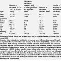
It is estimated that 80% of men and 93% of women with moderate to severe sleep apnea are undiagnosed [105]. Thus, identifying patients with OSA before surgery and anesthesia cannot rely on history alone. Prospectively identifying patients with OSA may be important because of data suggesting that they are at greater risk of perioperative morbidity [106]. For example, Gupta and colleagues [107] published a retrospective chart review in which they documented that patients with OSA were more likely to require unplanned admission to the intensive care unit (ICU) after joint replacement surgery than were patients without OSA. However, an important limitation of this study is that there was no reliable indication that it was the patients’ OSA (not other associated disease) that was responsible for the higher incidence of unplanned ICU admission. Blake and colleagues [108] showed that patients with OSA had more respiratory obstructive events per hour and more oxygen desaturation than controls without OSA during a 12-hour period on the first postoperative night. Liao and colleagues [109] published a retrospective review of postoperative complications in patients with OSA. Their data showed that patients with OSA had a higher incidence of postoperative complications compared with controls (39% vs 18%) primarily related to oxygen desaturation. The American Society of Anesthesiologists (ASA) guidelines for perioperative management of patients with OSA recommend that anesthesiologists should work to identify patients and consider obtaining sleep studies on patients at risk for OSA. However, the screening tool devised by the ASA task force has not been validated. Polysomnography remains the gold standard for the diagnosis of OSA. However, there are numerous barriers to obtaining sleep studies on patients before surgery. These studies are expensive and time-consuming and patients are often reluctant to comply with recommended testing. For example, Fidan and colleagues [110] screened 433 patients and recommended sleep studies for 41 of the patients based on symptoms. Only 18 of the patients agreed to undergo polysomnography.
Subjective clinical evaluation is probably the most common tool used to screen patients for OSA. However, this method of screening has serious limitations. Viner and colleagues [111] found that in patients with a high predicted probability of sleep apnea, subjective impression alone correctly identified only 52% of patients with sleep apnea and had a specificity of 70%. However, in patients with a low predicted probability of sleep apnea, the use of clinical data is sufficiently sensitive to permit about a 30% reduction in the number of unnecessary sleep studies. Hoffstein and Szalai [112] found that subjective impression of the examining clinician provided a sensitivity of 60% and a specificity of 63%. Clinical impression alone is not sufficient to reliably identify patients with or without sleep apnea.
The Berlin Questionnaire consists of 3 categories related to the risk of having sleep apnea. Patients can be classified into high risk or low risk based on their responses to individual questions about smoking, weight, snoring, observed apnea, and daytime hypersomnolence. This questionnaire has been validated in the primary care population with a sensitivity and specificity of 0.89 and 0.71 [113].
The sensitivity of the STOP questionnaire with an AHI greater than 5, greater than 15, and greater than 30 as cutoffs was 65.6%, 74.3%, and 79.5%. Specificity for this tool was 60%, 53%, and 48%, respectively. When BMI, age, neck circumference, and gender were incorporated, the sensitivities were increased to 83.6%, 92.9%, and 100% with specificity of 56.4%, 43%, and 37% [114].
More recently Ramachandran and colleagues [115] developed the Perioperative Sleep Apnea Prediction (P-SAP) score for the diagnosis of OSA, which incorporates airway screening tools normally used during a preoperative anesthetic assessment. The investigators identified independent clinical predictors of OSA including age greater than 43 years, male gender, obesity, snoring, diabetes type 2, hypertension, thick neck, Mallampati class 3 or 4, and reduced thyromental distance. A P-SAP score of 2 or greater resulted in high sensitivity (93%) at the expense of specificity (32%), whereas a P-SAP score of 6 or greater resulted in low sensitivity (23%) with high specificity (91%).
OSA in the perioperative period
The issue of whether patients suffer morbidity or mortality from acute episodes of airway obstruction is important because if they do not then our concerns that we may acutely worsen the risk of obstruction are misplaced. However, the available evidence suggests that this risk is all too real. For example, Gami and colleagues [75] examined death certificates for 112 persons who were diagnosed with OSA at the Mayo Clinic and who died suddenly from cardiac causes. They compared these patients with individuals in the general population who also died a sudden cardiac death. They found that in the general population the timing of sudden cardiac death peaked between 6 am and noon and was lowest during the hours of sleep, from midnight to 6 am. In contrast, the patients with diagnosed OSA were most likely to die during the hours of sleep (midnight to 6 am). In addition, the risk of dying between midnight and 6 am was directly correlated with OSA severity. This finding is not proof that OSA episodes can precipitate sudden cardiac death, but it is strong suggestive evidence.
Consistent with this study, Hanly and colleagues [116] reported that the occurrence of ST-segment depression during sleep in patients with OSA without a history of coronary artery disease was positively correlated with OSA severity, arousal index (number of arousals per hour), and time spent with O2 saturation less than 90%. Again, this study is not proof, but it is suggestive evidence that anything that acutely increases OSA severity, arousals, and/or desaturations may put patients at risk of morbid cardiac events. Our study of opioid effects in patients with OSA showed that opioid administration increased arousals, increased the amount of time spent with O2 saturations less than 90%, and increased OSA severity as measured by the AHI [117]. We found that opioids increase all of the risk factors that Hanley and colleagues found were associated with ST-segment depression in patients with OSA.
Although these findings provide only indirect evidence that OSA can acutely cause morbidity/mortality, there is one clear example of death caused by OSA. Specifically, Pearce and Saunders [118] published a case report in which a patient suffered cardiorespiratory arrest during a sleep study. The investigators attributed the arrest to the fact that the patient did not arouse sufficiently to reestablish a patent airway during an obstructive episode. (NB At the institution where this occurred, polysomnography was not monitored, ie, nobody was watching the woman who died. That is not the case at many institutions where sleep studies are continuously supervised and monitored by registered polysomnographic technologists.) The patient could not be resuscitated and postmortem examination revealed no clear cause of death. The coroner attributed death to OSA and entered this on the death certificate.
Preoperative Period
Berry and colleagues [119] used a blinded observer study design to examine the dose-dependent effects of a targeted propofol infusion on the frequency of airway obstruction in a group of patients with suspected sleep-disordered breathing and a group of control subjects. They found that all patients suspected of having sleep-disordered breathing experienced partial or complete obstruction at one or more propofol concentrations and that the frequency of obstruction was dose-related. In contrast, none of the control group experienced airway obstruction. The goal of these investigators’ study was to determine whether propofol sedation could be used as a provocative test for sleep apnea so they did not examine whether propofol sedation impaired arousal mechanisms in response to obstruction. However, because propofol can prevent arousal in response to surgery it is clear that there are propofol doses at which patients do not arouse and clear their obstructed airway. What that dose may be is unknown.
The effect of sedative doses of benzodiazepines on airway patency has also been investigated. Drummond [120] examined the effect of midazolam on airway patency and airway muscle tone in nonobese individuals of unknown OSA status. Midazolam was titrated to produce light sedation but with the retained ability to respond to a loud voice (median dose 5 mg). Tongue muscle tone decreased to 42% of baseline as measured by electromyography, and 10 of 12 patients developed airway obstruction. Drummond observed that muscle tone increased to 69% of baseline during obstruction but this increased muscle tone was insufficient to alleviate obstruction in any patient.
Opioids are another class of drugs commonly used for sedation. They carry the double risk of both generalized depression of the central nervous system and a potent depression of respiratory drive. Bernards and colleagues [117] examined the effect of remifentanil on sleep and respiration in patients with moderate OSA (AHI = 15–30) in a double-blind, randomized, single-dose, polysomnography study. These investigators found that in this group of patients receiving this remifentanil dose (0.075 μg/kg/h) remifentanil decreased the number of obstructive apneas, and did not impair arousal in response to obstruction. Arousals were increased. However, oxygen saturation was decreased on average and the number of events in which saturation decreased to less than 90% was markedly increased. The decrease in obstructive episodes was attributed to the near complete abolition of REM sleep, which is a previously described effect of opioids on human sleep pattern [121,123]. Decreased oxygen saturation was attributed not to a worsening of OSA but to opioid-mediated hypoventilation.
OSA represents a difficult challenge for the clinician caring for these patients in the perioperative period because there are virtually no data from which we can define appropriate clinical practices. For example, in 2003 the Clinical Practice Review Committee of the American Academy of Sleep Medicine (AASM) addressed the question of how best to ameliorate the risks of OSA in the perioperative period and concluded, “Scientific literature regarding the perioperative risk and best management techniques for obstructive sleep apnea patients is scanty and of limited quality. There is insufficient information to develop AASM standards of practice recommendation” [124]. Several years later (2006) the ASA Task Force on Perioperative Management of Patients with Obstructive Sleep Apnea addressed the same issue and essentially agreed with the AASM in concluding that there was insufficient scientific information to provide evidence-based recommendations for the care of patients with OSA [125]. The absence of data did not prevent the ASA from promulgating numerous recommendations, some of which are unhelpful and possibly dangerous (see later discussion).
Intraoperative Management
Airway management
Airway management is a paramount concern in the anesthetic management of all surgical patients. Because OSA is a disease of airway patency, it is reasonable to be concerned that these patients pose a greater risk of difficult airway management than do patients without OSA. However, given that a large proportion of patients with OSA are obese it is difficult to separate difficulties resulting from obesity from those resulting from OSA. A recent study by Neligen and colleagues [126] was able to distinguish OSA versus obesity-related reasons for difficult endotracheal intubation. In a group of 180 consecutive morbidly obese patients (BMI average 49.4 kg/m2) these investigators found a difficult laryngoscopy rate of 8.3% (Cormack and Lehane grade 3 or 4 view) and a difficult intubation rate of 3.3 % (3 or more intubation attempts). However, there was no correlation between the diagnosis or severity of OSA and difficulty with laryngoscopy or intubation. The only correlate with difficult laryngoscopy was neck circumference, but neck circumference did not correlate with difficult intubation. Only male gender and Mallampati score of 3 or 4 predicted difficult intubation. Thus, in this study of morbidly obese patients positioned on a ramp for intubation, OSA was not a predictor of difficult laryngoscopy or intubation.
Chung and colleagues [127] and Hiremath and colleagues [128] took a slightly different approach to addressing the issue of OSA and airway management. These investigators referred patients who were difficult to intubate for polysomnography to determine whether they had OSA. Both of these studies suffer from an unavoidable selection bias because not all patients who were difficult to intubate agreed to undergo polysomnography. However, of those who did agree to undergo polysomnography, both studies found that a significant percentage of patients who were difficult to intubate also had OSA (Chung and colleagues = 66%; Hiremath and colleagues = 28%). In both of these studies the average BMI (30 and 32 kg/m2) was in the obese range, thus it is not possible to determine whether intubation difficulties were related to obesity or OSA.
The currently available data suggest that patients with OSA may be at greater risk of difficult intubation but whether this results from obesity or OSA is unclear. However, it seems prudent to consider that airway difficulties may be more likely in patients with OSA and to plan accordingly. Thus, it seems reasonable to pay strict attention to head positioning (Isono and colleagues [129] have shown that the sniffing position is optimal for a patent airway in patients with OSA) and immediate availability of ancillary airway devices (eg, nasal and pharyngeal airways, laryngeal mask airway, fiberoptic bronchoscope).
Postoperative Care
The practice guidelines promulgated by the ASA task force on the care of patients with OSA addressed this issue. The guidelines state, “These patients should not be discharged from the recovery room to an unmonitored setting until they are no longer at risk for postoperative respiratory depression.” Given the chronic nature of their disease one is left to wonder whether patients with OSA are ever “no longer at risk of respiratory depression”, postoperative or otherwise. The ASA practice guidelines go on to define criteria by which risk of postoperative respiratory depression can be assessed: “Adequacy of post-operative respiratory function may be documented by observing patients in an unstimulated environment, preferably while they seem to be asleep, to establish that they are able to maintain their baseline oxygen saturation while breathing room air.” In evaluating this recommendation it is worth keeping in mind that these patients suffer from OSA not “preferably while they seem to be asleep” apnea. OSA is a state-dependent disease that is manifest only during sleep. Therefore, it is not reasonable to assume one can make any judgments about ventilatory adequacy in the absence of documented sleep, which is why the electroencephalography is recorded during polysomnographic diagnosis of OSA. In addition, observing a period of adequate ventilation and assuming, based on that observation, that the patient is no longer at risk is like assuming that a single normal blood sugar reading documents that a diabetic is no longer at risk of diabetic ketoacidosis. In addition, adequate ventilation at discharge from the recovery room tells one nothing about what happens when the patient begins self-administering opioids to treat postoperative pain. In addition, although the work by Bernards and colleagues [117] suggests that moderate opioid doses do not pose unique risks to patients with moderate OSA their study was performed during a single night in which remifentanil markedly suppressed REM sleep. Because many patients’ OSA is worse during REM sleep, REM suppression may increase their risk on following nights. Specifically, REM suppression is generally followed by REM rebound over subsequent nights [130,132]. The confluence of REM rebound, sleep deprivation, and ongoing opioid use may represent an important risk that does not occur until several days after patients leave the recovery room.