Right gastric artery
Right gastroepiploic artery
Esophageal branch of left gastric artery
Left gastric artery
Splenic artery
Left gastroepiploic artery
Branches of left and right gastric arteries
(Top) In “conventional” arterial anatomy of the stomach and duodenum (present in only 50% of the population), the left gastric artery arises from the celiac trunk, supplies the lesser curvature, and anastomoses with the right gastric artery, a branch of the proper hepatic artery. The greater curvature of the stomach is supplied by anastomosing branches of the gastroepiploic arteries, with the left arising from the splenic artery.
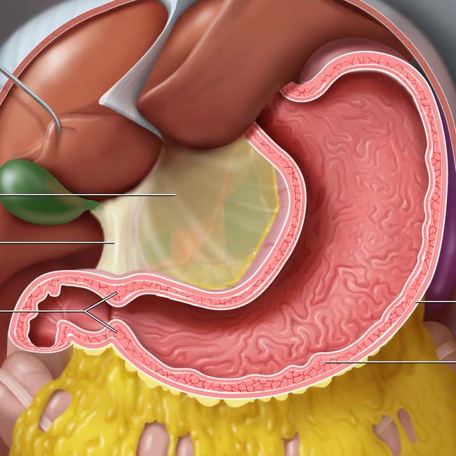
Hepatoduodenal ligament
Pyloric sphincter
Outer (longitudinal) muscle layer
Middle (circular) muscle layer
(Bottom) The lesser omentum extends from the stomach to the porta hepatis and can be divided into the broader, thinner hepatogastric ligament and the thicker hepatoduodenal ligament. Note the layers of gastric muscle, with the middle circular layer being the thickest.
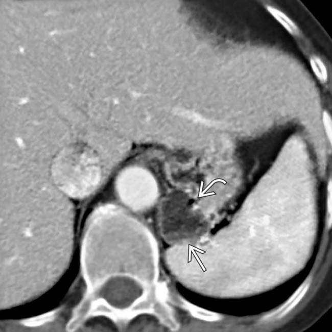
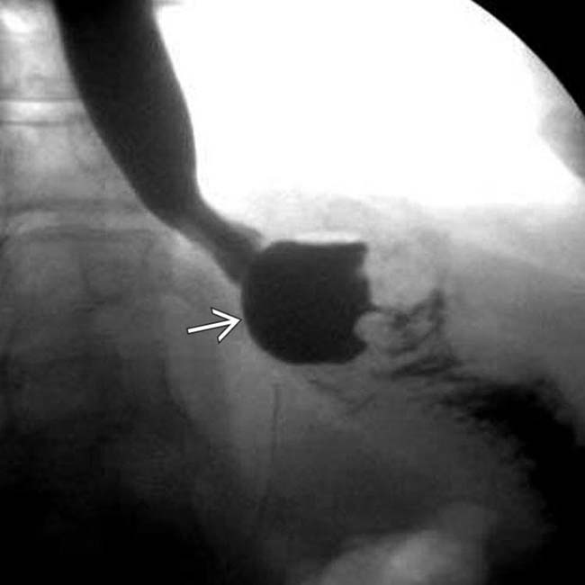
 that might be mistaken for an adrenal adenoma or other lesion. Its contiguity with the stomach and a tiny bubble of gas
  suggest the correct etiology of gastric diverticulum.
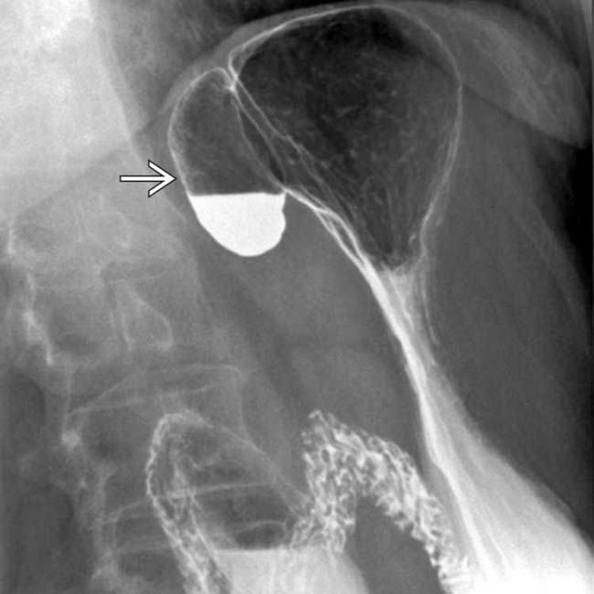
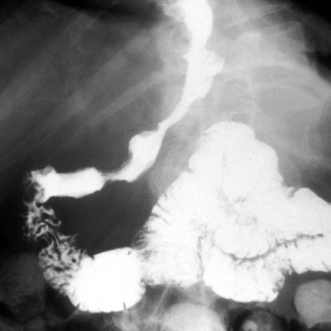
 with an air-fluid level.
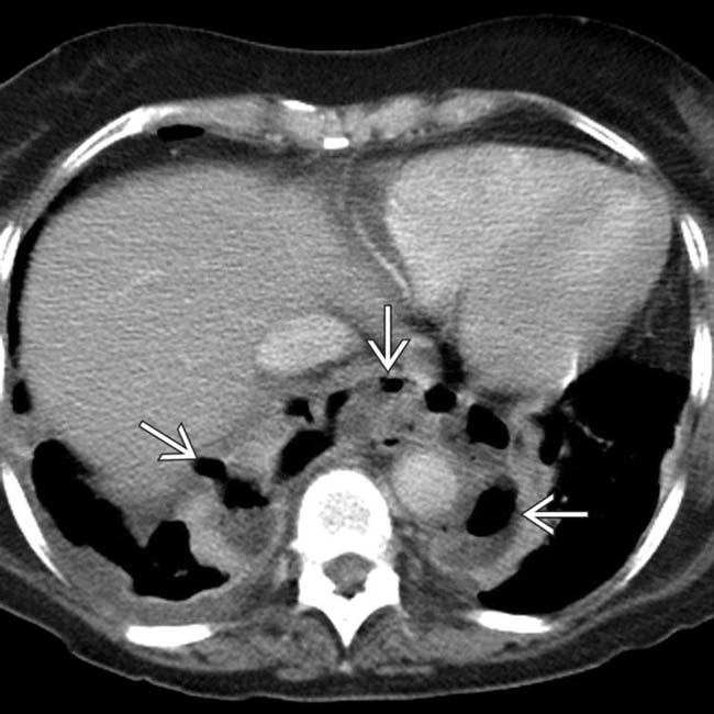
 within the mediastinum, suggestive, but not diagnostic of a leak or perforation.
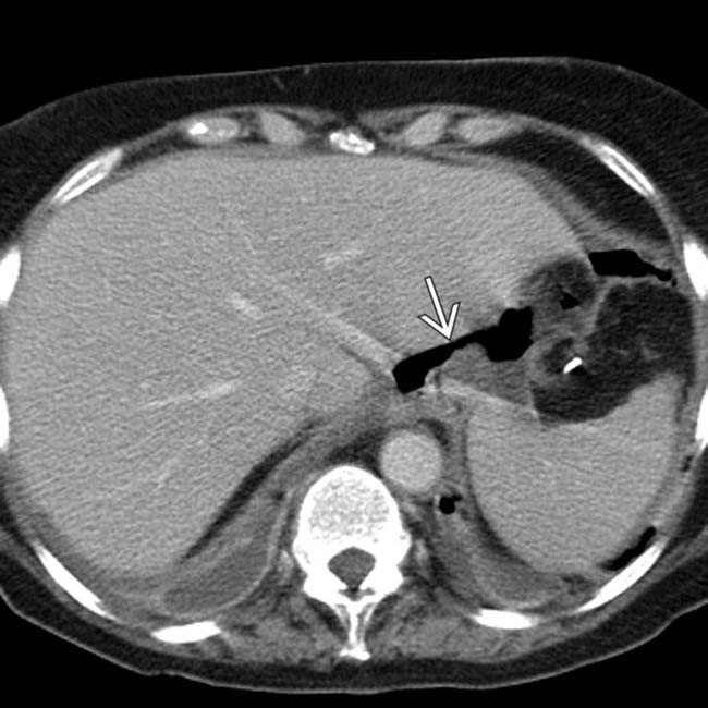
 . Bilateral pleural effusions are also noted.
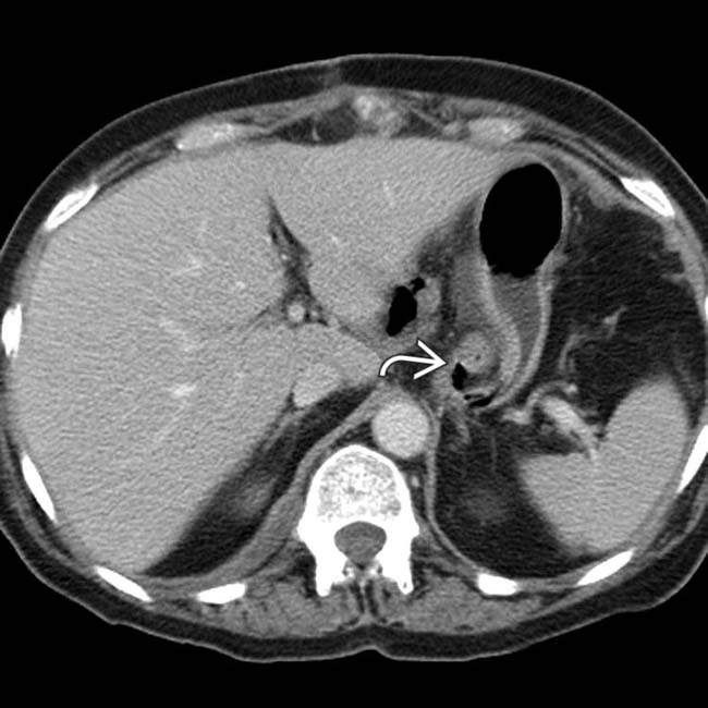
 , compressing the distal esophagus and proximal stomach.
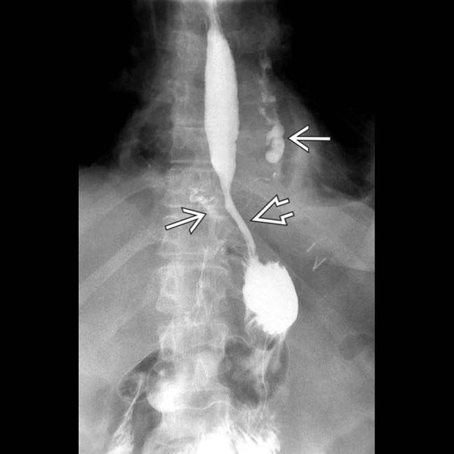
 from an intact fundoplication. Leak of contrast material
  into the mediastinum and upper abdomen, however, confirms perforation (leak) of the esophagus or the gastric wrap itself.
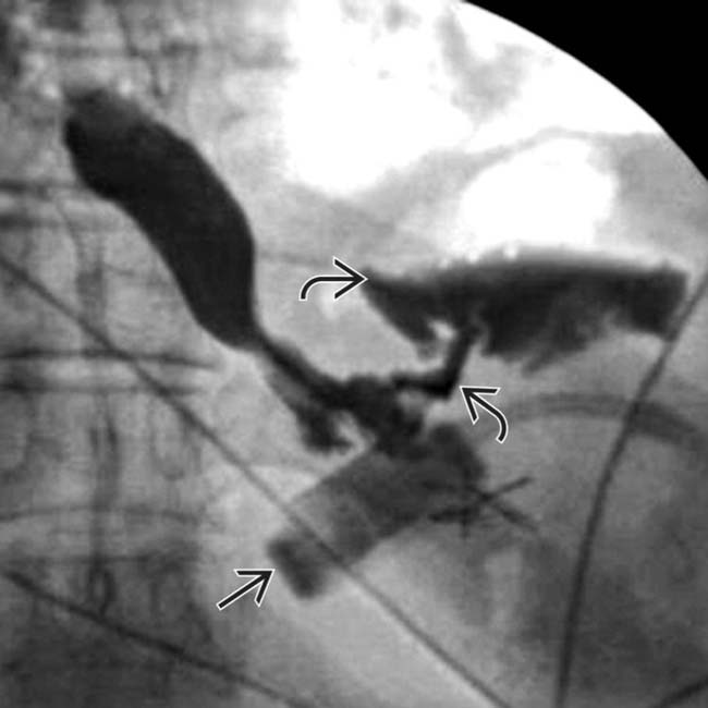
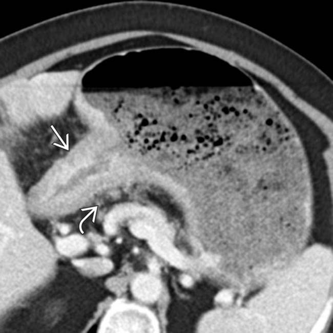
 around the proximal stomach with the correct orientation. A leak of water-soluble contrast medium and gas is evident
  .
.
 , with an air-fluid level and delayed emptying, signs of a stricture of the anastomosis between the gastric pouch and the Roux limb.
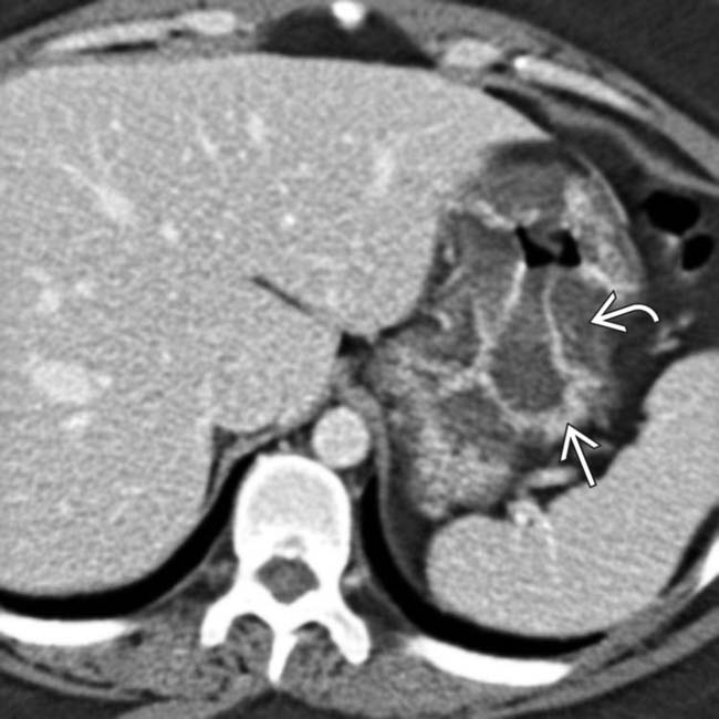
 . Note the enhancing mucosa as distinct from near water density submucosal edema
  .
.
 , and submucosal soft tissue density. Adjacent lymphadenopathy
  indicates spread beyond the stomach.
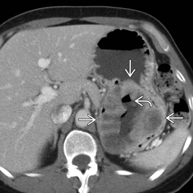
 , which is necrotic in its center and contains a gas-fluid level
  due to communication with the gastric lumen.