112
Idiopathic guttate
hypomelanosis
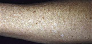
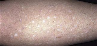
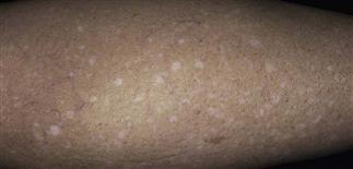
Idiopathic guttate hypomelanosis is characterized by 2- to 5-mm white spots with sharply demarcated borders.
Idiopathic guttate hypomelanosis is a sign of photodamage.
Idiopathic guttate hypomelanosis occurs in 50% of 50- to 70-year-olds.
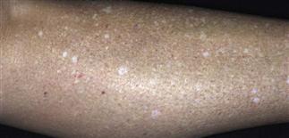
Macules are scattered on the exposed upper and lower extremities.
DESCRIPTION
Common asymptomatic dermatosis of unknown etiology consisting of small white macules on sun-exposed upper and lower extremities.
HISTORY
• Occurs in middle-aged and older people; in 50–70% of people over the age of 50. • More common in women. • Higher prevalence in family members; genetic predisposition likely. • Actinic damage has been incriminated as major cause, but a senile degenerative phenomenon may play a role. • Although asymptomatic, this condition is cosmetically distressing. • Lesions stable in size and remain fixed while the number of lesions increases with age.
PHYSICAL FINDINGS
• Macules are white and hypopigmented, 2–5 mm, with regular borders and smooth to slight xerotic scaling. • Macules are scattered on the exposed upper and lower extremities. • Patients have associated signs of photoaging, including atrophy, lentigines, and xerosis in the same areas.
TREATMENT
• Reassurance is all that is required for most patients. Encourage sun protection with clothing, as sunscreens are less effective than clothing. • White macules can be camouflaged with tinted make-up, such as Covermark or Dermablend. • Self-tanning creams that contain dihydroxyacetone darken the lesions, but the appearance is speckled and not pleasing. • A light spray with liquid nitrogen may partially fade the lesions, although there is the potential for worsening the dyspigmentation. • Intralesional triamcinolone (2.5 mg/mL) infiltrated into individual lesions may partially cause repigmentation of lesions, although there is risk of atrophy. • A qualified aesthetician skilled with spray tanning can provide excellent cover-up.


