I
iatrogenic Relating to a disorder induced by the treatment itself. Example: the development of amblyopia in the good eye following occlusion treatment.
idiopathic Relating to any primary pathological condition of unknown origin.
idoxuridine See antiviral agents.
illiterate chart See chart, illiterate.
illuminance Quotient of the luminous flux, F, incident on an element of surface divided by the area, A, of that element of surface. Symbol: E. Thus
< ?xml:namespace prefix = "mml" />

The units are in lux or footcandles. Syn. illumination.
See law of illumination, inverse square; photometer.
illuminance, retinal See retinal illuminance.
illuminants, CIE standard The colorimetric illuminants A, B, C and D defined by the CIE in terms of relative spectral energy (power distribution): standard illuminant A representing the full radiator at T = 2854 K; standard illuminant B representing direct sunlight with a correlated colour temperature of T = 4874 K; standard illuminant C representing daylight with a correlated colour temperature of T = 6774 K; standard illuminant D representing daylight with a correlated colour temperature of T = 6504 K (CIE).
See chromaticity diagram; lamp, Macbeth; light, white.
illumination 1. The action of brightening an object with light. 2. The science of the application of lighting. 3. Synonym for illuminance.
diffuse i. In slit-lamp examination, it is the illumination obtained with a wide slit and an out of focus beam or with a diffuser, thus providing an overall view of the structures of the eye.
direct i. In slit-lamp examination, the slit beam and the microscope are both focused sharply on the structure to be observed. Syn. focal illumination.
focal i. See illumination, direct.
indirect i. In slit-lamp examination, the slit beam is focused on a structure located adjacent to the structure to be observed.
inverse square law of i. See law of illumination, inverse square.
oscillation i. In slit-lamp examination, it is a technique in which the beam of light is oscillated to provide alternative direct and indirect illumination. It sometimes allows one to see slight changes more easily which otherwise would remain unnoticed under sustained illumination of either kind.
retinal i. See retinal illuminance.
retro-i. In slit-lamp examination, it is a method of illuminating a structure by using the light that is reflected by the iris or an opaque or senescent lens. This method is closely related to indirect illumination and often in corneal examination part of the cornea will simultaneously be under retro- and indirect illumination. Syn. transillumination.
See clouding, central corneal; oedema.
sclerotic scatter i. In slit-lamp examination, it is a method in which the beam of light is focused on the sclera near the limbus and the cornea remains uniformly dark in the absence of an opacity. However, an opacity in the cornea becomes easily visible as it scatters light.
See clouding, central corneal; oedema.
specular reflection i. In slit-lamp examination, it is a method in which the beam of light and the microscope are placed at equal angles from the normal to the corneal or lens surface to be viewed. This is a method for examining the quality of a surface. This method is particularly useful to observe the corneal endothelium.
See blebs, endothelial; cornea guttata; shagreen.
illuminometer See photometer.
illusion A false interpretation of an object or figure presented to the eye (visual illusion). Illusions can occur with each of the senses. See figure, ambiguous.
autokinetic visual i. The apparent motion of a luminous object fixated in the dark, or in a large blank field. It is not due to eye movements and the illusion disappears as soon as the ambient luminance increases so that other objects become visible. Syn. visual autokinesis.
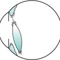
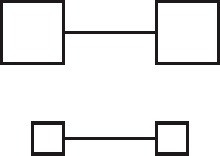
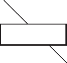
Baldwin’s i. 1. Illusion in which a line connecting two large squares appears shorter than a line connecting two smaller squares (Fig. I1). 2. Illusion in which a dot placed halfway between a large disc and a smaller disc appears to be nearer the large one.
café wall i. An illusion induced by a pattern of alternating columns of black and white rectangles (or squares) placed in such a way that the lines that they compose do not appear to be parallel. Syn. Munsterberg illusion. A variant of this illusion consists of hollow squares without alternating colour and is called a ‘hollow square illusion’.
Cornsweet i. See effect, Craik–O’Brien– Cornsweet.
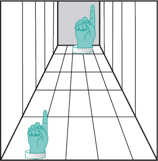
corridor i. Illusion in which images of equal size in a perspective figure of a corridor, appear to be of different sizes. The figure that seems further away appears larger than the one in the foreground (Fig. I2).
Craik–Cornsweet i. See effect, Craik–O’Brien–Cornsweet.
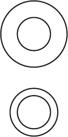
Delboeuf i. Illusion in which a circle surrounded by a slightly larger concentric circle appears larger than another circle of the same size surrounded by a much larger concentric circle (Fig. I3).
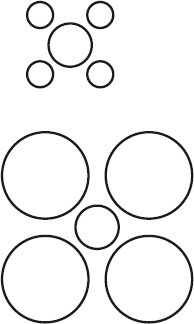
Ebbinghaus i. Illusion in which a circle usually appears larger when surrounded by smaller circles than by larger circles (Fig.I4).
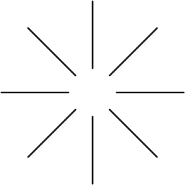
Ehrenstein’s brightness i. Illusion in which the erased area at the intersection of radial (or horizontal and vertical) lines appears to be brighter than the background and with an illusory contour (Fig. I5).
floating-finger i. Illusion noted when fixating a point in the distance while the forefingers of each hand are held horizontally about 30 centimetres in front of the eyes, with the fingertips nearly touching. A small, disembodied finger with two tips appears floating in between and can be shortened or lengthened by varying the distance between the fingertips. It is a peculiar illustration of physiological diplopia (Fig. I6).
frequency doubling i. Illusion in which a grating pattern appears to have twice as many black and white bars as it actually has.

This happens when a sinusoidal grating with a low spatial frequency (less than 4 c/deg) flickers in a counterphase fashion (i.e. light bars become dark and vice versa) at a high temporal frequency (more than 15 Hz). This type of stimulation is assumed to stimulate the non-linear mechanism within the magnocellular visual system.
See perimetry, frequency doubling.
Helmholtz i. See irradiation.
Hering’s i. Illusion in which a pair of parallel lines appear bent when placing diagonal lines across them. This illusion is most noticeable when radiating lines are crossing two parallel lines on opposite sides of the point of radiation. In this case, the two parallel lines appear to bend away from each other (Fig. I7).
Hermann’s i. See Hering–Hermann grid.
hole in the hand i. See test, hole in the hand.
hollow square i. See illusion, café wall.
horizontal-vertical visual i. Illusion in which the vertical line appears longer than the horizontal line when two lines of equal length are placed with the vertical line at the midpoint of the horizontal.
See illusion, top hat.
Jastrow i. Illusion in which two identical curved and tapering ring segments placed one above the other appear unequal in size, the band nearer the centres of curvature appearing to be the longest (Fig. I8).
Kundt’s i. Illusion occurring when one attempts to bisect a horizontal line with only one eye and the segment on the temporal side of the visual field is then larger than the other.
moon i. Illusion in which the moon appears much larger at the horizon than when viewed

high in the sky. In fact, the actual size of the moon remains constant as does its distance from the earth. One possible explanation is that at the horizon there are many other cues in the field of view (e.g. houses, mountains) that make the moon appear to be much closer than when it is high in the sky and thus should be larger.
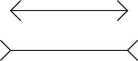
Müller–Lyer i. Illusion in which a line with outgoing fins on both ends appears longer than another of equal length but with arrowheads on both ends (Fig. I9).
Munsterberg’s i. See illusion, café wall.
oculogyral i. Illusion of apparent movement of viewed objects when the body is subjected to rotary acceleration. The initial apparent movement is opposite to that of the direction of rotation of the body and is followed by an apparent movement in the same direction.
Oppel–Kundt i. Illusion in which a divided, interrupted or filled area appears to be larger than an empty area of equal size.
optical i. See illusion, visual.
Orbison i. Illusion of a distorted geometric figure such as a square or a circle drawn on a background of radiating lines or concentric lines.
Poggendorff’s i. Illusion in which two visible portions of a diagonal line overlaid by a rectangle do not appear to be continuous (Fig. I10).
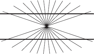
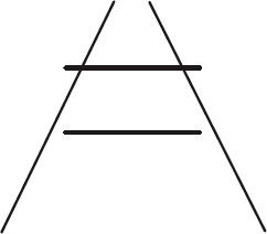
Ponzo i. Illusion in which two parallel lines of equal length do not appear equal when they are surrounded by two radiating straight lines, one on each side. The parallel line nearer the point of radiation appears to be longer (Fig. I11).
Schroeder’s staircase visual i. See Schroeder’s staircase.
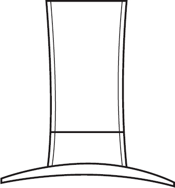
top hat i. Illusion in which a top hat drawn with equal vertical and horizontal dimensions appears to be much greater vertically than horizontally. It is closely related to the horizontal-vertical illusion (Fig. I12).
See illusion, horizontal-vertical visual.
visual i. Perception of an object or a figure that does not correspond to the actual physical characteristics of the stimulus. Syn. optical illusion; geometrical optical illusion.
waterfall i. See after-effect, waterfall.
Wundt’s i. Illusion in which a pair of parallel lines appear bent towards each other when crossed by lines radiating from two points, one on each side of the parallel lines.
See illusion, Hering’s.
Zollner’s i. Illusion in which a series of parallel lines appear to converge or diverge from
each other when crossed by short diagonal lines.
illusory contours See contour.
image A picture of an object formed by a lens, a mirror or other optical system.
aerial i. An image found in space and not on a screen, such as the image viewed in indirect ophthalmoscopy.
after-i. See after-image.
axial point i. The point of intersection of an image with the optical axis.
catadioptric i. Image formed by both reflecting and refracting surfaces.
See system, catadioptric.
catoptric i. Image formed by specular reflection, either from a mirror or by reflection at refracting surfaces such as the optical surfaces of the eye, which form the Purkinje–Sanson images.
corneal i. Catoptric image formed by either the anterior or posterior surface of the cornea. They are also called the first and second Purkinje–Sanson images.
dioptric i. An image formed by a refracting surface as distinguished from a catoptric image.
direct i. A virtual image such as the erect image seen in direct ophthalmoscopy.
double i. A pair of images obtained either optically through a doubling system or due to diplopia.
eidetic i. Visual perception arising from the imagination of the subject or what has previously been seen, and not from immediate retinal stimulation. That image may last from a few seconds to several minutes and appears to be located in front of the eyes.
entoptic i. Visual sensation arising from stimuli within the eye and perceived as in the external world. Examples: muscae volitantes; phosphene. Syn. entoptic phenomenon.
See angioscotoma; arcs, blue; entoptoscope, blue field; floaters; Haidinger’s brushes; spot, Maxwell’s.
erect i. Image that is not inverted with respect to the object such as a virtual image produced by a concave lens.
See images, Purkinje–Sanson.
extraordinary i. See birefringence.
false i. 1. The retinal image in the deviating eye in strabismus. It is less well defined than the true image. 2. See image, ghost. See image, true.
ghost i. 1. Unwanted image as may be formed by internal reflection in a lens or an optical system. These images are sometimes annoying to spectacle wearers, and even to observers as they detract from the appearance of the spectacle lens or hide the wearer’s eyes behind a veil. The intensity of ghost images is diminished by antireflection coatings. 2. The faint image seen in monocular diplopia. Syn. false image.
See Fresnel’s formula; lens flare; light, stray; mirror, front surface.
indirect i. A real image, such as the inverted image seen in indirect ophthalmoscopy.
inverted i. Image that is upside down and right for left with respect to its object. Syn. reversed image.
See images, Purkinje–Sanson.
i. jump See jump.
ocular i. 1. The retinal image. 2. The image formed by the refracting system of the eye, disregarding the presence or the position of the retina.
perceptual i.; psychic i. See image, visual.
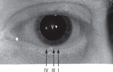
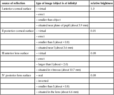
Purkinje–Sanson i’s. Catoptric images produced by reflection from the optical surfaces of the eye. The first image is reflected by the anterior surface of the cornea, the second image by the posterior surface of the cornea, the third image by the anterior surface of the crystalline lens and the fourth image by the posterior surface of the crystalline lens. Only the fourth image is inverted. The third is the largest but the first is by far the brightest (Fig. I13). During accommodation, the third image becomes smaller while the size of the fourth diminishes only a little. Purkinje–Sanson images are used to measure or calculate various optical dimensions of the eye, to establish angle alpha or lambda and to contribute to some diagnostic tests of strabismus (e.g. Hirschberg’s method; Krimsky’s method). Syn. Purkinje images.
See axis, optical; ophthalmophakometer; phacoscope.
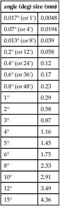
Table I1
Approximate relationship between the retinal image size of an emmetropic eye with a power of 60 D and the angular subtense of a distant object
| angle (deg) size (mm) | |
| 0.017° (or 1’) | 0.0048 |
| 0.07° (or 4’) | 0.0194 |
| 0.013° (or 8’) | 0.039 |
| 0.2° (or 12’) | 0.058 |
| 0.4° (or 24’) | 0.12 |
| 0.6° (or 36’) | 0.17 |
| 0.8° (or 48’) | 0.23 |
| 1° | 0.29 |
| 2° | 0.58 |
| 3° | 0.87 |
| 4° | 1.16 |
| 5° | 1.45 |
| 6° | 1.75 |
| 8° | 2.33 |
| 10° | 2.91 |
| 12° | 3.49 |
| 15° | 4.36 |
real i. An image that can be formed on a screen.
See focus, principal; object, real.
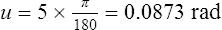
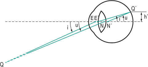
retinal i. Image formed on the retina by the optical system of the eye. The size of the retinal image h’ of a distant object subtending angle u in an emmetropic eye is equal to

where h’ is in metres, u in radians and the power of the eye F in dioptres. The formula is only valid for small angles (Fig. I14). Example: a distant object subtends an angle of 5° viewed by an emmetropic eye of power 60 D (π is equal to 3.1416)
reversed i. See image, inverted.
i. shell The curved surface containing either all the sagittal or all the tangential foci corresponding to a given object plane.
See astigmatism, oblique.
stabilized retinal i. See stabilized retinal image.
Table I2
Purkinje-Sanson images (all figures are calculated and rounded off and all distances are referred to the anterior corneal pole)
| source of reflection | type of image (object is at infinity) | relative brightness |
| I anterior corneal surface | – virtual | 1.0 |
| – erect | ||
| – smaller than object | ||
| – situated near plane of pupil (about 3.9 mm) | ||
| II posterior corneal surface | – virtual | 0.01 |
| – erect | ||
| – smaller than I (about × 0.8) | ||
| – situated near I (about 3.6 mm) | ||
| III anterior lens surface | – virtual | 0.08 |
| – erect | ||
| – larger than I (about × 2.0) | ||
| – situated in vitreous (about 10.7 mm) | ||
| IV posterior lens surface | – real | 0.08 |
| – inverted | ||
| – smaller than I (about × 0.8) | ||
| – situated in the lens (about 4.6 mm) |
true i. The retinal image in the normally fixating eye in strabismus.
virtual i. One from which refracted or reflected rays appear to have come. This image can be seen but it is not an actual image and cannot be formed on a screen. Examples: the image seen in a plane mirror; the image seen in the cornea.
See focus, principal; object, virtual.
visual i. 1. Perceived image formed by the whole visual system. It includes the physiological and psychological processing. Syn. perceptual image; psychic image. 2. A mental picture based on the recollection of a previous visual experience.
See aniseikonia; visualization.
imagery 1 . Process of recalling past visual experiences. 2. Synonym for visualization. See image, visual.
imbalance, muscular Generic term referring to a defect in the oculomotor system, as in heterophoria or strabismus.
Imbert–Fick law See law, Imbert–Fick.
immersion lens See lens, immersion.
immune system See system, immune.
immunosuppressants Drugs that prevent or reduce the immune response. They are used in the treatment of a variety of severe inflammations such as uveitis, scleritis, keratoconjunctivitis sicca, Behçet’s syndrome, sympathetic ophthalmia, and to prevent corneal graft rejection. They include the corticosteroids (e.g. prednisolone), ciclosporin (cyclosporine), tacrolimus, and cytotoxic agents (e.g. azathioprine, chlorambucil, cyclophosphamide, methotrexate). It must be noted that immunosuppressants render the patient more susceptible to infection because immunity is reduced.
implant, intraocular lens (IOL) See lens, intraocular.
impression cytology See cytology, impression.
impression, eye A negative form or replica of the anterior part of the eye. A substance with rapid gelling properties is held in contact with the eye until gelled. This impression (or mould) is then used in the preparation of a positive model called a cast (or casting) of the anterior part of the eye: it is made by filling the impression with a material containing a plaster of Paris base which hardens to artificial stone. Using this cast a shell of a scleral contact lens is produced with optimum shape of the back surface. Syn. impression; impression moulding; mould; ocular impression.
impression tonometer See tonometer, impression.
in vitro Term referring to a measurement or a process taking place in a test tube. Example: the measurement of the cholesterol content of the crystalline lens done in a test tube.
in vivo Term referring to a measurement or a process taking place in the living body. Example: the effect of a contact lens on the cornea.
inadequate stimulus See stimulus, adequate.
incandescence Emission of visible radiation by thermal excitation (CIE).
See lamp, incandescent electric; luminescence.
incidence 1. The intersection of a ray of light with an optical surface. 2. The number of new cases of a specific disease or condition occurring during a specific period of time (e.g. 1 year) divided by the population at risk during that period. Example: the incidence of keratoconus in Olmsted County, Minnesota was found to be 2 cases per 100 000 population a year.
See prevalence.
incidence, angle of See angle of incidence.
incidence, plane of See plane of incidence.
incident ray See ray, incident.
inclusion conjunctivitis See conjunctivitis, adult inclusion.
incoherent See coherent sources.
incomitance Condition in which the manifest or latent angle of deviation of the lines of sight of the two eyes differs according to which eye is fixating or in which direction the eyes are looking. This condition is usually attributed to a paresis or paralysis of one or more of the extraocular muscles. Syn. nonconcomitance. See anisophoria; concomitance; strabismus, incomitant; strabismus, paralytic.
incongruity, retinal See retinal correspondence, abnormal.
incongruous diplopia; scotoma See under the nouns.
incyclophoria See cyclophoria.
incyclovergence See excyclovergence.
indentation, scleral A clinical procedure used in conjunction with indirect ophthalmoscopy in which some slight pressure is applied to the sclera to bring the peripheral retina into view. Retinal detachments and tears, for example, are more easily seen with this procedure. Pressure is usually applied with an instrument called an indentor. The technique is contraindicated in patients with elevated intraocular pressure.
See ophthalmoscope, indirect.
indentation tonometer See tonometer, impression.
indentor See indentation, scleral.
index myopia See myopia, lenticular.
index of refraction The ratio of the speed of light in a vacuum or in air, c, to the speed of light in a given medium, v. Symbol: n. Hence,

The speed of light in a given medium depends upon its wavelength. Consequently, the index of refraction varies accordingly, being greater for short wavelengths (blue) than for longer wavelengths (red). The index of refraction forms the basis of Snell’s law, which quantitatively determines the deviation of light rays traversing a surface separating two media of different refractive indices. Syn. refractive index. Plural: indices.
See dispersion; law of refraction; lens, gradient-index; lens, high index; light, speed of; refractometer.
index of refraction, absolute The ratio of the speed of light in a vacuum to the speed of light in a given medium.
Table I3
Refractive indices of some transparent media at selected wavelengths
| spectral line | G | F | D | C | A |
| origin | calcium | hydrogen | sodium | hydrogen | oxygen |
| wavelength (nm) | 430.8 | 486.1 | 589.3 | 656.3 | 759.4 |
| aqueous or vitreous humour | 1.3440 | 1.3404 | 1.3360 | 1.3341 | 1.3317 |
| crystalline lens | 1.4307 | 1.4259 | 1.4200 | 1.4175 | 1.4144 |
| spectacle crown | 1.5348 | 1.5293 | 1.5230 | 1.5204 | 1.5163 |
| dense flint | 1.6397 | 1.6290 | 1.6170 | 1.6122 | 1.6062 |
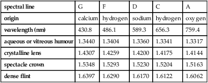
Table I4
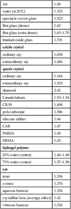
Index of refraction n of various media for sodium light (λ = 589.3)
| Air | 1.00 |
| water (at 20°C) | 1.333 |
| spectacle crown glass | 1.523 |
| flint glass (dense) | 1.62 |
| flint glass (extra dense) | 1.65–1.70 |
| titanium oxide glass | 1.701 |
| calcite crystal | |
| ordinary ray | 1.658 |
| extraordinary ray | 1.486 |
| quartz crystal | |
| ordinary ray | 1.544 |
| extraordinary ray | 1.553 |
| diamond | 2.42 |
| Canada balsam | 1.53–1.54 |
| CR-39 | 1.498 |
| polycarbonate | 1.586 |
| silicone rubber | 1.44 |
| CAB | 1.47 |
| PMMA | 1.49 |
| HEMA | 1.43 |
| hydrogel polymer | |
| 20% water content | 1.46–1.48 |
| 75% water content | 1.37–1.38 |
| eye | |
| tears | 1.336 |
| cornea | 1.376 |
| aqueous humour | 1.336 |
| crystalline lens (average effect) | 1.42 |
| vitreous humour | 1.336 |
index of refraction, relative The ratio of the speed of light in air (or other medium of reference) to the speed of light in a given medium.
indirect ophthalmoscope See ophthalmoscope, indirect.
indirect vision See vision, peripheral.
indomethacin See antiinflammatory drug.
induced astigmatism See astigmatism, induced.
induced prism See prism, induced.
induction The production of an effect by indirect or asynchronized stimulation.
colour i. The modification or generation of colour perception without direct stimulation of the corresponding cones.
See after-image; Benham’s top; Bidwell’s ghost.
spatial i. Modification of perception as a result of a simultaneous stimulation in another part of the visual field.
See summation.
temporal i. Modification of perception as a result of a previous stimulus and in some cases a later stimulus, as in metacontrast.
See summation.
industrial vision See vision, industrial.
infantile esotropia syndrome See strabismus, infantile.
infantile glaucoma See glaucoma, congenital.
infection An invasion of the body by diseaseproducing microorganisms (e.g. bacteria, virus, fungus, parasite). Treatment typically includes anti-infective drugs, such as antibiotic, antifungal or antiviral agents.
See inflammation.
inferior oblique muscle; orbital fissure; rectus muscle See under the nouns.
inferior tarsal muscle See muscles, Muller’s palpebral.
infinity, optical In optics, it is the region from which a point on an object sends rays of light which are considered to be parallel onto an optical system. Consequently it forms a clear image in the focal plane of that system. In clinical optometry, 6 metres is usually regarded as infinity.
inflammation A complex reaction that occurs in response to injury, infection, irritation, toxicity or hypersensitivity. The reaction is characterized by redness, heat, pain and swelling to different degrees. Treatment depends on the cause.
See antiinflammatory drug; infection.
infraduction See depression.
infranuclear Situated under a nucleus. It refers here to nerve fibres situated below the abducens, oculomotor or trochlear cranial nerve nuclei.
infraorbital canal See canal, infraorbital.
infrared (IR) Radiant energy of wavelengths between the extreme red wavelengths of the visible spectrum and a wavelength of a few millimetres. The wave band comprising radiations between 780 and 1400 nm is referred to as IR-A. Excessive exposure to these radiations can cause visual loss (e.g. eclipse blindness) and cataract. The waveband comprising radiations between 1400 and 3000 nm is referred to as IR-B. Excessive exposure to these radiations can cause cataract and corneal opacity. The wave band comprising radiations between 3000 and 1 × 10⁶ nm (or 1 mm) is referred to as IR-C. Excessive exposure to these radiations can cause cataract (heat-ray cataract).
See blindness, eclipse; lens, absorptive; optometer, infrared.
infravergence Movement of one eye downward relative to the other. Syn. deorsumvergence. See supravergence; vergence.
infraversion See version.
inheritance The acquisition of traits, characteristics and disorders from parents to their children by transmission of genetic information. Genes come in pairs: one originating from the father, the other from the mother. If an individual presents only the hereditary characteristics determined by one gene of the pair on an autosomal chromosome, that gene is called dominant. Conditions caused by such genes are said to show autosomal dominant inheritance. For instance, for a rare autosomal dominant disease, if one parent is affected, then on average about 50% of their children will also be affected, irrespective of the children’s sex. Examples: Marfan’s syndrome, congenital stationary night blindness, neurofibromatosis 1 and 2, von Hippel–Lindau disease. If the individual does not present the hereditary characteristics unless both genes in a pair are of the same type, then the gene is called recessive. Conditions caused by such genes are said to show autosomal recessive
Table I5
Divisions of the infrared spectrum
| IR-A (near) | 780–1400 nm |
| IR-B (middle) | 1400–3000 nm |
| IR-C (far) | 3000–1000000 nm |
inheritance. For a rare autosomal recessive disease, if a child is affected, then on average about 25% of their siblings will also be affected, irrespective of their sex. Examples: Laurence–Moon–Biedl syndrome, Tay–Sachs disease, oculocutaneous albinism, galactokinase deficiency.
Thirdly, inheritance may be controlled by genes on one of the sex chromosomes, most often the X chromosome. A recessive mutation on the single X chromosome carried by a male will cause a disease, whereas in the female, a recessive X chromosome mutation would have to be carried on both of her X chromosomes. Therefore in X-linked recessive inheritance (sex-linked recessive inheritance) males are affected more often than females. Examples: colour blindness, ocular albinism, choroideremia. A fourth type of inheritance considered in ophthalmic practice is mitochondrial (maternal) inheritance in which the inheritance of a trait encoded in the mitochondrial DNA is transmitted through the female line (mother to son or mother to daughter). Examples: Leber’s hereditary optic atrophy; Kearns–Sayre syndrome.
See acquired; chromosome; colour vision, defective; gene; hereditary.
inhibition, lateral Action of one neuron (e.g. in the retina) on the neighbouring neuron, the effect of which is to depress or prevent activity in the latter. This mechanism accounts for the increased contrast perception observed at the border of a black and white pattern. In the retina this is produced by the lateral connections of the amacrine and horizontal cells that interconnect the various retinal cells. Syn. lateral antagonism.
See field, receptive; Hering-Hermann grid; Mach’s bands.
injection 1 . A state of visible hyperaemia due to dilatation and engorgement of the small blood vessels. 2. The act of introducing a drug into the body.
ciliary i. Redness (almost lilac) around the limbus of the eye caused by dilatation of the deeper small blood vessels located around the cornea. It occurs in inflammation of the cornea, iris and ciliary body, and in angle-closure glaucoma. Each of these conditions is associated with loss of vision and usually pain. Syn. ciliary flush.
See decongestant, ocular; eye, red; plexus, pericorneal.
conjunctival i. Redness (bright red or pink) of the conjunctiva fading towards the limbus due to dilatation of the superficial conjunctival blood vessels occurring in conjunctival inflammations. There is no loss of vision but ocular discomfort and no pain. See decongestant, ocular; eye, red; ophthalmopathy, thyroid; plexus, pericorneal.
intravitreal i. Injection into the eye posterior to the limbus and directed towards the vitreous. It may be used to administer medication, corticosteroids (e.g. triamcinolone), an antiviral agent (e.g. ganciclovir) in extremely severe ocular inflammations, usually of a purulent nature, to inject antibiotics (e.g. amikacin, ceftazidime, vancomycin) immediately after vitrectomy, or to inject anti-VEGF drugs in the treatment of wet age-related macular degeneration.
peribulbar i. Injection of a local anaesthetic (e.g. bupivacaine, lidocaine, procaine) around the globe (either single or multiple injections) to produce anaesthesia of the globe and periocular tissues, as well as paralysis of the extraocular muscles. Peribulbar injection may also be used to administer medication (e.g. corticosteroids) in posterior segment inflammation. Syn. peribulbar block.
retrobulbar i. Injection of a local anaesthetic into the muscle cone behind the eye to produce anaesthesia of the globe and periocular tissues, as well as paralysis of the extraocular muscles (akinesia). It is used less commonly than peribulbar block. Syn. retrobulbar block.
subconjunctival i. A method of administering medication (e.g. antibiotics, corticosteroids, mydriatics) postoperatively, or in acute anterior segment inflammations. An area of conjunctiva away from the limbus is lifted to form a bleb and an injection is made into it.
sub-Tenon’s i. Injection of a local anaesthetic near or beyond the equator using a cannula, which has been inserted under the conjunctiva and Tenon’s capsule a few mm from the limbus and slid posteriorly to produce anaesthesia of the globe as well as paralysis of the extraocular muscles. A sub-Tenon’s injection may also be used to administer medication (e.g. corticosteroids) in posterior segment inflammation.
injury See irrigation.
inner nuclear layer; plexiform layer See retina.
innervation, reciprocal See law of reciprocal innervation, Sherrington’s.
insertion See muscles, extraocular.
instability, binocular A condition in which there is a difficulty in the maintenance of clear, single binocular vision. The patient tends to lose the line of text while reading or there is an apparent movement of the text. Clinical features include reduced fusional reserve, unstable heterophoria and unequal visual acuities.
See convergence, relative.
Intacs Trade name of an intracorneal implant consisting of two tiny half-ring segments, which are inserted into the cornea to reshape its curvature and correct ametropia. The method is presently used to flatten the cornea by a given amount (the thicker the ring segments the flatter the cornea) in order to correct low myopia. It is an outpatient procedure carried out under local anaesthesia, takes less than half an hour and is reversible. The ring segments are made of clear biocompatible plastic inserted into the stroma and around the optical zone of the cornea.
intensity, luminous Quotient of the luminous flux leaving the source, propagated in an element of solid angle containing the given direction, divided by the element of solid angle. Symbol: I. Unit: candela (CIE).
intercilium See glabella.
interface, optical A plane or surface forming a common boundary between two optical media.
interference Modification of light intensity arising from the joint effects of two or more coherent trains of light waves superimposed at the same point in space and arriving at the same instant. The waves may either reinforce each other, being in phase (constructive interference) or cancel each other, being out of phase (destructive interference).
See coherent sources; experiment, Young’s; holography; phase; tomography, optical coherence.
interference filter See filter, interference.
interference fringes The alternate light and dark bands produced when two or more coherent rays of light are superimposed on a surface.
interferometer 1. Instrument designed to measure the wavelength of light, the refractive index of a medium, as well as the flatness, thickness, the quality of optical surfaces, etc. The interferometer is based on the phenomenon of interference between two coherent beams of light. 2. Name given to several types of clinical maxwellian view system used to measure visual acuity.
See maxwellian view system, clinical.
interferometer, partial coherence See biometry of the eye.
intermittent strabismus See strabismus, intermittent.
intermuscular membrane See membrane, intermuscular.
internal hordeolum; ophthalmoplegia See under the nouns.
internal limiting membrane See membrane of the retina, internal limiting.
internal rectus muscle See muscle, medial rectus; muscles, extraocular.
internuclear ophthalmoplegia See ophthalmoplegia, internuclear.
interocular Situated between the eyes.
interocular distance See distance, interocular.
interocular transfer (IOT) Refers to a change in threshold in one eye which had been occluded, similar to, but of lower magnitude than that in the fixating eye, in response to a visual stimulation. Example: an elevation of contrast threshold with both eyes following adaptation to high contrast gratings of a given spatial frequency in one eye. The presence of interocular transfer indicates the existence of binocular cortical neurons.
interpalpebral fissure See aperture, palpebral.
interposition See perception, depth.
interpupillary distance See distance, interpupillary.
interpupillometer See pupillometer.
interstitial keratitis See keratitis, interstitial.
intertrabecular spaces See meshwork, trabecular.
interval, achromatic See interval, photochromatic.
interval, astigmatic; focal See Sturm, interval of.
interval, photochromatic Range of low luminances between the absolute threshold of light perception and the threshold of hue. The length of this interval varies with wavelength, being nearly nil around 650 nm (Fig. I15). Syn. achromatic interval.
See Purkinje shift; theory, duplicity.
interval of Sturm See Sturm, interval of.
intorsion See torsion.
intracapsular cataract extraction See cataract extraction, intracapsular.
intraocular Within the eye.

intraocular lens implant See lens, intraocular.
intraocular muscles; pressure See under the nouns.
intrinsic light See light, idioretinal.
intrinsic muscles See muscles, intraocular.
inverse square law of illumination See law of illumination, inverse square.
inverted retina See retina, inverted.
invisible spectrum See spectrum, invisible.
involuntary eye movements See movements, fixation.
involutional ectropion; entropion See under the nouns.
iodine See antiseptic.
iodopsin A photosensitive pigment found in the retinal cones of the chicken. Its maximum absorption is around 562 nm.
IOL Master See biometry of the eye.
ipsilateral A term relating to the same side. See contralateral; geniculate bodies, lateral.
iridaemia Haemorrhage from the iris. Note: also spelt iridemia.
iridectomy The surgical removal of part of the iris. The main reasons for iridectomy are to reduce the intraocular pressure, to enlarge an abnormally small pupil, to excise an iris tumour and, in cataract extraction, to prevent possible blockage of the angle of the anterior chamber. Nowadays, laser iridotomy is preferred in the treatment of angle-closure glaucoma because the incision obtained by this technique can be carried out as an outpatient procedure with only topical anaesthesia, although the cornea must not be hazy (Fig. I16).
See iridotomy.
irideremia Absence of all or part of the iris. Strictly speaking a total absence of the iris is called aniridia.
See aniridia.
iridescent Presenting a rainbow-like play of colours as in soap bubbles.
iridiagnosis See iridodiagnosis.
iridis rubeosis See rubeosis iridis.
iridocorneal angle See angle of the anterior chamber.
iridocorneal endothelial syndrome See syndrome, ICE.
iridocyclitis Inflammation of both iris and ciliary body. The ciliary body is almost always involved with an inflammation of the iris. The clinical picture of iridocyclitis is practically the same as iritis. The condition is often associated with ankylosing spondylitis or sarcoidosis.
See anisocoria; arthritis, rheumatoid; heterochromia; syndrome, Behçet’s; uveitis.
iridocyclitis, Fuchs’ heterochromic A chronic, idiopathic, non-granulomatous anterior uveitis characterized by heterochromia, often complicated by cataract which then leads to blurred vision, the main complaint. It is occasionally bilateral; in this case there is no heterochromia. The condition occurs in about 4% of all cases of uveitis. Keratic precipitates are usually present, being small, round or stellate, grey-white in colour and scattered throughout the posterior surface of the cornea. They do not conglomerate or become pigmented and filaments may be seen in between. Glaucoma may develop. Treatment may involve topical steroids as well as cataract or glaucoma therapy. Syn. Fuchs’ uveitis syndrome; Fuchs’ heterochromic cyclitis; heterochromatic iridocyclitis.
See keratic precipitates.
iridodiagnosis Diagnosis of systemic diseases through observation of changes in form and colour of the iris. The validity of this method is questionable. Syn. iridiagnosis.
See iridology.
iridodialysis A tearing away of the iris from its attachment to the ciliary body. It usually occurs as a result of blunt trauma to the eye.
iridology The study of the iris (colour, shape, etc.), normal and abnormal.
See iridodiagnosis.
iridodonesis A tremulous condition of the iris. It usually occurs in aphakic eyes, when the eye is subluxated or when there has been an injury to the eye. Syn. tremulous iris.
See luxation of the lens; phacodonesis.
iridoplegia Paralysis of the sphincter muscle of the iris resulting in a dilated pupil. The iridoplegia can be partial as in Argyll Robertson pupil, or complete in which case the pupil does not react to light or to a near object. It may be due to trauma, drugs (e.g. cocaine instilled in the eye) or a systemic disease (e.g. neurosyphilis).
iridoschisis A condition in which the anterior stroma of the iris atrophies and separates from the posterior layer. It mostly affects the inferior iris in elderly patients. In advanced cases the ruptured anterior fibres float in the aqueous humour. It often accompanies angle-closure glaucoma.
iridotomy Creation of an opening in the iris to allow aqueous humour to flow from the posterior to the anterior chamber. It is commonly performed with a neodymium-yag or argon laser (laser iridotomy) in angle-closure glaucoma, especially that caused by pupillary block.
See iridectomy; pupillary block.
iris The anterior part of the vascular tunic of the eye, which is situated in front of the crystalline lens and behind the cornea. It has the shape of a circular membrane with a perforation in the centre (the pupil) and is attached peripherally to the ciliary body. The iris forms a curtain dividing the space between the cornea and the lens into the anterior and posterior chambers of the eye. The anterior surface of the iris is divided into two portions: the largest peripheral ciliary zone and the inner pupillary zone. The two zones are separated by a zigzag line, the collarette. The iris consists of four layers which are, starting in the front: (1) the layer of fibrocytes and melanocytes; (2) the stroma in which are embedded the following structures: (a) the sphincter pupillae muscle which constricts the pupil and is supplied mainly by parasympathetic fibres via the third cranial nerve, (b) the vessels which form the bulk of the iris, and (c) the pigment cells; (3) the posterior membrane consisting of plain muscle fibres which constitute the dilator muscle which is supplied mainly by sympathetic motor fibres, via the long ciliary nerves; (4) the posterior epithelium which is highly pigmented.
Sensory fibres from the iris are contained in the nasociliary branch of the ophthalmic nerve. The blood supply is provided by the ciliary arteries. The colour of the iris is blue in babies belonging to the white races and changes colour after a few months of life as pigment is deposited in the anterior limiting layer and the stroma. Iris colour is inherited; brown as a dominant trait and blue as a recessive trait. Iris patterns are unique for each individual and can be used as a type of identification. The function of the iris and pupil is to regulate the amount of light admitted into the eye, to optimize the depth of focus and to mitigate ocular aberrations. See cell, clump; corectopia; Fig. C 13 ; Fuchs, crypts of; heterochromia; inheritance; iridectomy; iridodialysis; iridology; iritis; melanin; membrane, pupillary; polycoria; reflex, pupil light.
Table I6
Differential diagnosis* between acute conjunctivitis, acute iritis and angle-closure glaucoma
| acute conjunctivitis | acute iritis (anterior uveitis) | angle-closure glaucoma | |
| signs | |||
| injection | conjunctival | ciliary | conjunctival and ciliary |
| pupil | normal | contracted | semi-dilated and fixed |
| intraocular pressure | normal | normal or low, occasionally | high |
| increased | |||
| cornea | normal | KP | oedematous |
| anterior chamber | normal depth | normal depth, aqueous | shallow |
| flare | |||
| iris | normal | faded | faded |
| view of fundus | clear | misty | almost invisible |
| symptoms | |||
| pain | irritation | moderate to servere | very severe and radiating |
| photophobia | slight | marked | slight |
| lacrimation | watery, purulent | watery | watery |
| or mucopurulent | |||
| vision | normal | slightly reduced | much reduced, haloes |
| onset | gradual | rapid | sudden |
| systemic complications | none | malaise or fever | nausea and vomiting |
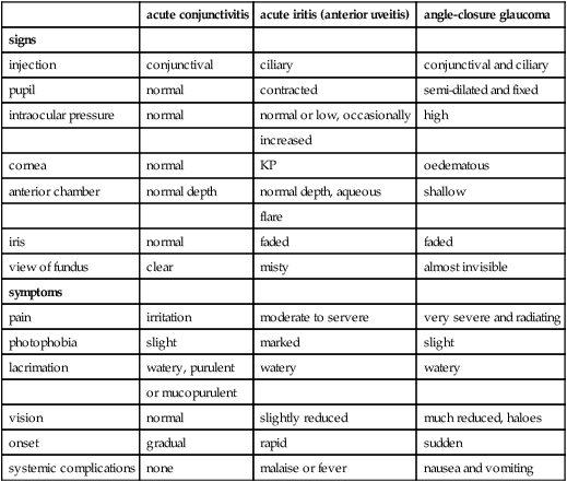
*This is a guide, as individual cases vary according to the cause and severity of the disease.
iris bombé A condition occurring in posterior annular synechia in which an increase of aqueous humour contained in the posterior chamber causes a forward bulging of the iris. It may provoke an attack of angle-closure glaucoma as the iris may block the drainage angle (Fig. I17).
iris coloboma; melanoma; naevus; nodules See under the nouns.
iris naevus syndrome See syndrome, Cogan–Reese.
iris neovascularization See neovascularization, iris.
iris, plateau An anatomical anomaly in which the iris lies in a plane rather than bulging anteriorly. This is due to the fact that the root (or ciliary margin) of the iris is inserted more anteriorly into the ciliary body than is usual. On dilatation of the pupil the peripheral iris expands against the trabecular meshwork. It can predispose the eye to angle-closure glaucoma.

iris processes Fine bands of tissue extending from the anterior surface of the iris, bridging the angle of the anterior chamber from the root of the iris to the scleral spur or more often to the trabecular meshwork into which they usually merge. The processes are found in only about half of the population.
iris, prolapse of the Protrusion of a portion of the iris into a corneal wound. It results from either trauma, a severe corneal ulcer or an operation. In some cases an anterior synechia may develop as the iris remains fixed in the wound by scar tissue.
iris, tremulous See iridodonesis.
iritis Inflammation of the iris. The acute form is usually characterized by pain, photophobia, ciliary injection, exudates in the anterior chamber (aqueous flare), keratic precipitates, oedema, constricted and sluggish pupil, discoloration of the iris, posterior synechia, lacrimation and loss of vision. In some cases there may be hypopyon and an increase in intraocular pressure due to blocking of the angle of the anterior chamber. Iritis is most often associated with cyclitis (anterior uveitis). The majority of cases are idiopathic or the result of trauma or medication. Treatment includes mydriatics (to prevent synechia) and topical corticosteroid drops. It is essential to differentiate acute iritis from angle-closure glaucoma because of the possible harm of using a mydriatic in the latter.
See iridocyclitis; photophobia; syndrome, Fuchs’; uveitis.
Irlen lens See syndrome, Meares–Irlen.
Irlen’s syndrome See syndrome, Meares–Irlen.
iron deposits See line, iron; siderosis.
irradiation 1 . Application of electromagnetic radiations to an object. 2. A phenomenon in which a bright area against a black background appears larger than a darker area of equal size against the same background. Syn. Helmholtz illusion.
irregular astigmatism See astigmatism, irregular.
irrigation The act of washing or cleansing a cavity or a surface with a stream of water or other solution (e.g. physiological saline) as in chemical or thermal burns or other superficial injuries to the eye, or to dislodge small foreign bodies on the cornea or in the conjunctival sac.
See corneal abrasion; eversion, lid.
Irvine–Gass syndrome See oedema, cystoid macular.
ischaemia Insufficient blood supply for the need of a part of the body, usually as a result of a disease of the blood vessels supplying that part. Note: also spelt ischemia.
ischaemia of the retina Lack of blood in the retina due either to arterial narrowing or profuse haemorrhage from any part of the body. See arteritis, temporal.
ischaemic optic neuropathy; ocular syndrome See under the nouns.
iseikonia Condition in which the size and shape of the ocular images of the two eyes are equal, as distinguished from aniseikonia.
iseikonic lens See lens, aniseikonic.
Ishihara test See plates, pseudoisochromatic.
island of vision See vision, island of.
isoacuity area 1 . An area surrounding the fixation point in which visual acuity is approximately constant. The width of this area varies with the test target, being smallest with resolution of two dots and largest with Landolt rings or gratings. 2. An area of the visual field in which visual acuity is more or less constant.
isoametropia Condition in which the ametropia is similar in the two eyes. Syn. isometropia.
isoametropic amblyopia See amblyopia.
isochromatic Possessing the same colour.
isocoria Having two pupils of equal size. See anisocoria.
isoluminant Possessing the same luminance. Example: red and green bars of a grating which have the same luminance. If such a grating is moving sideways, many observers will barely perceive its motion or fail to notice it altogether. Syn. equiluminant.
isometropia See isoametropia.
isophoria Constancy of the heterophoria in various directions of gaze.
See heterophoria.
isopia Identical vision in the two eyes.
isopter In the determination of visual fields, it is the contour line representing the limits of equal retinal sensitivity to a given test target. See field, visual; perimeter.
isotonic solution See solution, isotonic.
isotropic Having the same properties of refraction in all directions.
See anisotropic; birefringence.
itraconazole See antifungal agent.
ivermectin See onchocerciasis.