Hypoparathyroidism

Published on 22/03/2015 by admin

Filed under Pediatrics

Last modified 22/04/2025

Print this page

rate 1 star rate 2 star rate 3 star rate 4 star rate 5 star
Your rating: none, Average: 0 (0 votes)

This article have been viewed 1386 times

Chapter 565 Hypoparathyroidism

Etiology

Hypocalcemia is common between 12 and 72 hr of life, especially in premature infants, in infants with asphyxia, and in infants of diabetic mothers (early neonatal hypocalcemia) (Chapter 100) (Table 565-1). After the 2nd to 3rd day and during the 1st wk of life, the type of feeding also is a determinant of the level of serum calcium (late neonatal hypocalcemia). The role played by the parathyroid glands in these hypocalcemic infants remains to be clarified, although functional immaturity of the parathyroid glands is invoked as one pathogenetic factor. In a group of infants with transient idiopathic hypocalcemia (1-8 wk of age), serum levels of parathyroid hormone (PTH) are significantly lower than those in normal infants. It is possible that the functional immaturity is a manifestation of a delay in development of the enzymes that convert glandular PTH to secreted PTH; other mechanisms are possible.

Table 565-1 ETIOLOGIC CLASSIFICATION OF HYPOCALCEMIA

PARATHYROID HORMONE DEFICIENCY

PARATHYROID HORMONE RECEPTOR DEFECTS (PSEUDOHYPOPARATHYROIDISM)

MITOCHONDRIAL DNA MUTATIONS

MAGNESIUM DEFICIENCY

EXOGENOUS INORGANIC PHOSPHATE EXCESS

VITAMIN D DEFICIENCY

APECED, autoimmune polyendocrinopathy-candidiasis-ectodermal dystrophy; cAMP, cyclic adenosine monophosphate; PTH, parathyroid hormone.

Aplasia or Hypoplasia of the Parathyroid Glands

Aplasia or hypoplasia of the parathyroid glands is often associated with the DiGeorge/velocardiofacial syndrome (see Fig. 564-1). This syndrome occurs in 1/4,000 newborns. In 90% of patients, the condition is caused by a deletion of chromosome 22q11.2. Approximately 25% of these patients inherit the chromosomal abnormality from a parent. Neonatal hypocalcemia occurs in 60% of affected patients, but it is transitory in the majority; hypocalcemia can recur or can have its onset later in life. Associated abnormalities of the 3rd and 4th pharyngeal pouches are common; these include conotruncal defects of the heart in 25%, velopharyngeal insufficiency in 32%, cleft palate in 9%, renal anomalies in 35%, and aplasia of the thymus with severe immunodeficiency in 1%. This syndrome has also been reported in a small number of patients with a deletion of chromosome 10p13, in infants of diabetic mothers, and in infants born to mothers treated with retinoic acid for acne early in pregnancy.

Autoimmune Hypoparathyroidism

An autoimmune mechanism for hypoparathyroidism is strongly suggested by the finding of parathyroid antibodies and by its frequent association with other autoimmune disorders or organ-specific antibodies. Autoimmune hypoparathyroidism is often associated with Addison disease and chronic mucocutaneous candidiasis. The association of at least 2 of these 3 conditions has been tentatively classified as autoimmune polyglandular disease type I. It is also known as autoimmune polyendocrinopathy, candidiasis, and ectodermal dystrophy (APCED). This syndrome is inherited in an autosomal recessive fashion and is not related to any single HLA-associated haplotype. One third of patients with this syndrome have all 3 components; 66% have only 2 of 3 conditions. The candidiasis almost always precedes the other disorders (70% of cases occur in children <5 yr of age); the hypoparathyroidism (90% after 3 yr of age) usually occurs before Addison disease (90% after 6 yr of age). A variety of other disorders occur at various times and include alopecia areata or totalis, malabsorption disorder, pernicious anemia, gonadal failure, chronic active hepatitis, vitiligo, and insulin-dependent diabetes. Some of these associations might not appear until adult life. Autoimmune thyroid disease is a rare concomitant finding.

Affected siblings can have the same or different constellations of disorders (hypoparathyroidism, Addison disease). The disorder is exceptionally prevalent among Finns and Iranian Jews. The gene for this disorder is designated AIRE (autoimmune regulator); it is located on chromosome 21q22. It appears to be a transcription factor that plays an essential role in the development of immunologic tolerance. Patients with Addison disease as part of polyendocrinopathy syndrome type I have demonstrated adrenal-specific autoantibody reactivity directed against the side-chain cleavage enzyme.

Idiopathic Hypoparathyroidism

The term idiopathic hypoparathyroidism should be reserved for the small residuum of children with hypoparathyroidism for whom no causative mechanism can be defined. Most children in whom onset of hypoparathyroidism occurs after the first few years of life have an autoimmune condition. Autoantibodies to the extracellular domain of the calcium-sensing receptor have been identified in some patients with acquired hypoparathyroidism. One should always consider incomplete forms of DiGeorge syndrome or an activating calcium-sensing receptor mutation in the differential diagnosis.

Clinical Manifestations

There is a spectrum of parathyroid deficiencies with clinical manifestations varying from no symptoms to those of complete and long-standing deficiency. Mild deficiency may be revealed only by appropriate laboratory studies. Muscular pain and cramps are early manifestations; they progress to numbness, stiffness, and tingling of the hands and feet. There may be only a positive Chvostek or Trousseau sign or laryngeal and carpopedal spasms. Convulsions with or without loss of consciousness can occur at intervals of days, weeks, or months. These episodes can begin with abdominal pain, followed by tonic rigidity, retraction of the head, and cyanosis. Hypoparathyroidism is often mistaken for epilepsy. Headache, vomiting, increased intracranial pressure, and papilledema may be associated with convulsions and might suggest a brain tumor.

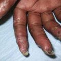
In patients with long-standing hypocalcemia, the teeth erupt late and irregularly. Enamel formation is irregular, and the teeth may be unusually soft. The skin may be dry and scaly, and the nails might have horizontal lines. Mucocutaneous candidiasis, when present, antedates the development of hypoparathyroidism; the candidal infection most often involves the nails, the oral mucosa, the angles of the mouth, and less often, the skin; it is difficult to treat.

Cataracts in patients with long-standing untreated disease are a direct consequence of hypoparathyroidism; other autoimmune ocular disorders such as keratoconjunctivitis can also occur. Manifestations of Addison disease, lymphocytic thyroiditis, pernicious anemia, alopecia areata or totalis, hepatitis, and primary gonadal insufficiency may also be associated with those of hypoparathyroidism.

Permanent physical and mental deterioration occur if initiation of treatment is long delayed.

Treatment

Emergency treatment of neonatal tetany consists of intravenous injections of 5-10 mL or 1-3 mg/kg of a 10% solution of calcium gluconate (elemental calcium 9.3 mg/mL) at the rate of 0.5-1 mL/min while the heart rate is monitored and a total dose not to exceed 20 mg of elemental calcium/kg. Additionally, 1,25-dihydroxycholecalciferol (calcitriol) should be given. The initial dosage is 0.25 µg/24 hr; the maintenance dosage ranges from 0.01-0.10 µg/kg/24 hr to a maximum of 1-2 µg/24 hr. Calcitriol has a short half-life and should be given in 2 equal divided doses; it has the advantages of rapid onset of effect (1-4 days) and rapid reversal of hypercalcemia after discontinuation in the event of overdosage (calcium levels begin to fall in 3-4 days). Calcitriol is supplied as an oral solution.

An adequate intake of calcium should be ensured. Supplemental calcium can be given in the form of calcium gluconate or calcium glubionate (Neo-Calglucon) to provide 800 mg of elemental calcium daily, but it is rarely essential. Foods with high phosphorus content such as milk, eggs, and cheese should be reduced in the diet.

Clinical evaluation of the patient and frequent determinations of the serum calcium levels are indicated in the early stages of treatment to determine the requirement for calcitriol or vitamin D2. If hypercalcemia occurs, therapy should be discontinued and resumed at a lower dose after the serum calcium level has returned to normal. In long-standing cases of hypercalcemia, repair of cerebral and dental changes is not likely. Pigmentation, lowering of blood pressure, or weight loss can indicate adrenal insufficiency, which requires specific treatment. Patients with autosomal dominant hypocalcemic hypercalciuria can develop nephrocalcinosis and renal impairment if treated with vitamin D.

Differential Diagnosis

Magnesium deficiency must be considered in patients with unexplained hypocalcemia. Concentrations of serum magnesium <1.5 mg/dL (1.2 mEq/L) are usually abnormal. Familial hypomagnesemia with secondary hypocalcemia has been reported in about 50 patients, most of whom developed tetany and seizures at 2-6 wk of age. Administration of calcium is ineffective, but administration of magnesium promptly corrects both calcium and magnesium levels. Oral supplements of magnesium are necessary to maintain levels of magnesium in the normal range. Two genetic forms have been described. One is caused by an autosomal recessive gene on chromosome 9, resulting in a specific defect in absorption of magnesium. The other is caused by an autosomal dominant gene on chromosome 11q23, resulting in renal loss of magnesium.

Hypomagnesemia also occurs in malabsorption syndromes such as Crohn disease and cystic fibrosis. Patients with autoimmune polyglandular disease type I and hypoparathyroidism can also have concurrent steatorrhea and low magnesium levels. Therapy with aminoglycosides causes hypomagnesemia by increasing urinary losses.

It is not clear how low levels of magnesium lead to hypocalcemia. Evidence suggests that hypomagnesemia impairs release of PTH and induces resistance to the effects of the hormone, but other mechanisms also may be operative.

Poisoning with inorganic phosphate leads to hypocalcemia and tetany. Infants administered large doses of inorganic phosphates, either as laxatives or as sodium phosphate enemas, have had sudden onset of tetany, with serum calcium levels <5 mg/dL and markedly elevated levels of phosphate. Symptoms are quickly relieved by intravenous administration of calcium. The mechanism of the hypocalcemia is not clear (Chapter 52.6).

Hypocalcemia can occur early in the course of treatment of acute lymphoblastic leukemia. Hypocalcemia is usually associated with hyperphosphatemia resulting from destruction of lymphoblasts.

Episodic symptomatic hypocalcemia occurs in the Kenny-Caffey syndrome, which is characterized by medullary stenosis of the long bones, short stature, delayed closure of the fontanel, delayed bone age, and eye abnormalities. Idiopathic hypoparathyroidism and abnormal PTH levels have been found. Autosomal dominant and autosomal recessive modes of inheritance have been reported. Mutations of the TBCE gene (1q 43-44) perturb microtubule organization in diseased cells.

Bibliography

Alimohammadi M, Björklund P, Hallgren A, et al. Autoimmune polyendocrine syndrome type 1 and NALP5, a parathyroid autoantigen. N Engl J Med. 2008;358:1018-1028.

Bowl MR, Nesbit MA, Harding B, et al. An interstitial deletion-insertion involving chromosome 2p25.3 and Xq27.1, near SOX3, causes X-linked recessive hypoparathyroidism. J Clin Invest. 2005;115(10):2822-2831.

Brown EM. The calcium-sensing receptor (CaR) and its disorders. Hormones. 2002;1:10-21.

Ding C, Buckingham B, Levine M. Familial isolated hypoparathyroidism caused by a mutation in the gene for the transcription factor GCMB. J Clin Invest. 2001;108:1215-1220.

Gavalas NG, Kemp EH, Krohn KJ, et al. The calcium-sensing receptor is a target of autoantibodies in patients with autoimmune polyendocrine syndrome type 1. J Clin Endocrinol Metab. 2007;92(6):2107-2114.

Glaudemans B, van der Wijst J, Scola RH, et al. A missense mutation in the Kv1.1 voltage-gated potassium channel-encoding gene KCNA1 is linked to human autosomal dominant hypomagnesemia. J Clin Invest. 2009;119(4):936-942.

Kahn KT, Uma R, Farag TI, et al. Kenny-Caffey syndrome in six Bedouin sibships: autosomal recessive inheritance is confirmed. Am J Med Genet. 1997;69:126-132.

Mannstadt M, Bertrand G, Muresan M, et al. Dominant-negative GCMB mutations cause an autosomal dominant form of hypoparathyroidism. J Clin Endocrinol Metab. 2008;93(9):3568-3576.

Marx SJ. Hyperparathyroid and hypoparathyroid disorders. N Engl J Med. 2000;343:1863-1875.

Oskarsdottir S, Persson C, Eriksson BO, et al. Presenting phenotype in 100 children with the 22q11 deletion syndrome. Eur J Pediatr. 2005;164(3):146-153.

Parvari R, Hershkovitz F, Grossman N, et al. Mutation of TBCE causes hypoparathyroidism-retardation-dysmorphism and autosomal recessive Kenny-Caffey syndrome. Nat Genet. 2002;32:448-452.

Perheentupa J. Autoimmune polyendocrinopathy-candidiasis-ectodermal dystrophy. J Clin Endocrinol Metab. 2006;91(8):2843-2850.

Tengan CH, Kiyomoto BH, Moraes CT, et al. Mitochondrial encephalomyopathy and hypoparathyroidism associated with a duplication and a deletion of mitochondrial deoxyribonucleic acid. J Clin Endocrinol Metab. 1998;83:125-129.

Thomas BR, Bennett JD. Symptomatic hypocalcemia and hypoparathyroidism in two infants of mothers with hyperparathyroidism and familial benign hypercalcemia. J Perinatol. 1995;15:23-26.

Yagi H, Furutani Y, Hamada H, et al. Role of TBX1 in human del22q11.2 syndrome. Lancet. 2003;362(9393):1366-1373.