Hypoglycemia

Published on 06/06/2015 by admin

Filed under Pediatrics

Last modified 22/04/2025

Print this page

rate 1 star rate 2 star rate 3 star rate 4 star rate 5 star
Your rating: none, Average: 0 (0 votes)

This article have been viewed 2366 times

72 Hypoglycemia

Hypoglycemia is often the initial manifestation of a disorder of energy metabolism and regulation. The accurate diagnosis of the underlying condition facilitates institution of disease-specific therapy that can prevent long-term complications such as seizures, developmental delay, permanent brain damage, and even death. In children, the most common cause of persistent hypoglycemia is hyperinsulinism, a disorder affecting approximately 1 in 20,000 children in the United States.

Etiology and Pathogenesis

In children, as in adults, blood glucose levels are maintained within a narrow range in the postabsorptive and fed state. Thus, normal fasting blood glucose levels should be above 70 mg/dL. A lower level of 50 mg/dL is recommended for diagnostic purposes only. The classic diagnostic triad of hypoglycemia, also known as “Whipple’s triad,” consists of a blood glucose level less than 50 mg/dL, symptoms of hypoglycemia, and resolution of symptoms with normalization of the blood glucose level.

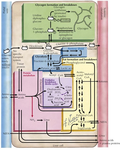
To recognize the different causes of hypoglycemia, it is helpful to understand the fundamentals of energy homeostasis (Figure 72-1). In the fed state, as glucose levels increase, so do insulin levels. Insulin stimulates glucose uptake into cells for use as a source of energy and metabolic intermediate or for storage (see Chapter 4, Figure 4-1). Insulin has other effects that influence energy metabolism; in the liver, insulin inhibits glycogenolysis, gluconeogenesis, and ketogenesis. In the fasted state, insulin secretion is suppressed, allowing glycogenolysis and gluconeogenesis to commence, followed by fatty-acid oxidation, which leads to ketogenesis. In the absence of glucose, the brain uses ketones (e.g., β-hydroxybutyrate and acetoacetate) as energy sources. Insulin secretion from pancreatic β-islet cells is suppressed in the fasted state, and there is increased secretion of counterregulatory hormones that maintain blood glucose levels by stimulating glycogenolysis and gluconeogenesis, such as glucagon, cortisol, growth hormone, and epinephrine. Disorders that impair insulin regulation and counterregulatory hormone secretion, as well as storage or production of glucose, can result in hypoglycemia.

Clinical Presentation

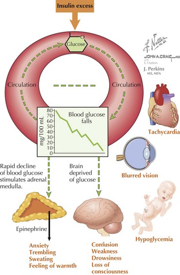
The symptoms of hypoglycemia can result from two different mechanisms. An adrenergic response (e.g., fight or flight) typically is triggered in response to a rapid decrease in blood glucose, as occurs with postprandial hypoglycemia (PPH). By contrast, the slower decline in blood glucose that occurs with fasting hypoglycemia may not trigger an obvious adrenergic response but can manifest as loss of consciousness caused by neuroglycopenia (Figure 72-2). In general, hypoglycemia in infants and children is fasting hypoglycemia. Hypoglycemia in neonates can present with irritability, shakiness, difficulty feeding, hypothermia, pallor, hypotonia, and seizures. In children, symptoms include sweatiness, unsteadiness, headache, hunger, nausea, weakness, tachycardia, change in mentation, and seizures. If symptoms suggestive of hypoglycemia are present, it is imperative, if possible, to send a blood specimen for a glucose level to the laboratory to confirm that hypoglycemia is at the root of the symptoms.

Differential Diagnosis

The differential diagnosis of hypoglycemia is expansive and includes not only disorders of carbohydrate metabolism but also disorders of fat oxidation, hormone deficiencies, and medication-induced hypoglycemia (Box 72-1). It is perhaps most useful to separate these disorders into those associated with hyperinsulinism and those associated with appropriately suppressed levels of insulin. Not included in this classification is transient neonatal hypoglycemia, typically seen within the first 6 hours of life, caused by immaturity of fasting mechanisms and poor glucose stores in premature infants and breastfed infants. In these cases, the hypoglycemia improves with feedings and typically resolves within the first day of life.

Box 72-1 Differential Diagnosis of Hypoglycemia in Infants and Children

ATCH, adrenocorticotropic hormone; F-1,6-Pase, fructose-1,6-biphosphatase; GDH, glutamate dehydrogenase; GSD 0, glycogen synthase deficiency; GSD 1a, glucose-6-phosphatase deficiency; GSD 1b, glucose-6-phosphate translocase deficiency; GSD 3; debrancher enzyme deficiency; GSD 6, glycogen phosphorylase deficiency; GSD 9, phosphorylase kinase deficiency; KATP, adenosine triphosphate–sensitive potassium; PPH, postprandial hypoglycemia; SCHAD, short-chain 3-hydroxyacyl-CoA dehydrogenase.

Hypoglycemia Secondary to Excessive and Inappropriate Insulin

Permanent Hypoglycemia (Congenital Hyperinsulinism)

The most common cause of persistent hypoglycemia in infants and children is congenital hyperinsulinism (CHI). In general, infants with CHI are large for gestational age and develop severe hypoglycemia shortly after birth. They require large amounts of intravenous (IV) glucose to maintain blood glucose above 70 mg/dL. During hypoglycemia (blood glucose <50 mg/dL), these infants have inappropriately normal or elevated serum insulin levels (although insulin levels may not be detected in all assays), suppressed free fatty acids and β-hydroxybutyrate, and a glycemic response to glucagon. Additional laboratory tests that may help distinguish specific forms of hyperinsulinism include ammonia (glutamate dehydrogenase hyperinsulinism), 3-hydroxy-butyrylcarnitine (short chain 3-hydroxacyl Co-A dehydrogenase hyperinsulinism), and abnormally processed transferrin (congenital disorders of glycosylation). Mutations in at least six genes have been associated with hyperinsulinism: the sulfonylurea receptor 1 (SUR-1), potassium inward rectifying channel (Kir6.2), glucokinase (GK), glutamate dehydrogenase (GDH), short-chain 3-hydroxyacyl-CoA dehydrogenase (SCHAD), and ectopic expression on the β-cell plasma membrane of SLC16A1 (encodes monocarboxylate transporter 1 [MCT1]). Ectopic expression of MCT-1 on the plasma membrane of pancreatic β cells leads to exercise-induced hyperinsulinism.

The most common and severe form of CHI is caused by inactivating mutations of the adenosine triphosphate–sensitive potassium (KATP) channel, made up of SUR-1 and Kir6.2. Inactivating mutations in the KATP channel result in constitutive closure of the channel, allowing membrane depolarization and calcium influx into the β cell, resulting in constitutive insulin secretion from the β cell. In about half of cases, the baby has inherited a defective allele from each parent, resulting in diffuse hyperinsulinism. In the remaining cases, a loss of function mutation inherited from the father in combination with loss of heterozygosity with loss of tumor suppressor genes imprinted in the maternal allele results in focal hyperinsulinism.

Other Causes of Hyperinsulinism

Hypoglycemia Caused by Nonhyperinsulinemic States

A large number of disorders are associated with hypoglycemia with appropriately suppressed insulin levels. Accordingly, serum (and urine) levels of ketones are elevated in children with these conditions (except in cases caused by fatty acid oxidation [FAO] or defective ketone production), which distinguishes them from patients with hyperinsulinism.

Adrenal Insufficiency (Addison’s Disease)

Patients with primary adrenal insufficiency resulting in the loss of glucocorticoid production may present with hypoglycemia (see Chapter 70). Additionally, these patients can present with hyponatremia, hyperkalemia, and dehydration caused by a loss of mineralocorticoid production. Patients may also appear hyperpigmented secondary to the effects of excess ACTH. The diagnosis can be made by checking 8 AM cortisol and ACTH levels or by performing a high-dose ACTH stimulation test.

Evaluation and Management

Glucose can be measured in whole blood or serum (i.e., plasma). In the past, blood glucose values were given in terms of whole blood, but most laboratories now measure and report serum glucose levels. By contrast, point-of-use glucometers typically measure and report whole blood glucose concentrations. Serum has a higher water content than blood cells and consequently contains more dissolved glucose than does whole blood. A calculated serum glucose level can be estimated from the whole-blood glucose by multiplying the blood glucose level by 1.15.

Additional caveats are worth noting. Blood samples that are not processed promptly can have erroneously low glucose levels owing to glycolysis by red and white blood cells (i.e., at room temperature, the decline of whole blood glucose can be 5-7 mg/dL/hr). In addition, hospital bedside glucose monitors and similar home glucose monitors are less precise than clinical laboratory methods and can be expected to have an error range of 10% to 15%. For practical reasons, the authors use the same threshold of 70 mg/dL if using plasma glucose or whole blood, but for diagnostic purposes, all measurements below 60 mg/dL should be verified with a plasma level in the clinical laboratory.

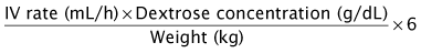
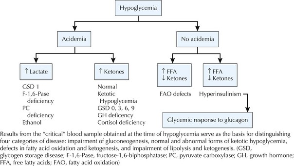
In severely symptomatic or clinically unstable patients with hypoglycemia, the first step should be administration of a 2 mL/kg bolus of 10% dextrose in water IV followed by a continuous infusion of 10% dextrose in an age-appropriate saline concentration at a glucose infusion rate of 6 to 8 mg/kg/min (Box 72-2). Whenever hypoglycemia is suspected in a clinically stable patient, the first step should be to send a “critical” blood sample to the laboratory to confirm hypoglycemia and to obtain the diagnostic tests necessary to establish the underlying cause of hypoglycemia (Box 72-3). If the patient is stable after the critical sample is sent, a glucagon stimulation test will help to refine the differential diagnosis (Figure 72-3).

The initial management, regardless of the etiology, is essentially always the same: dextrose-containing IV fluids. Long-term management of children with hypoglycemia depends on the cause of the hypoglycemia.

Treatment of Hyperinsulinism

The mainstay therapy for hyperinsulinism is diazoxide. Diazoxide suppresses insulin secretion by its action in the KATP channel. Because a functional KATP channel is required for diazoxide to exert an effect, most patients with KATP hyperinsulinism do not respond to diazoxide. A notable exception is a child with a dominant inhibitor mutation in a KATP gene, which causes a milder form of diffuse hyperinsulinism. The dosage of diazoxide is 5 to 15 mg/kg/day given orally and divided into two equal doses. The side effects of diazoxide include sodium and fluid retention and hypertrichosis. If it occurs, fluid retention can be managed with concomitant diuretic therapy.

Second-line medical therapy for infants unresponsive to diazoxide is octreotide. Octreotide is a long-acting somatostatin analog that inhibits insulin secretion distal to the KATP channel by inducing hyperpolarization of β cells, direct inhibition of voltage-dependent calcium channels, and more distal events in the insulin secretory pathway. Octreotide is administered either subcutaneously every 6 to 8 hours or via continuous infusion at 5 to 20 µg/kg/d. The initial response to octreotide is good in most cases of hyperinsulinism, but tachyphylaxis develops after a few doses, rendering therapy inadequate for long-term use. Potential side effects of octreotide include nausea, diarrhea, gallstones, necrotizing enterocolitis, and suppression of growth hormone.

Glucagon can also be given as a continuous IV infusion of 1 mg/d to help maintain euglycemia in the hospital setting. Children with focal hyperinsulinism are excellent candidates for surgery if the lesion can be identified. Patients with diffuse hyperinsulinism require surgical resection of the pancreas to ameliorate or resolve the hypoglycemia if they do not respond to medical therapy (e.g., octreotide).