Hypertensive Disorders of Pregnancy

Published on 10/03/2015 by admin

Filed under Obstetrics & Gynecology

Last modified 22/04/2025

Print this page

rate 1 star rate 2 star rate 3 star rate 4 star rate 5 star
Your rating: none, Average: 0 (0 votes)

This article have been viewed 1714 times

Chapter 14 Hypertensive Disorders of Pregnancy

The hypertensive disorders of pregnancy are major contributors to maternal and perinatal morbidity and mortality. In the mother, they can cause multiorgan system dysfunction including renal failure, hepatic failure, central nervous system (CNS) hemorrhage and stroke, pulmonary edema, placental abruption and disseminated intravascular coagulation (DIC). Fetal and neonatal complications include growth restriction, prematurity, and perinatal death. The Centers for Disease Control and Prevention (CDC) have reported that preeclampsia/eclampsia is the third leading cause of maternal mortality in the United States, primarily due to CNS hemorrhage. The combined incidence of hypertensive disorders in pregnancy varies depending on the population being studied and on the criteria used but is reported to range from 12% to 22%, whereas the preeclampsia/eclampsia syndrome occurs in about 5% to 8% of pregnancies.

image Classification and Definitions

The general classification of hypertensive disorders recommended by the Working Group Report on High Blood Pressure in Pregnancy (2000) and adopted by the American College of Obstetricians and Gynecologists (ACOG) in 2002 is listed in Box 14-1. Toxemia should not be used because it represents the entire spectrum of hypertensive disorders of pregnancy and may also refer to isolated proteinuria.

Blood pressure readings vary depending on maternal position and the gestational age of the pregnancy. Maternal blood pressure tends to be lower in the left lateral decubitus position and higher in the sitting position. In the supine position, some pregnant women have elevated pressures, whereas others have supine hypotension due to compression of the vena cava by the uterus. In addition to positional variations, arterial blood pressure normally declines during the first and second trimesters of pregnancy and rises to prepregnant levels in the third trimester.

The diagnosis of hypertension should be reserved for patients with a systolic blood pressure of greater than or equal to 140 mm Hg or a diastolic pressure of greater than or equal to 90 mm Hg. Blood pressure measurements should be taken in the sitting position after the woman has rested at least 10 minutes. Arterial pressures may also be taken in the lateral decubitus position, but the measurements should be corrected to the level of the right atrium. In the hospitalized patient, either sitting or lateral decubitus measurements may be used, but consistency is advised. The length of the blood pressure cuff should be at least 1.5 times the circumference of the upper arm, and the fifth Korotkoff sound (disappearance) should be used for determining diastolic pressure.

PREECLAMPSIA/ECLAMPSIA

Preeclampsia is a syndrome unique to pregnancy, characterized by the new onset of hypertension and proteinuria in the latter half of gestation. Preeclampsia is classically considered to be a disease affecting the first pregnancy, but it also occurs in multiparas, especially if there are predisposing risk factors such as twins, diabetes mellitus, chronic hypertension, or a change in husband/partner. When it arises in the early second trimester (14 to 20 weeks), a hydatidiform mole or choriocarcinoma should be considered.

The following two criteria are essential for the diagnosis of preeclampsia: (1) the development of hypertension (systolic blood pressure ≥ 140 mm Hg or diastolic blood pressure ≥ 90 mm Hg), in a woman whose blood pressures were previously normal, after the 20th week of pregnancy; (2) the development of new-onset proteinuria after the 20th week of gestation. Proteinuria is defined as more than or equal to 0.3 g protein in a timed 24-hour urine collection. This usually correlates with a urinalysis report of 30 mg/dL (1+ on dipstick) or greater on a clean-catch urine sample.

In the past, a 30-mm Hg rise in systolic blood pressure or a 15-mm Hg rise in diastolic pressure was considered a sign of preeclampsia. Because of the previously described physiologic rise in blood pressure during the third trimester and the frequent lack of accurate prepregnant blood pressures for use as a baseline, this rise is no longer considered diagnostic if the blood pressure remains under 140/90 mm Hg. Despite this, rising blood pressures should be of concern because they may precede the development of the full preeclampsia syndrome. Similarly, preeclampsia is often preceded by, or associated with, the development of generalized edema. Dependent edema (edema of the lower extremities) is very common in normal pregnancies. Hand and facial edema are more likely to be associated with preeclampsia, but if unaccompanied by hypertension and proteinuria, they are not diagnostic of the preeclampsia syndrome.

Preeclampsia is divided into mild and severe forms, depending on the severity of the hypertension, the amount of proteinuria, and the degree to which other organ systems are affected. Box 14-2 lists specific criteria for the diagnosis of severe preeclampsia. If any of the symptoms, signs, or laboratory abnormalities listed in Box 14-2 is present in a woman with preeclampsia, it is very likely that she has severe disease, which is associated with much greater maternal and perinatal morbidity.

A variant of severe preeclampsia with particularly high morbidity is the HELLP syndrome. This syndrome occurs in preeclamptic women with evidence of hemolysis, elevated liver enzymes, and low platelets (thrombocytopenia). In contrast to more typical presentations of preeclampsia, the patient with HELLP syndrome is more likely to be multiparous, older than 25 years, and at less than 36 weeks’ gestation. Hypertension may be initially absent in 20% of the patients, whereas 30% will have mild elevations in blood pressure, and 50% will have severe elevations.

image Preeclampsia/Eclampsia

ETIOLOGY

Preeclampsia is called a “disease of theories,” because genetic, immunologic, vascular, hormonal, nutritional, and behavioral factors have all been proposed as causes. No single, definitive “cause” has been identified, and the origins of the disease are considered to be multifactorial. Because of the resolution of the preeclampsia after delivery, most attention has been focused on the placenta and the uterine-placental-fetal interface.

Placental ischemia, or hypoxia, appears to be central to the development of the disease and has been attributed to failure of the cytotrophoblasts to adequately invade the uterine spiral arteries and establish the low-resistance uteroplacental circulation characteristic of normal pregnancy. Placental ischemia could also be due to underlying maternal vascular disease such as might occur in chronic hypertension, or to immunologically mediated placental vascular damage (see Chapter 6). Alternatively, ischemia could be caused by increased metabolic demand in the setting of a multiple gestation or a large singleton fetus.

It is postulated that uteroplacental ischemia results in oxidative stress leading to the production and release of toxins that enter the circulation and cause widespread inflammation, endothelial dysfunction, and activation of the coagulation system. The nature of these toxins has not yet been identified but may involve production of reactive oxygen and reactive nitrogen species. This is supported by the observation that preeclampsia is increased in pregnant women with underlying conditions such as obesity and diabetes that are associated with chronic inflammation and dyslipidemia. A hypoxic placenta may also shed microparticles derived from apoptosis of syncytiotrophoblasts, which can then lead to widespread endothelial injury. Antiangiogenic factors have also been shown to cause systemic hypertension, vascular injury, and activation of the coagulation system. Preeclamptic women have an imbalance in angiogenic and antiangiogenic proteins. Circulating levels of the proangiogenic proteins vascular endothelial growth factor (VEGF) and placental growth factor (PlGF) are decreased, whereas levels of the antiangiogenic proteins soluble fms-like tyrosine kinase 1 (sFlt1) and soluble endoglin are markedly increased. In animal models, overexpression of sFlt1 results in a preeclampsia-like syndrome.

Endothelial dysfunction leads to an imbalance between different classes of locally produced vasoconstrictors and vasodilators. Preeclampsia is associated with a disturbance in prostaglandin production, with a decrease in the ratio of the vasodilators prostaglandin E2 (PGE2) and prostacyclin to the vasoconstrictor PGF series and thromboxanes. Endothelial changes also appear to involve a relative deficiency in the production of nitric oxide, a vasodilator and inhibitor of platelet aggregation, along with increased production of endothelin-I. Endothelin-I is an extremely potent vasoconstrictor and activator of platelets. This shift in the production of locally acting vasoactive substances could enhance vasoconstriction in response to circulating pressor hormones.

The net effect of these processes would be widespread vasoconstriction leading to hypoxic and ischemic damage in different vascular beds, systemic hypertension, the HELLP syndrome (see later) or DIC, and worsening placental ischemia. The relative severity of the signs and symptoms of preeclampsia in any given individual would vary on the basis of which specific organ systems were most affected.

PATHOPHYSIOLOGY

Although the cause of preeclampsia is unknown, one of the primary underlying pathophysiologic abnormalities is generalized vasospasm. Cardiac output in untreated pregnant patients with preeclampsia is not significantly different from that of normal subjects in the last trimester of pregnancy, but systemic vascular resistance is significantly elevated.

Renal blood flow and glomerular filtration rate (GFR) are significantly lower than in patients with a normal pregnancy. The decrease in renal blood flow results from constriction of the afferent arteriolar system. This afferent vasoconstriction may eventually lead to damage to the glomerular membranes, thereby increasing their permeability to proteins and leading to proteinuria. The renal vasoconstriction and decrease in GFR also account for oliguria.

The cerebral vascular resistance is high in patients with preeclampsia and eclampsia. In hypertensive patients without convulsions, cerebral blood flow may remain within normal limits as a result of autoregulatory phenomena. In patients with convulsions, however, cerebral blood flow and oxygen consumption are significantly lower. Likewise, there is decreased blood flow and increased vascular resistance in the uteroplacental circulation, and color-flow Doppler studies suggest changes that are consistent with the development of increased vascular resistance.

Activation of the coagulation system (thought to be due at least in part to endothelial injury) is often clinically apparent with severe disease. The coagulation system abnormalities may be manifested by the HELLP syndrome (see later) or may accompany placental abruption. In addition, women with preexisting thrombophilias, either acquired or inherited, are at increased risk for developing preeclampsia.

PATHOLOGY

Three major pathologic lesions are classically associated with preeclampsia and eclampsia: (1) lack of decidualization of the myometrial segments of the spiral arteries; (2) glomerular capillary endotheliosis; and (3) ischemia, hemorrhage, and necrosis in many organs, presumably secondary to arteriolar constriction.

Under normal circumstances, the invasion of trophoblast results in the replacement of the muscular and elastic layers of the spiral arteries by fibrinoid and fibrous tissue, resulting in large, tortuous, low-resistance channels that extend through the myometrium. In preeclampsia, this change is mostly limited to the decidual segments of the vessels and may result in a 60% reduction in the diameter of the myometrial segment of a spiral artery. The extent of placental infarction is increased in almost all preeclamptic pregnancies.

The typical renal lesion of preeclampsia/eclampsia is glomerular capillary endotheliosis, which is best seen by electron microscopy. This disorder is manifested by marked swelling of the glomerular capillary endothelium and deposits of fibrinoid material in and beneath the endothelial cells. On light microscopy, the glomerular diameter is increased, with protrusion of the glomerular tufts into the neck of the proximal tubules and variable degrees of endothelial and mesangial cellular swelling.

Arteriolar vasospasm of relatively short duration (1 hour) can cause hypoxia and necrosis of sensitive parenchymal cells. Vasospasm of longer duration (3 hours) can lead to infarction of vital organs, such as the liver, placenta, and brain. In the liver, periportal necrosis and hemorrhage may occur, with subcapsular hematoma and hepatic rupture being rare complications. In the brain, focal areas of hemorrhage and necrosis may occur. In the retina, the clinical window to the arterial vasculature, vasospasm may be visualized on ophthalmoscopic examination. Retinal hemorrhage is considered an extremely ominous sign because it may signal similar phenomena in other vital organs.

image Evaluation and Management of Preeclampsia

There are three important questions the clinician must ask when managing a woman with preeclampsia. First, is the disease process mild or severe? Second, is there evidence of fetal compromise (i.e., growth restriction, oligohydramnios, or heart rate abnormalities)? Third, is the fetus mature enough for a reasonably uncomplicated course after delivery?

Delivery is the only definitive cure for preeclampsia, so it is always beneficial for the mother but may result in the delivery of a very preterm neonate. The goal of management is to decrease or prevent the maternal complications of severe preeclampsia, while minimizing the neonatal complications arising from prematurity. A woman with mild preeclampsia, without evidence of fetal compromise, whose disease does not appear to be progressing, will generally not be delivered unless the gestational age is 37 weeks or older, whereas a woman with severe preeclampsia or eclampsia should usually be delivered after a period of stabilization, regardless of the gestational age of the fetus.

The initial maternal assessment involves a complete medical history, physical examination, and laboratory evaluation. The history should focus on whether there is any past history of elevated blood pressures or renal disease, either before pregnancy or during previous pregnancies. The patient should be carefully questioned regarding symptoms of severe preeclampsia or its complications, including headache, visual changes, nausea, vomiting, abdominal or epigastric pain, and vaginal bleeding. Her medical record should be reviewed to determine when in the current pregnancy blood pressures started to rise and when proteinuria developed.

The physical examination should focus on the assessment of blood pressure, weight gain, edema, fundal height, and reflexes, and on a qualitative assessment of urinary protein excretion with a dipstick. In addition, findings consistent with severe preeclampsia such as epigastric or right upper quadrant tenderness, uterine tenderness, petechiae due to low platelets, and signs of pulmonary edema should be sought. If there is severe headache or visual symptoms, an ophthalmic examination may be indicated. The initial laboratory studies recommended are outlined in Box 14-3.

A careful fetal evaluation is also indicated. This should begin with an accurate determination of fetal gestational age based on clinical and sonographic data, if available. A fetal ultrasound should be performed to evaluate fetal growth, amniotic fluid index and the umbilical artery Doppler resistance index, or S/D ratio. A nonstress test (NST) should also be done to determine whether there is evidence of acute fetal compromise.

It is generally advisable to hospitalize patients with a presumed diagnosis of preeclampsia to determine the disease’s severity and maternal and fetal stability. After the initial evaluation, if the mother’s disease is mild and if there is no evidence of fetal compromise, management consists of rest and observation. There is no evidence that chronic antihypertensive therapy or diuretic therapy prevents the progression of mild preeclampsia to severe preeclampsia or improves maternal or fetal outcomes. Depending on the special circumstances surrounding each case, management can be carried out in the hospital or in some cases as an outpatient. The mother will require frequent reassessment of symptoms, blood pressure, and qualitative urine protein excretion along with weekly laboratory tests. The fetus needs to be followed with monitoring of fetal activity, heart rate reactivity, and amniotic fluid volume. The patient should be delivered before she reaches 38 weeks, if she develops signs or symptoms of worsening disease, or if there is evidence of fetal compromise.

If the initial evaluation is consistent with the diagnosis of severe preeclampsia, the patient should remain hospitalized for the remainder of the pregnancy. After 32 to 34 weeks’ gestation, stabilization and delivery are appropriate for most patients. For those patients younger than 32 weeks with severe preeclampsia, the decision regarding delivery needs to be individualized after carefully weighing the risks to the neonate of prematurity vs. the potential maternal and fetal risks of continuing the pregnancy. Both the mother and fetus require very close monitoring with maternal laboratory parameters and fetal assessment testing repeated daily or more often if necessary. In some instances, stabilization of the patient with bed rest, along with medical control of severe hypertension and corticosteroids for fetal lung maturity, will moderate the disease process and allow delivery to be delayed in the hope of advancing gestational age. Deterioration in clinical status (e.g., uncontrollable hypertension, deteriorating renal function, pulmonary edema, evidence of HELLP or coagulopathy, CNS symptoms, abruption, or abnormal fetal testing) requires delivery.

SEIZURE PROPHYLAXIS

Because of the risk for seizures and their attendant morbidity and even mortality, a great deal of attention must be given to the level of CNS irritability. Peripheral reflexes, particularly of the patella and ankle, are most extensively used as determinants of heightened instability. In patients with preeclampsia, severe headaches and sustained clonus can be prodromal symptoms or signs of eclampsia.

Seizure prophylaxis with magnesium sulfate should be instituted in most patients with preeclampsia during the intrapartum period and continued for about 24 hours after delivery, although the benefits in patients with mild preeclampsia are unproved. Patents with severe preeclampsia should have seizure prophylaxis instituted on admission and continued during the initial period of stabilization. If it is determined that they will not be delivered, the magnesium infusion can be stopped, restarted intrapartum, and continued for 24 hours postpartum or until there is evidence of resolution of the disease. Randomized controlled trials have confirmed that magnesium sulfate is the agent of choice for the prevention and treatment of eclamptic seizures because it is efficacious and is associated with low neonatal morbidity. Both intramuscular (IM) and intravenous (IV) routes are effective for prophylaxis, but the IM injections can be very painful.

Table 14-1 outlines the protocols for magnesium administration, and Table 14-2 reviews the relationship among serum magnesium concentrations, clinical response, and signs of toxicity. Therapeutic levels are generally accepted to be in the range of 4.8 to 9.6 mg/dL, but levels should not be allowed to rise above 7 to 8 mg/dL to avoid toxicity. The magnesium ion is excreted exclusively through the kidneys, so careful monitoring of urine output is essential. A magnesium overdose can have severe, even fatal, consequences. Magnesium should be given by a controlled infusion pump with a fail-safe mechanism to prevent errors in administration (i.e., inadvertent bolus infusion). Serial assessments of urine output, deep tendon reflexes, and respirations are important for detecting signs of magnesium toxicity. These clinical assessments should be supplemented with serial measurements of serum magnesium levels every 6 hours and arterial O2 saturation through pulse oximetry. In a patient who has oliguria or a serum creatinine concentration of 1.1 or greater, maintenance infusion rates should be halved and serial magnesium levels measured every 2 hours. Magnesium toxicity can occur even in a patient with apparently normal renal function. Magnesium toxicity is treated by stopping infusion and administering calcium gluconate, 10 mL of a 10% solution, intravenously, and initiating resuscitative measures if necessary.

TABLE 14-1 ANTICONVULSIVE MAGNESIUM SULFATE THERAPY

Type of Treatment Intravenous Intramuscular
Prophylactic loading 4 g over 15-20 min 5 g in each buttock in 100 mL fluid
Maintenance 2 g/hr controlled IV infusion 5 g/4 hr infusion

TABLE 14-2 CLINICAL CORRELATES OF SERUM MAGNESIUM SULFATE LEVELS

Clinical Response Serum Levels (mg/dL)
Loss of patellar reflex 8-12
Warmth and flushing 9-12
Somnolence 10-12
Slurred speech 10-12
Paralysis and respiratory difficulty 15-17
Cardiac arrest 30-35

Therapeutic range: 4.8-9.6.

ANTIHYPERTENSIVE THERAPY

Arterial blood pressure greater than or equal to 160 mm Hg systolic or 105 mmHg diastolic must be treated promptly. In the setting of severe preeclampsia, blood pressures reaching these levels represent a hypertensive emergency. In some preeclamptic women, even elevations in the systolic blood pressure in the 150- to 159-mm Hg range require urgent treatment, especially those whose previous systolic blood pressures were in the 90- to 100-mm Hg range and who now have the HELLP syndrome or eclampsia. The goal of antihypertensive therapy in severe preeclampsia is to lower blood pressure carefully to prevent CNS hemorrhage. In general the blood pressure should not be lowered to “normal levels” or less than 130/80 mm Hg. Caution must always be exercised to not lower the arterial pressure too much or too rapidly because either may result in decreased uteroplacental blood flow and fetal distress, which may necessitate emergent cesarean delivery in an unstable mother.

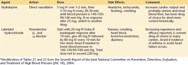
The safest, most efficacious drugs for the acute control of severe hypertension complicating preeclampsia are labetalol and hydralazine. Although hydralazine has theoretical advantages over labetalol in that it is a direct vasodilator and does not induce bronchospasm, rapid bolus infusions are potentially more likely to induce precipitous hypotension. In general, either is acceptable, and their use will be determined by individual circumstances. Table 14-3 details the dosage, duration of action, and potential complications of these two drugs.

Oral nifedipine has been used successfully, starting at a dose of 10 mg orally and repeated in 20 to 30 minutes if necessary to a maximal dose of 30 mg. Nifedipine should be used cautiously to avoid hypotension, particularly when used in conjunction with magnesium sulfate. Because of the potential for a precipitous drop in blood pressure, short-acting nifedipine is generally not advised in this setting. Intravenous sodium nitroprusside has the advantage of providing minute-to-minute control of blood pressure but may cause fetal cyanide toxicity with prolonged administration, so the use of this medication is generally limited to the postpartum period.

MANAGEMENT OF ECLAMPSIA

Eclampsia is a true obstetric emergency, and all physicians involved in the care of pregnant women should be prepared to recognize the occurrence of an eclamptic seizure and begin initial resuscitative and stabilization efforts. The management of these patients should be carried out by a team of physicians and well-trained nurses in an isolated labor room, with minimal noise and not too much light. As with any seizure condition, the initial requirement is to protect the patient from injury, clear the airway, and give oxygen by face mask to relieve hypoxia. Blood pressure and pulse oximetry should be recorded every 10 minutes with the patient in the lateral position. A 16- to 18-gauge IV line should be placed for drawing blood and administering drugs and fluids. An indwelling catheter should be placed in the bladder and laboratory tests obtained as outlined in Box 14-3.

Pharmacologic stabilization consists of preventing recurrent convulsions and controlling hypertension. Randomized, controlled trials have confirmed that magnesium sulfate is the most efficacious drug for preventing recurrent eclamptic seizures and has the best safety profile for the mother and fetus. The administration of IV magnesium sulfate for the treatment of eclamptic seizures is similar to its prophylactic use as outlined in Table 14-1, except that the loading dose is generally increased from 4 to 6 g. The maintenance dose remains 2 g/hour if renal function appears normal. If diazepam (Valium) is used in addition to magnesium sulfate, personnel skilled in intubation should be readily available in case maternal respiratory depression occurs. In general, it is desirable to avoid polypharmacy.

Eclamptic seizures often induce a fetal bradycardia that usually resolves after maternal stabilization and correction of hypoxia. It is very important to stabilize the mother before any attempt is made to deliver the infant. Induction of labor or cesarean birth during the acute phase may aggravate the course of the disease. Once hypoxia is corrected, convulsions controlled, and the diastolic blood pressures brought down to the 90- to 100-mm Hg range, delivery should be expedited, preferably by the vaginal route.

MANAGEMENT OF CHRONIC HYPERTENSION

The major goals are to control hypertension and to detect the development of superimposed preeclampsia in the mother and IUGR in the fetus. In the patient with uncomplicated hypertension whose blood pressures are well controlled and who does not show signs of superimposed preeclampsia or fetal growth restriction, the outcome for both the mother and fetus should be good.

When a woman with chronic hypertension is first seen during the pregnancy, it is important to review previous records to determine whether she has essential hypertension or a secondary cause of high blood pressure. If no previous evaluations have been done, it may be appropriate to rule out some of the more common endocrine, renal, or cardiovascular causes of hypertension. Baseline laboratory tests similar to those outlined in Box 14-3, with the addition of an electrocardiogram (ECG), are useful. The purpose of these tests is to establish a baseline should the patient later develop superimposed preeclampsia as well as to look for evidence of end-organ dysfunction.

It is important to review the antihypertensive medications being taken and to discontinue any that are potentially teratogenic. There is little evidence that lowering blood pressures below the 140/90 mm Hg range benefits the pregnancy. In fact, lowering the blood pressure too much may result in decreased uterine perfusion pressure and iatrogenic fetal growth restriction. In many women, blood pressures will decrease to normal in the second trimester, and no antihypertensive medication will be needed.

As a general rule, the safest antihypertensive medication should be used at the lowest possible dose needed to keep blood pressures about 130/80 to 140/90 mm Hg. Methyldopa is considered to be the safest antihypertensive medication in pregnancy, and calcium channel blockers and labetalol are also considered to be safe. Angiotensin-converting enzyme inhibitors and angiotensin II receptor blockers should be avoided at all stages of pregnancy because of potential fetal toxicity. β-Blockers should be used with caution because they may cause fetal growth restriction and may affect the interpretation of the NST. In addition to pharmacologic control of blood pressure, the foundations of conservative management also include increased periods of rest in the lateral decubitus position. The risks and benefits of moderate exercise (i.e., walking) have not been well defined.

Because these pregnancies have a high incidence of IUGR, both early and serial sonographic examinations are indicated. The early ultrasound (before 12 weeks) is primarily for dating, and the 16- to 20-weeks ultrasound is for the assessment of fetal anomalies. Serial ultrasonic examinations (every 3 to 4 weeks after 24 to 28 weeks) are of great assistance in detecting growth restriction. Depending on the clinical circumstances, periodic fetal monitoring with NST and amniotic fluid assessment, supplemented by umbilical artery Doppler studies if there is evidence of IUGR or preeclampsia, may start as early as 26 to 28 weeks and should be commenced by 32 to 34 weeks in all hypertensive patients. Maternal detection of daily fetal kick counts in the third trimester or earlier are an important method of assessing fetal well-being.

A significant increase in hypertension or the development of proteinuria in a previously nonproteinuric patient with chronic hypertension are likely signs of superimposed preeclampsia. The incidence of superimposed preeclampsia varies from 15% to 25%. These patients should undergo repeat laboratory evaluation, as outlined in Box 14-3. Management should follow that outlined for severe preeclampsia.

The timing of delivery in the chronic hypertensive patient depends on the clinical circumstances. For patients without evidence of fetal growth restriction in whom the blood pressure is well controlled and no signs of superimposed preeclampsia are present, a full-term gestation may be allowed, provided that fetal well-being is normal. Any progression beyond the 40th week should be very carefully considered and probably avoided. The presence of growth restriction or blood pressure deterioration or the advent of proteinuria may dictate earlier delivery. If delivery is desirable but not imperative before 37 weeks, confirmation of fetal lung maturity should be obtained. The route of delivery should be vaginal in the absence of obstetric reasons for cesarean delivery.

image Sequelae and Outcome

Uncomplicated mild preeclampsia at term in the primigravid patient carries essentially no long-term maternal sequelae. Such patients are at no greater risk for subsequent development of hypertensive cardiovascular disease than any other individual. However, patients with a pregnancy complicated by severe preterm preeclampsia are at higher risk for cardiovascular disease in later life and at very high risk for recurrent preeclampsia (up to 40%) in a subsequent pregnancy. Women with gestational hypertension also seem to have a higher incidence of developing chronic hypertension later in life. The female offspring of preeclamptic women experience an increased risk for preeclampsia in their own pregnancies, providing evidence of a genetic basis to the disease.

Pregnancy does not appear to affect the long-term prognosis in a patient with chronic hypertension. Some of the more serious complications of preeclampsia, such as cerebrovascular accidents and renal failure, may have long-term maternal sequelae. Overall, the mortality rate in women with hypertensive disease of pregnancy varies according to the severity of the disease, socioeconomic level, and quality of care received. Although at present there is no proven way of preventing preeclampsia, accessible, high-quality prenatal care should prevent most of the severe complications associated with the disease.

Fetal and neonatal sequelae are more difficult to determine because some of the morbidity and mortality associated with these hypertensive syndromes are related to IUGR, prematurity, and acute and chronic fetal distress. All of these may have long-term CNS effects.