Endometrial Hyperplasia and the Differential Diagnosis for Thick Endometrium

Published on 10/03/2015 by admin

Filed under Obstetrics & Gynecology

Last modified 22/04/2025

Print this page

rate 1 star rate 2 star rate 3 star rate 4 star rate 5 star
Your rating: none, Average: 0 (0 votes)

This article have been viewed 2864 times

Endometrial Hyperplasia and the Differential Diagnosis for Thick Endometrium

Synonyms/Description

Endometrial proliferation

Etiology

Endometrial hyperplasia refers to abnormal proliferation of endometrial glands and stroma, representing a spectrum of endometrial abnormalities ranging from benign overgrowth to precancerous tissue. Endometrial hyperplasia can cause a diffusely thickened endometrium or, less commonly, focal thickening within the cavity.

Ultrasound Findings

The sonographic appearance of endometrial hyperplasia is a heterogeneous thickening of the endometrial echo (lining). Endometrial hyperplasia may be circumferential, involving most of the endometrium or focal and nodular. In premenopausal patients, optimal evaluation of the endometrium is in the early follicular (proliferative) phase when the lining is at its thinnest. Later in the menstrual cycle the endometrium becomes topographically irregular, and the appearance of endometrial hyperplasia may be indistinguishable from the normal thickening that occurs during the luteal (secretory) phase. There are no established values for the normal width of the endometrial echo in premenopausal women. The sonographic texture of the endometrial echo is an important feature, and focal irregularities may be further delineated with sonohysterography.
In postmenopausal patients with bleeding, the normal width of the endometrium in longitudinal view should measure less than or equal to 4 mm and appear linear, with no focal irregularities. Some authors report that a measurement less than 5 mm is normal; hence there is disagreement as to whether the upper limit of normal should be 4 or 5 mm; however, the American College of Obstetrics and Gynecology states less than or equal to 4 mm is normal. It is important to evaluate the endometrium in its entirety. If part of the endometrium is obscured by fibroids, polyps, or adenomyosis or if the margins are indistinct, saline infusion sonohysterography can be used for further evaluation. There is no accepted normative data for the width of the endometrium in nonbleeding postmenopausal patients. The sonographic appearance of the endometrial echo and color flow are important factors in detecting the presence of endometrial disease.
Tamoxifen is a selective estrogen receptor modulator used in the treatment and prevention of breast cancer. The estrogen receptor agonist activity in the uterus caused by Tamoxifen has been associated with an increased risk of endometrial polyps, hyperplasia, and cancer when used in postmenopausal women. In addition, patients on Tamoxifen can have a very indistinct endometrial/myometrial border that often results in the overestimation of the endometrial echo width. This is often caused by microcystic formation in the subendometrial region, which results in an irregular endometrial-myometrial junction. These microcysts are glandular cystic atrophy. Sonohysterography is very useful to delineate the true appearance of the endometrial surface itself.

Differential Diagnosis

The differential diagnosis for a thickened endometrium is extensive, but generally includes endometrial hyperplasia, polyps, fibroids (submucous), endometrial cancer, retained products of conception, and adenomyosis. Patients with endometrial hyperplasia typically have a circumferentially thickened endometrium. Unfortunately, endometrial hyperplasia and cancer are indistinguishable sonographically and require tissue sampling.
Sonohysterography is crucial to differentiating a focal lesion such as a polyp from a global process such as hyperplasia or malignancy. Polyps are echogenic focal lesions, typically with a feeder vessel, and often detectable without sonohysterography. Submucous fibroids are typically rounded structures, more echolucent than the surrounding endometrium and displacing the endometrial echo. Adenomyosis may make the endometrial-myometrial junction indistinct, necessitating a sonohysterogram to clarify. If a patient has had a recent pregnancy, retained products of conception should be considered. Color flow Doppler may show extensive vascularity, further confirming the diagnosis. Please see the individual sections for Endometrial Carcinoma; Polyps, Endometrial; Adenomyosis; and Retained Products of Conception for more detail on each.

Clinical Aspects and Recommendations

Clinically there is a great difference between endometrial evaluation in premenopausal and postmenopausal patients. In premenopausal patients who are still cycling, it is essential that sonographic evaluation be performed in the early follicular phase, when the endometrium is thinnest. In postmenopausal patients who are not on hormone therapy, there is no “cycling,” and sonographic evaluation may be carried out at any time. The value of sonography in patients suspected of having endometrial hyperplasia is the high negative predicative value of a thin, distinct endometrial echo when present. When a thin echo is not present, saline infusion sonohysterography can help to differentiate between global abnormalities, which can be sampled blindly, and focal abnormalities (polyps, focal tissue growth), which should be sampled under direct visualization (hysteroscopically).

Figures

image

Figure E3-1 Endometrial hyperplasia. A, A diffusely thickened endometrium. B and C, Images from the sonohysterogram showing that the thickening is global.

image

Figure E3-2 Differential diagnosis. A, A thickened endometrium. B, A 3-D image from the sonohysterogram showing a focal lesion that was a large polyp.


image

Figure E3-3 Differential diagnosis. A, A thickened and blotchy endometrium with a hint of a focal lesion. B, The 3-D coronal view shows the polyp without the need for a sonohysterogram.


image

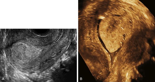
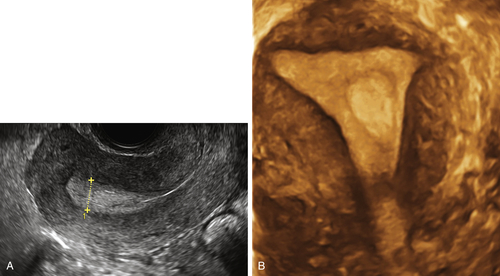
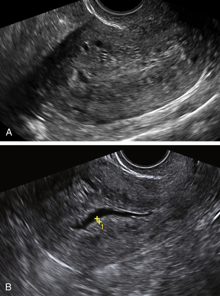
Figure E3-4 Differential diagnosis. A, A thick and irregular endometrium with many cystic spaces obscuring the endometrial-myometrial junction. This patient was on Tamoxifen, and the ultrasound image was not sufficient to evaluate the endometrium. B, The sonohysterogram, which revealed that most of the cystic areas are in the subendometrial region.

image

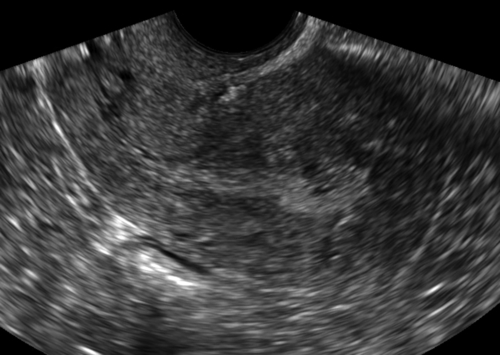
Figure E3-5 Differential diagnosis. Longitudinal view of the endometrium of a postmenopausal patient with bleeding. Note the focal nodular thickening of the endometrium at the fundus (retroverted uterus), with slight irregularity and loss of definition of the endometrial-myometrial junction. The pathology revealed papillary serous adenocarcinoma of the endometrium.


Suggested Reading

Ballard P., Tetlow R., Richmond I., Killick S., Purdie D.W. Errors in the measurement of endometrial depth using transvaginal sonography in postmenopausal women on tamoxifen: random error is reduced using saline instillation sonography. Ultrasound Obstet Gynecol. 2000;15:321–326.

Goldstein R.B., Bree R.L., Benson C.B., Benacerraf B.R., Bloss J.D., Carlos R., Fleischer A.C., Goldstein S.R., Hunt R.B., Kurman R.J., Kurtz A.B., Laing F.C., Parsons A.K., Smith-Bindman R., Walker J. Evaluation of the woman with postmenopausal bleeding: Society of Radiologists in Ultrasound-Sponsored Consensus Conference statement. J Ultrasound Med. 2001;20:1025–1036.

Goldstein S.R. Modern evaluation of the endometrium. Obstet Gynecol. 2010;116:168–176.

Goldstein S.R. Significance of incidentally thick endometrial echo on transvaginal ultrasound in postmenopausal women. Menopause. 2011;18:434–436.

Goldstein S.R. Sonography in postmenopausal bleeding. J Ultrasound Med. 2012;31:333–336.

Mills A.M., Longacre T.A. Endometrial hyperplasia. Semin Diagn Pathol. 2010;27:199–214.

Montgomery B.E., Daum G.S., Dunton C.J. Endometrial hyperplasia: a review. Obstet Gynecol Surv. 2004;59(5):368–378.