Hyperfunction of the adrenal cortex

Published on 02/03/2015 by admin

Filed under Basic Science

Last modified 22/04/2025

Print this page

rate 1 star rate 2 star rate 3 star rate 4 star rate 5 star
Your rating: none, Average: 0 (0 votes)

This article have been viewed 2477 times

49

Hyperfunction of the adrenal cortex

Hyperfunction of the adrenal cortex can be conveniently discussed in terms of the overproduction of the three main products:

Cortisol excess

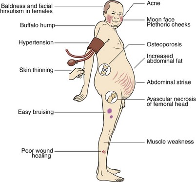
Prolonged exposure of body tissues to cortisol or other glucocorticoids gives rise to the clinical features that collectively are known as Cushing’s syndrome (Fig 49.1), after the American neurosurgeon Harvey Cushing. It most often results from prolonged use of steroid medications (iatrogenic). Much less frequently, it is caused by tumours that secrete either cortisol or ACTH (see below); these can sometimes be very difficult to diagnose.

In any investigation of Cushing’s syndrome the clinician should ask two questions:

Confirming the diagnosis

Iatrogenic Cushing’s syndrome is usually obvious – the patient is on steroid medications. The steroid may have been taken orally, inhaled or applied topically. Iatrogenic Cushing’s syndrome is not usually a diagnostic dilemma and will not be considered further here.

Cortisol, secreted in excess by the adrenal cortex, will rapidly exceed the available capacity of the plasma binding protein, cortisol binding globulin. Unbound cortisol is filtered readily into the urine. ‘Urinary free cortisol’ in a 24-hour collection, or assessed as a cortisol : creatinine ratio in an early morning urine sample, is one of the initial screening tests in a patient with suspected adrenocortical hyperfunction. The latter measurement can be made on a small aliquot of urine. Repeatedly high early morning urine cortisol : creatinine ratios are evidence enough to proceed with further investigations of the patient. If the test is negative on three occasions, Cushing’s syndrome may be excluded from the differential diagnosis.

Cortisol concentrations measured at 08:00 and 22:00 normally show a circadian rhythm with the evening sample having a lower value than that in the morning. This difference is usually not apparent in the patient with Cushing’s syndrome. It is essential that the patients are not stressed when such measurements are made.

Failure of 1 mg of dexamethasone taken at 23:00 to suppress the serum cortisol level at 08:00 the following morning, or failure to suppress urinary cortisol secretion overnight (as measured by an early morning urine cortisol : creatinine ratio), is another pointer towards the presence of Cushing’s syndrome.

Failure of the serum cortisol to rise after insulin-induced hypoglycaemia is also a characteristic feature of Cushing’s syndrome. Since patients with cortisol overproduction will be insulin-resistant, adequate hypoglycaemia may not be achieved with 0.15 units of insulin/kg body weight. A higher dose may have to be used. In normal individuals a fall of blood glucose concentration to less than 2.2 mmol/L is accompanied by a rise in serum cortisol of more than 200 nmol/L. A patient who shows such a response in the IST is unlikely to have a pathological excess of cortisol production.

Determining the cause

The possible causes of Cushing’s syndrome are illustrated in Figure 49.2. These include:

Classically, ACTH is not detectable in the plasma of patients with adrenal tumours. In patients with pituitary-dependent Cushing’s syndrome (known somewhat confusingly as Cushing’s disease) the plasma ACTH may be within the reference range or modestly elevated. The plasma ACTH level is often very high in patients with ectopic ACTH production.

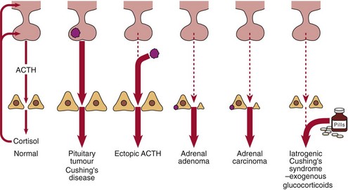
In patients with pituitary-dependent Cushing’s disease the serum or urinary cortisol will be partially suppressed after 2 days of dexamethasone, 2.0 mg q.i.d. (Fig 49.3). Failure to suppress suggests either ectopic ACTH production or the autonomous secretion of cortisol by an adrenal tumour. The presence of hypokalaemia is a tell-tale sign of ectopic ACTH production.
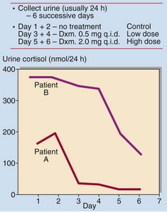
The cause of Cushing’s syndrome will determine the therapeutic options, and it is therefore essential that a definitive diagnosis is made. CT scans or magnetic resonance imaging of the pituitary may be helpful in detecting a pituitary adenoma in patients with Cushing’s disease. Selective venous sampling with ACTH measurement is sometimes carried out to locate the ACTH source in difficult cases.

Androgen excess

Adrenocortical tumours, particularly adrenal carcinomas, may produce excess androgens (DHA, androstenedione and testosterone) causing hirsutism and/or virilization in females (see pp. 100–101). This may not necessarily be accompanied by cortisol excess, and signs of Cushing’s syndrome may be absent. Patients with congenital adrenal hyperplasia (p. 95) may also present with signs of increased androgen production.

Aldosterone excess

Primary hyperaldosteronism (Conn’s syndrome) is rare. In most cases, the disease is due to a single adrenocortical adenoma. Patients may present with polydipsia and polyuria, symptoms of neuromuscular abnormalities such as weakness, paraesthesiae and tetany, and hypertension. All symptoms other than hypertension are attributable to potassium depletion.

Preliminary investigations must include determination of serum and urine electrolytes over several days, with adequate sodium intake. Serum potassium will be low, and urinary potassium excretion will be elevated. Documented, careful collection of specimens for assay of aldosterone, renin or ‘plasma renin activity’, may be made on 2 consecutive days after 8 hours recumbency, and again with the patient ambulatory, to confirm the diagnosis.

The diagnosis of hyperaldosteronism may be made in the hypokalaemic patient if the serum aldosterone level exceeds the upper limit of normal or if the level is persistently inappropriate to the serum potassium. In primary hyperaldosteronism, where the excess aldosterone arises from an adrenal adenoma, the levels of plasma renin will be low.

Secondary hyperaldosteronism is common and is associated with renal, heart or liver disease.