Etiology
A hydrosalpinx is a dilated fallopian tube filled with fluid. A normal tube is not visible sonographically; however, when a tube becomes obstructed, it distends and fills with fluid, giving it a sausage-like appearance. Infection is a major cause of hydrosalpinx, also called pelvic inflammatory disease (PID), in which the tube fills with pus in the acute phase of the disease (pyosalpinx). In ectopic pregnancy, the tube can distend and fill with blood (hematosalpinx) and have a similar appearance to a pyosalpinx. Tubal torsion presents with acute pain and a tender, distended tube. Scarring and adhesions in the pelvis can obstruct the tubes and allow them to fill with fluid, but this is more often found in asymptomatic patients since it is not an acute process. This can also be seen in cases of severe endometriosis, or previous pelvic surgery. Rarely, tuberculosis and other infections can attack the pelvis and result in hydrosalpinx.
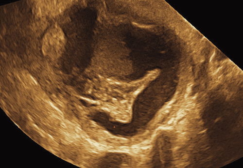
Ultrasound Findings
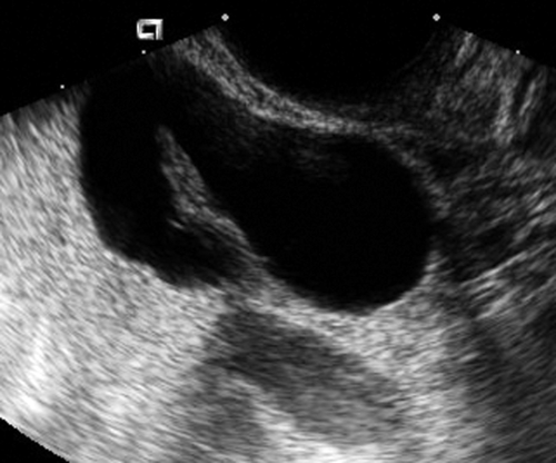
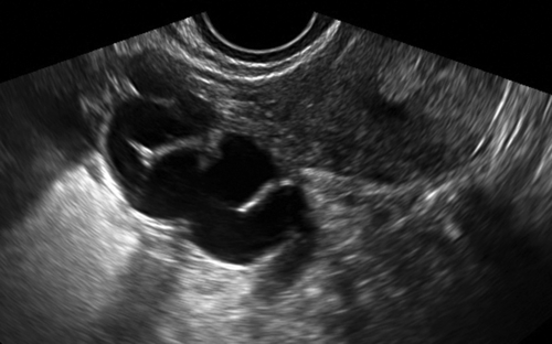
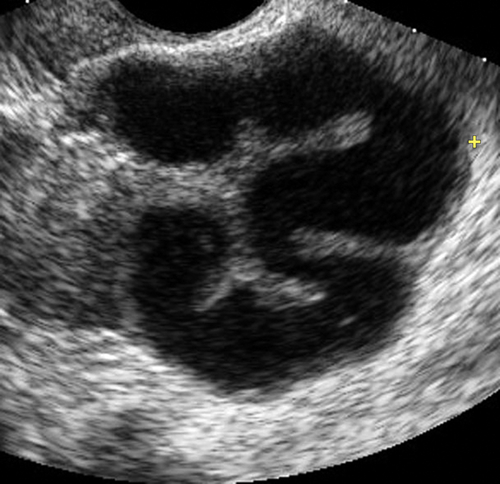
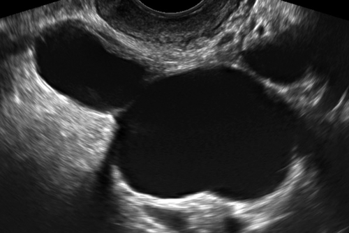
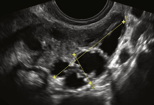
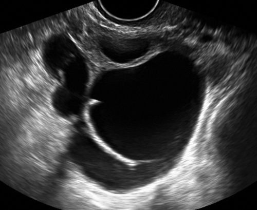
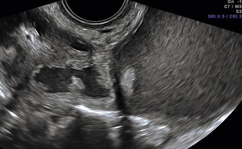
A hydrosalpinx has a typical sonographic appearance. Evaluation of the adnexa reveals a cystic, sausage-shaped, serpiginous, tubular mass. The distended tube often has a characteristic “spoke-wheel” pattern made up by the incomplete septae of the tube. The tube can become redundant and fold upon itself, giving the appearance of a multiseptate cystic mass. If the adjacent ovary is not clearly visualized, it is easy to suspect ovarian pathology. If the hydrosalpinx is the result of pelvic inflammatory disease, the tubal wall can occasionally be thick and irregular, making the correct diagnosis more difficult. In the study by Sokalska and colleagues (from the IOTA multicenter study), the sensitivity for the sonographic diagnosis of hydrosalpinx was 86%.
Hydrosalpinges are often accompanied by free pelvic fluid, especially in acute pelvic inflammatory disease or ectopic pregnancy. The presence of a solid mass within a dilated tube, along with a negative pregnancy test, may suggest fallopian tube carcinoma, although abundant vascularity would also usually be present.
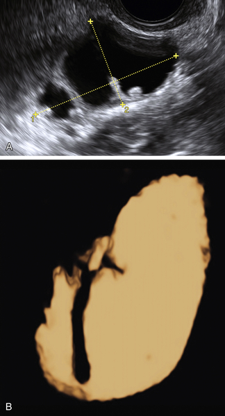
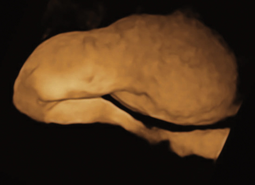
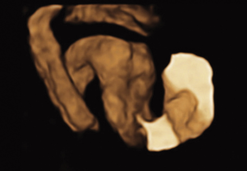
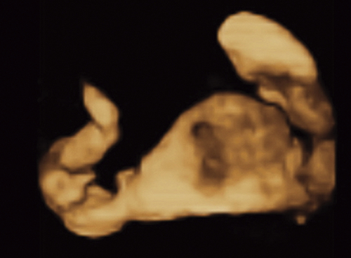
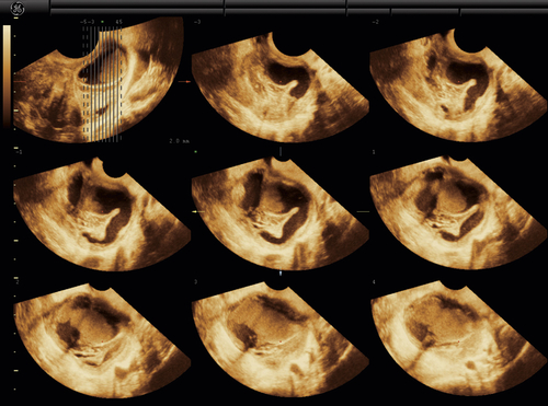
Three-dimensional ultrasound is very helpful for demonstrating the course of the dilated tubes. Once a volume is obtained, the complete cast of the convoluted fallopian tube is easily seen using the inverse mode. This technique enables the practitioner to view a cast of the entire cystic tube previously hidden within the volume. The whole tube is then demonstrated, even if the tube traverses multiple planes.
Differential Diagnosis
Most hydrosalpinges are tubular fluid collections that do not conform to any single plane. Therefore with 2-D ultrasound, they can have the appearance of a multiseptate cystic mass. The “spoke-wheel” walls and a separate ovary suggest the correct diagnosis; however, too often the findings are nonspecific. Using standard 2-D sonography, the differential diagnosis includes an ovarian neoplasm or a peritoneal inclusion cyst. Volume (3-D) ultrasound with inverse mode is essential to connecting the cystic spaces into a tubal architecture if the mass is a hydrosalpinx. The patient’s symptoms also play an important part in making the correct diagnosis. If a patient presents with a cystic adnexal mass and severe pain, the differential diagnosis would include pelvic inflammatory disease (signs of infection), an ectopic pregnancy (positive pregnancy test), or tubal torsion. Chronic pain or cyclic pain might indicate endometriosis. Ovarian neoplasms are often asymptomatic, but so are chronic hydrosalpinges.
A right-sided adnexal mass can be confused with an appendiceal mucocele. A dilated ureter
can occasionally mimic a hydrosalpinx although the ureter typically peristalses.
Clinical Aspects and Recommendations
Most hydrosalpinges are found incidentally and are asymptomatic. Treatment, if indicated, focuses on the etiology. If the hydrosalpinx is filled with anechoic fluid, has thin walls, and the patient is asymptomatic, treatment is not typically required. Salpingectomy may be indicated for infertile patients or those with prior ectopic pregnancies desiring to conceive. Acute pelvic inflammatory disease will be treated with antibiotics, often intravenously.