Human T-Lymphotropic Viruses (1 and 2)

Published on 22/03/2015 by admin

Filed under Pediatrics

Last modified 22/04/2025

Print this page

rate 1 star rate 2 star rate 3 star rate 4 star rate 5 star
Your rating: none, Average: 0 (0 votes)

This article have been viewed 1298 times

Chapter 269 Human T-Lymphotropic Viruses (1 and 2)

Epidemiology

HTLV-1 infects 10-20 million persons globally. It is endemic in southwestern Japan (where >10% of adults are seropositive), areas of the Caribbean including Jamaica and Trinidad (up to 6%), and in parts of sub-Saharan Africa (up to 5%). Lower seroprevalence rates are found in South America (up to 2%) and Taiwan (0.1-1%). There is microclustering with marked variability within geographic regions.

The seroprevalence of HTLV-1 and HTLV-2 in the USA in the general population is 0.01-0.03% for each virus, with higher rates with increasing age. HTLV-1 infection correlates greatest with birth in endemic areas or sexual contact with persons from endemic areas. HTLV-2 infection correlates with intravenous illicit drug use. A prevalence of approximately 18% was found in a study of illicit drug users in the USA, often with concomitant HIV infection.

HTLV-1 and HTLV-2 are transmitted as cell-associated viruses by vertical transmission from mother to child and horizontal transmission through genital secretions, contaminated blood products, and intravenous illicit drug use. Vertical HTLV-1 transmission occurs primarily via breast-feeding from infected mothers, with a 3-fold increased risk of transmission with breast-feeding for >6 mo. Intrauterine and intrapartum transmission account for <5% of vertical transmission. In Japan, approximately 20-25% of children born to infected mothers become infected, and >90% of HTLV-1-infected children have HTLV-1-infected mothers. HTLV-2 may also be transmitted via breast-feeding but has a slightly lower reported transmission rate via breast milk of approximately 14%.

Clinical Manifestations

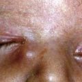
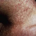
The lifetime risk of disease associated with HTLV-1 infection is estimated at 10%, and is highest following vertical transmission. HTLV-1 is associated with ATL and several nonmalignant conditions, including the neurodegenerative disorder HTLV-1–associated myelopathy (HAM), also known as tropical spastic paraparesis (TSP) and sometimes termed HAM/TSP. The geographic epidemiology of ATL and HAM are similar. HTLV-1–associated arthropathy mimics rheumatoid arthritis, including a positive rheumatoid factor. Treatment is with anti-inflammatory agents. HTLV-1–associated uveitis may be unilateral or bilateral, more common among women, and resolves spontaneously, though it often recurs within 1-3 yr. Topical corticosteroids hasten recovery. Infective dermatitis associated with HTLV-1 is a chronic and recurrent eczematous disease occurring during childhood and adolescence. HTLV-1 infection predisposes to disseminated and recurrent Strongyloides stercoralis infection, increased risk of developing tuberculosis disease following latent infection, and severe scabies.

Adult T-Cell Leukemia/Lymphoma (ATL)

The age distribution of ATL peaks at approximately 50 yr, underscoring the long latent period of HTLV-1 infection. HTLV-1-infected persons remain at risk for ATL even if they move to an area of low HTLV-1 prevalence, with a lifetime risk for ATL of 2-4%. Most cases of ATL are associated with monoclonal integration of HTLV-1 provirus into the cellular genome of CD4 lymphocytes resulting in unchecked proliferation of CD4 T cells. There is a spectrum of disease that is categorized into different forms: acute, lymphomatous, chronic, primary cutaneous smoldering, and primary cutaneous tumoral. The acute form of ATL comprises 55-75% of all cases. Smoldering, subclinical lymphoproliferation may spontaneously resolve, in approximately half of cases, or progress to chronic leukemia or lymphomatous or even acute ATL. Chronic, low-grade, HTLV-1-associated lymphoproliferation (pre-ATL) may persist for years with abnormal lymphocytes with or without peripheral lymphadenopathy before progressing to the acute form. Acute ATL is characterized by hypercalcemia, lytic bone lesions, lymphadenopathy that spares the mediastinum, hepatomegaly, splenomegaly, cutaneous lymphomas, and opportunistic infections. Leukemia may develop with circulating polylobulated malignant lymphocytes, called flower cells, possessing mature T-cell markers. Cyclophosphamide, doxorubicin, vincristine, and prednisolone (CHOP) is often 1st line therapy for ATL, although chemotherapy is not curative and relapses are common. The median survival after diagnosis is 6-13 mo.

HTLV-1-Associated Myelopathy (HAM)

HAM is more common in women than in men and has a relatively short incubation period after HTLV-1 infection, of 1-4 yr compared with 40-60 yr for ATL. HAM occurs in up to 4% of persons with HTLV-1 infection, usually developing during middle age. It is characterized by infiltration of mononuclear cells into the gray and white matter of the thoracic spinal cord, leading to severe white matter degeneration and fibrosis. HTLV-1 is found near but not directly within the lesions, suggesting that reactive inflammation is a major mechanism of disease. The cerebrospinal fluid typically shows a mildly elevated protein and a modest monocytic pleocytosis, along with anti-HTLV-1 antibodies. Neuroimaging studies are normal or show periventricular lesions in the white matter. Clinical manifestations include gradual onset of slowly progressive, symmetric neurologic degeneration of the corticospinal tracts and, to a lesser extent, the sensory system that leads to lower extremity spasticity or weakness, lower back pain, and hyperreflexia of the lower extremities, with an extensor plantar response. The bladder and intestines may become dysfunctional, and men may become impotent. Some patients develop dysesthesias of the lower extremities, with diminished sensation to vibration and pain. Upper extremity function and sensation, cranial nerves, and cognitive function are usually preserved. Treatment regimens have included corticosteroids, danazol, interferon, plasmapheresis, high-dose vitamin C, and antivirals, all with minimal effects.

Bibliography

Centers for Disease Control and Prevention. Recommendations for counseling persons infected with human T-lymphotropic virus, types I and II. MMWR. 1993;42(RR-9):1-13.

Eshima N, Iwata O, Iwata S, et al. Age and gender specific prevalence of HTLV-1. J Clin Virol. 2009;45(2):135-138.

Hollsberg P, Hafler DA. Pathogenesis of diseases induced by human lymphotropic virus type I infection. N Engl J Med. 1993;328:1173-1182.

Kaplan JE, Abrams E, Schaffer N, et al. Low risk of mother-to-child transmission of human lymphotropic virus type II in non-breast-fed infants. J Infect Dis. 1992;166:892-895.

Levin MC, Jacobson S. HTLV-1 associated myelopathy/tropical spastic paraparesis (HAM/TSP): a chronic progressive neurologic disease associated with immunologically mediated damage to the central nervous system. J Neurovirol. 1997;3:126-140.

Manns A, Hisada M, La Grenade L. Human T-lymphotrophic virus type 1 infection. Lancet. 1999;353:1951-1958.

Proietti FA, Carneiro-Proietti BF, Catalan-Soares BC, et al. Global epidemiology of HTLV-1 infection and associated diseases. Oncogene. 2005;24:6058-6068.

Taylor GP, Matsuoka M. Natural history of adult T-cell leukemia/lymphoma and approaches to therapy. Oncogene. 2005;24:6047-6057.

Van Dyke RB, Heneine W, Perrin ME, et al. Mother-to-child transmission of human T-lymphotropic virus type 2. J Pediatr. 1995;127:924-928.

Verdonck K, Gonzalez E, Van Dooren S, et al. Human T-lymphotropic virus 1: recent knowledge about an ancient infection. Lancet Infect Dis. 2007;7:266-281.