[level-membership-for-hematology-oncology-and-palliative-medicine-category]
20 Human immunodeficiency virus (HIV) related malignancies
Introduction
Malignancies have a significant impact on the morbidity and mortality of people with human immunodeficiency virus (HIV) infection. 30–40% of HIV-infected patients develop malignancies during the course of their illness. Some of the malignancies are AIDS-defining conditions whereas others appear to be more common in HIV patients (Box 20.1). Although AIDS-defining malignancies may be caused mainly by progressive immunosuppression, the exact relationship between immunosuppression and non-AIDS-defining malignancies is yet to be established. The most common cancers in the general population such as breast, prostate and colon do not appear to increase in HIV infection.
Pathogenesis
AIDS-defining malignancies
Kaposi’s sarcoma
The clinical manifestation of KS varies from a mild to a fulminant course. Skin lesions appear mainly on the lower extremities, face and genitalia. Skin lesions are typically multifocal and papular (Figure 20.1), but can appear plaque-like or as a fungating mass. Extracutaneous lesions appear in the oral cavity (most commonly palate followed by gingiva), larynx, gastrointestinal tract and lung. Gastrointestinal lesions can be asymptomatic or can cause weight loss, abdominal pain, nausea, vomiting and bleeding. Pulmonary KS may be symptomatic with cough, dyspnoea, haemoptysis etc. or present with asymptomatic radiological features of infiltrates, isolated nodules, pleural effusion and hilar or mediastinal lymphadenopathy.
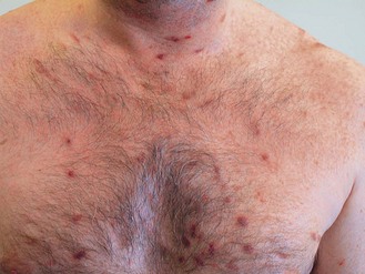
Figure 20.1 Kaposi’s sarcoma.
From Clutterbuck: Specialist Training in Sexually Transmitted Infections and HIV, with permission.
In the post HAART era, tumour burden and systemic illness are useful to categorize patients into two prognostic groups (Box 20.2). Diagnosis of KS is confirmed by biopsy.
Box 20.2
Staging of Kaposi’s sarcoma
| Good risk (all of the following) | Poor risk (any of the following) | |
|---|---|---|
| Tumour (T) | Confined to skin and/or lymph nodes and/or non-nodular disease in palate |
* B symptoms – unexplained fever, night sweats, >10% involuntary weight loss or diarrhoea persisting more than 2 weeks
Indications for systemic chemotherapy (Box 20.3) include:
Non-Hodgkin’s lymphoma
Optimal treatment of HIV-related lymphoma is not known. Patients are generally treated similarly to those who are seronegative (p. 302). The overall prognosis is poor with a less than 1 year median survival.
Primary CNS lymphoma is 15 times more common than in the general population. The histology is large cell or immunoblastic type of B cell origin. Treatment involves radiotherapy combined with steroids, which extend the median survival from 2–3 months to 6–8 months (p. 306).
Non-AIDS-defining malignancies
Anal cancer
The high-risk group includes patients who practice receptive anal intercourse, men who have sex with men and anal co-infection with HPV or syphilis. The clinical presentation and treatment is similar to that of the seronegative population (p. 170). Patients with high-grade anal intra-epithelial neoplasia (AIN), invasive anal margin cancer and those with severe drug reactions require surgical management. Although the response to treatment is equivalent to seronegative patients with anal cancer, there is a high risk of severe radiation toxicity.
Liver cancer
Hepatocellular cancer is increased by 8-fold in the HIV population. Tumours tend to be more symptomatic, advanced at presentation and have an aggressive course. Resection or transplantation is an option for early stage whereas systemic chemotherapy is used in advanced cancer (p. 147). Survival is not influenced by CD4 count.
Hodgkin’s disease
Patients present early in HIV infection with unfavourable histologic subtypes (mixed cellularity, lymphocyte depleted). There is a 5–15 fold increase in risk with a high rate of EBV co-infection (75–100%). Clinical presentation with B symptoms and extranodal disease are common. They are treated as seronegative patients (p. 298) and the median survival is 12–18 months.
Spano JP, Costagliola D, Katlama C, et al. AIDS-related malignancies: state of the art and therapeutic challenges. J Clin Oncol. 2008;26:4834-4842.
Deeken JF, Pantanowitz L, Dezube BJ. Targeted therapies to treat non-AIDS-defining cancers in patients with HIV on HAART therapy: treatment considerations and research outlook. Curr Opin Oncol. 2009;21:445-454.
[/level-membership-for-hematology-oncology-and-palliative-medicine-category][not-level-membership-for-hematology-oncology-and-palliative-medicine-category]
20 Human immunodeficiency virus (HIV) related malignancies
Introduction
Malignancies have a significant impact on the morbidity and mortality of people with human immunodeficiency virus (HIV) infection. 30–40% of HIV-infected patients develop malignancies during the course of their illness. Some of the malignancies are AIDS-defining conditions whereas others appear to be more common in HIV patients (Box 20.1). Although AIDS-defining malignancies may be caused mainly by progressive immunosuppression, the exact relationship between immunosuppression and non-AIDS-defining malignancies is yet to be established. The most common cancers in the general population such as breast, prostate and colon do not appear to increase in HIV infection.
Pathogenesis
AIDS-defining malignancies
Kaposi’s sarcoma
The clinical manifestation of KS varies from a mild to a fulminant course. Skin lesions appear mainly on the lower extremities, face and genitalia. Skin lesions are typically multifocal and papular (Figure 20.1
[/not-level-membership-for-hematology-oncology-and-palliative-medicine-category]
