Chapter 91 How Do You Make a Diagnosis of an Infected Arthroplasty?
Infection after a total hip or knee arthroplasty is an uncommon but serious complication with significant impact. Although the postoperative infection rate has decreased from 9% to 1% to 2%, large centers still treat a significant number of patients with infected arthroplasties every year. With the aging population and also younger patients demanding joint replacement procedures, the problem is likely to increase over the years to come.
EVIDENCE
History and Clinical Examination
No study has investigated solely the sensitivity and specificity of history and clinical examination to accurately diagnose a prosthetic infection. However, Spangehl and colleagues1 correctly diagnosed 27 of 35 infected total hip arthroplasties with history and examination alone (Level I). All 35 infections were later confirmed with positive intraoperative cultures. Acute onset of pain, systemic illness, and sinus formation were the most commonly found signs and symptoms. No studies comment on the value of history and examination in correctly diagnosing an infected knee arthroplasty. History and physical examination are useful because they give you an initial index of suspicion or a pretest likelihood of infection. Further diagnostic testing simply changes the pretest likelihood of disease. As such, a patient who presents with a low likelihood of infection based on history and physical examination, will require much more stringent criteria to clearly diagnose infection. However, a patient who presents with florid signs and symptoms of infection will require much less stringent criteria for the confirmation of infection. This is an application of Bayes’ theorem in probability theory, and this is why it is important to understand how to apply the various tests that are discussed in the context of the initial encounter with the patient, namely, the history and physical examination.
Preoperative Hematologic Tests
White Blood Cell Count.
Obtaining a white blood cell count (WBC) is a routine preoperative investigation that is of little diagnostic value. In a prospective study (Level I evidence), Spangehl and colleagues1 evaluated the diagnostic accuracy to correctly diagnose an infection in 202 hips. With a value of more than 11.0 = 109 WBC/L considered to be a positive result indicating infection, they found a sensitivity of 0.20 and a specificity of 0.96. The positive predictive value, however, was only 0.54, and the negative predictive value 0.85. Di Cesare and coworkers2 (Level IV) undertook a prospective, case–control study of 58 patients undergoing reoperation. Seventeen were diagnosed as having a prosthetic infection. The sensitivity of WBCs was 0.47, and the specificity was 1.00. The positive predictive value was 1.00, and the negative predictive value was 0.82. In a retrospective case series (Level IV evidence), Canner and coauthors3 note a low prevalence of increased WBC counts in patients with an infected total joint arthroplasty. Therefore, based on the evidence, WBC count is of limited use, except when the pretest probability is high.
Erythrocyte Sedimentation Rate and C-Reactive Protein.
The erythrocyte sedimentation rate (ESR) is a measure of erythrocyte rouleaux formation, which occurs whenever an inflammatory condition is present. C-reactive protein (CRP) is an acute-phase protein synthesized in the liver. As with all acute-phase reactants, CRP level is increased in many inflammatory, infectious, and some neoplastic conditions. Spangehl and colleagues1 (Level I) prospectively analyzed the ESR and CRP level in 171 and 142 revision hip arthroplasties. ESR had a sensitivity of 0.82, specificity of 0.85, positive predictive value of 0.58, and negative predictive value of 0.95. CRP had a sensitivity of 0.96, specificity of 0.92, positive predictive value of 0.74, and negative predictive value of 0.99. If both tests were negative (ESR <30 mm/hr, CRP <10 mg/L), the probability of infection was zero. When both tests were positive, the probability of infection was 0.83. Virolainen and researchers4 (Level IV) also conclude that combined ESR and CRP are of value in the preoperative evaluation. For ESR, Di Cesare and coworkers2 (Level IV) found a sensitivity of 1.0 and specificity of 0.56. CRP had a sensitivity of 0.95 and specificity of 0.76.
Greidanus and investigators5 (Level I) evaluated 145 patients presenting for revision total knee arthroplasty for the presence of infection using the ESR and CRP. The ESR and CRP were obtained at the time of clinical assessment before definitive revision total knee arthroplasty. All patients had undergone preoperative aspiration for culture and had intraoperative periprosthetic tissue sent for bacterial culture. A diagnosis of infection was established for 45 of 151 knees that underwent revision total knee arthroplasty (prevalence, 0.298). The ESR (sensitivity, 0.93; specificity, 0.83; likelihood ratio positive, 5.81; accuracy, 0.86) and CRP (sensitivity, 0.91, specificity, 0.86; likelihood ratio positive, 6.89; accuracy, 0.86) had excellent diagnostic test performance. Combination testing of ESR and CRP together increases overall sensitivity to 0.95 for the diagnosis of infection. In this study, using receiver operating characteristics curves, the optimal cut point for ESR was found to be 22.5 mm/hr, and that of CRP was found to be 13.5 mg/L.
Serum Interleukin-6.
Interleukin-6 (IL-6) is a factor produced by monocytes and macrophages. It functions as a hepatocyte-stimulating factor and induces the production of acute-phase reactants. Wirtz and coworkers6 established that it returns to normal after total joint arthroplasties. Di Cesare and coworkers2 (Level III) selected 58 patients who underwent a revision procedure and measured the IL-6 levels before and after surgery. Seventeen of the 58 patients were infected as determined after surgery with intraoperative cultures. The sensitivity of IL-6 was 1.0, the specificity was 0.95, the positive predictive value was 0.89, and the negative predictive value was 1.00. However, this study was done on a small and selected sample, and further large studies (Levels I and II) are needed to determine the value of IL-6 in accurately detecting infection before surgery.
Polymerase Chain Reaction.
Polymerase chain reaction (PCR) is a molecular technique that allows the production of large quantities of DNA from one DNA molecule. Through this amplification, it enables the detection of rare RNA and DNA sequences. Various genetic diseases and infections can be diagnosed with PCR. Panousis and investigators7 (Level II) conducted a prospective study of 92 cases undergoing a revision arthroplasty (76 hips, 16 knees) to evaluate the efficacy of PCR sampled intraoperatively from joint aspirate before capsulotomy to detect a prosthetic infection. The authors used a combination of ESR, CRP, preoperative aspiration, and intraoperative cultures to determine the presence or absence of infection. PCR yielded a high false-positive rate. The sensitivity was 92%, the specificity was 74%, the positive predictive value was 34%, and the negative predictive value was 98%.
NUCLEAR MEDICINE TECHNIQUES
In a prospective study of 50 painful total hip arthroplasties, Reinartz and colleagues8 (Level III) compared the value of technetium-99 triple-phase bone scan (TPBS) with 18-fluorodeoxyglucose (FDG) positron emission tomography (PET) in differentiating between infection and aseptic loosening. The sensitivity and specificity for the technetium scan to differentiate between a prosthetic infection and aseptic loosening was 0.68 and 0.68, and for the PET scan was 0.94 and 0.95. The sensitivity and specificity for the technetium scan to detect a pathologic process was 0.79 and 0.88, respectively, and of the PET 0.94 and 0.97, respectively. In contrast, Stumpe and coauthors9 (Level II) compared PET, TPBS, and conventional radiography in the ability to differentiate between infection and aseptic loosening in 35 patients with painful hip replacements. The authors conclude that FDG-PET and TPBS had a similar diagnostic accuracy. Pill and investigators10 (Level II) compared FDG-PET with 111indium-white blood cell imaging in 92 patients with painful total hip arthroplasties. The specificity of both investigations to differentiate between infection and aseptic loosening was comparable (93% and 95%). However, FDG-PET yielded a significantly higher sensitivity (95% vs. 50%).
Preoperative Invasive Test
Preoperative Fluid Aspiration and Tissue Sampling.
Spangehl and colleagues1 analyzed the diagnostic accuracy of image-guided aspiration and tissue sampling in 180 patients undergoing a revision hip arthroplasty. Patients who were taking antibiotics were excluded from this study. The results of the initial aspiration showed a sensitivity of 0.86, a specificity of 0.94, a positive predictive value of 0.67, and a negative predictive value of 0.98. In five hips, repeat aspirations were performed because the initial results were inconsistent with the clinical presentation. This improved the diagnostic accuracy of the aspiration. This suggests that aspiration alone is not sufficient for the diagnosis because of the risk for false-positive and -negative results. For this reason, in low-probability cases, the authors suggest that if the ESR and the CRP are not suggestive of infection, an aspiration is not necessary. If, however, the pretest probability is high, an aspiration in addition to the ESR and the CRP is required, particularly if the ESR and CRP are normal. In all cases, an aspiration is required to confirm the diagnosis of infection if the ESR or the CRP level is increased.
Williams and colleagues11 (Level II) compared open capsular biopsy with needle aspiration in 273 consecutive total hip arthroplasties before revision surgery. Sensitivity and specificity of the 2 modalities were comparable (0.83 vs. 0.80 and 0.90 vs. 0.94, respectively). The authors conclude that both methods are reliable in differentiation between septic and aseptic loosening, but open biopsy offers no advantages over needle aspiration.
Sadiq and coworkers12 (Level II) report on the diagnostic accuracy of open core biopsy to detect a prosthetic infection in 159 total hip and knee arthroplasties before a revision arthroplasty. This showed a sensitivity of 88%, a specificity of 91%, and an accuracy of 89% considering the final diagnosis. This suggests no advantage over less invasive techniques.
Trampuz and investigators13 (Level II) analyzed the synovial fluid leukocyte count and differential obtained by arthrocentesis in 133 patients with a painful total knee arthroplasty. Using intraoperative cultures, they diagnosed aseptic failure in 99 patients and a prosthetic infection in 34 patients. A leukocyte count of more than 1.7 = 103 μL had a sensitivity of 94% and a specificity of 88%. A differential of more than 65% neutrophils had a sensitivity of 97% and a specificity of 98%. Notably, the absolute cell count is much lower than that seen in septic arthritis of an unreplaced joint. The differential count is of much greater importance. Whenever the neutrophil proportion is more than 65%, the likelihood of infection is high.
Intraoperative Tests
Frozen Section.
Frozen sections are particularly helpful when clinical suspicion is high but preoperative tests were negative or equivocal. For example in patients with inflammatory disease (rheumatoid arthritis), ESR or CRP can be high in the absence of infection. Preoperative aspiration can also be false negative or false positive. Spangehl and colleagues1 evaluated frozen sections in 202 hips using the criteria of Mirra.14 They considered a specimen infected if any single high-power field contained at least five stromal neutrophils. The analysis of the frozen section compared with the final histologic result had a sensitivity of 0.80, a specificity of 0.94, a positive predictive value of 0.74, and a negative predictive value of 0.96.
Using more stringent criteria (more than 10 polymorphonuclear leucocytes per high-power field), Bandit and coworkers15 studied 121 revision total joint arthroplasties. They compared a positive frozen section with intraoperative cultures as the gold standard. For frozen section, the sensitivity was 0.6, the specificity was 0.93, the positive predictive value was 0.67, and the negative predictive value was 0.93. Therefore, using more stringent criteria lowers the sensitivity, making it possible to miss certain infections, without significantly increasing the specificity. Therefore, we recommend the use of more than 5 polymorphonuclear leucocytes per high-power field as the cut point instead of more than 10.
Intraoperative Cultures.
In the absence of other factors, intraoperative cultures remain the gold standard to which all other investigations are compared. Unless the patient is already taking antibiotics, they should provide the definitive diagnosis in most patients with infection at the site of joint replacement. Spangehl and colleagues1 analyzed specimens from 180 hips undergoing revision hip replacement. They took at least three intraoperative samples from obviously inflamed tissues. Infection was diagnosed if more than one third of all specimens was positive (2/3 or 3/3 samples); the sensitivity was 0.94, specificity was 0.97, positive predictive value was 0.77, and negative predictive value was 0.99. Atkins and researchers16 (Level II) performed a prospective study of 297 revision hip and knee arthroplasties. The authors conclude that if three or more specimens out of five were positive, the sensitivity was 66% and the specificity was 99.6%.
No one diagnostic test is available today to make the definitive diagnosis of a chronically infected arthroplasty. Diagnosis starts with a thorough history and examination. Patients with an infected arthroplasty often describe pain unrelated to activity and not relieved by rest. Often the pain has abeen present since the initial operation, although different in character. There may be a history of a slow-healing wound after surgery or prolonged redness and swelling. Serial radiographs can show rapid loosening over months rather than years, as would be typical for aseptic loosening. Scalloping and osteolysis can be present but are more often absent.
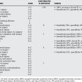
Serologic investigations are performed in every patient with pain at the site of an arthroplasty. White blood cell count, CRP level, and ESR are routine. In the absence of any clinical suspicion and with a normal CRP level and a normal ESR, an infection can be safely excluded and no other tests are required. An aspiration with or without a synovial biopsy is warranted if either the CRP level or ESR is increased, and/or if there is clinical suspicion for an infection. If the first aspirate is negative, a repeat aspiration is warranted. It is also important to wait as long as possible after antibiotics are stopped to reduce the potential for a false-negative result, although in such a situation, a negative aspirate does not necessarily exclude the diagnosis of infection, and more imaging such as a serial bone/indium scan or a PET scan may be warranted. At the end of the day, the surgeon has to exercise substantial judgment in the interpretation of the battery of tests and has to apply Bayes’ theorem before a definitive diagnosis may be made. An intraoperative frozen section is potentially helpful to aid in the diagnosis of such difficult cases. Intraoperative cultures (at least three samples from different locations taken with clean instruments) are mandatory in all revision cases. Some authors recommend withholding prophylactic antibiotics before obtaining these cultures; however, there is no high-level evidence to warrant such a practice. In straightforward cases where infection has been ruled out, and the probability of infection is low, the advantage of withholding antibiotics is not high enough to overshadow the increased risk for postoperative infection related to the lack of prophylactic antibiotics before the skin incision. For this reason, when we believe that infection has been ruled out, we administer prophylactic antibiotics. Of course, in cases where infection cannot be easily conclusively ruled out and the presurgical probability of infection is still reasonably high, we prefer to withhold preoperative antibiotics and to give them only after the intraoperative cultures have been taken. This way, if there is an unexpected positive culture results, the patient can be treated with 6 weeks of intravenous antibiotics after surgery17 (Level V). Table 91-1 provides a summary of the evidence.
| STATEMENT | LEVEL OF EVIDENCE/GRADE OF RECOMMENDATION | REFERENCES |
|---|---|---|
CRP, C-reactive protein; ESR, erythrocyte sedimentation rate; FDG, 18-fluorodeoxyglucose; PCR, polymerase chain reaction; PET, positron emission tomography; TPBS, triple-phase bone scan.
1 Spangehl MJ, Masri BA, O’Connell JX, et al. Prospective analysis of preoperative and intraoperative investigations for the diagnosis of infection at the sites of two hundred and two revision total hip arthroplasties. J Bone Joint Surg Am. 1999;81A:672-682.
2 Di Cesare PE, Chang E, Preston CF, et al. Serum interleukin-6 as a marker of periprosthetic infection following total hip and knee arthroplasty. J Bone Joint Surg Am. 2005;87A:1921-1927.
3 Canner GC, Steinberg ME, Heppenstall RB, et al. The infected hip after total hip arthroplasty. J Bone Joint Surg Am. 1984;66A:1393-1399.
4 Virolainen P, Lahteenmaki, Hiltunen A, et al. The reliability of diagnosis of infection during revision arthroplasties. Scand J Surg. 2002;91:178-181.
5 Greidanus NV, Masri BA, Garbuz DS, et al. Use of erythrocyte sedimentation rate and c-reactive protein level to diagnose infection before revision total knee arthroplasty. a prospective evaluation. J Bone Joint Surg Am. 2007;89:1409-1416.
6 Wirtz DC, Heller KD, Miltner O, et al. Interleukin 6: A potential inflammatory marker after total joint replacement. Int Orthop. 2000;24:194-196.
7 Panousis K, Grigoris P, Butcher I, et al. Poor predictive value of broad range PCS for the detection of arthroplasty infection in 92 cases. Acta Orthop Scand. 2005;76:341-346.
8 Reinartz P, Mumme T, Hermanns B, et al. Radionuclide imaging of the painful hip arthroplasty. J Bone Joint Surg Br. 2005;87-B:465-470.
9 Stumpe KD, Notzli HP, Zanetti M. FDP PET for differentiation of infection and aseptic loosening in total hip replacements: Comparison with conventional radiography and three phase bone scintigraphy. Radiology. 2004;231:333-341.
10 Pill SG, Parvizi J, Tang PH, et al: Comparison of fluorodeoxyglucose positron emission tomography and 111indium-white blood cell imaging in the diagnosis of periprosthetic infection of the hip. J Arthroplasty 21(suppl 2):91–97, 200.
11 Williams JL, Norman P, Stockley I. The value of hip aspiration versus tissue biopsy in diagnosing infection before exchange hip arthroplasty surgery. J Arthroplasty. 2004;19:582-586.
12 Sadiq S, Wootton JR, Morris CA, et al. Application of core biopsy in revision arthroplasty for deep infection. J Arthroplasty. 2005;20:196-201.
13 Trampuz A, Hanssen AD, Osmon DR, et al. Synovial fluid leukocyte count and differential for the diagnosis of prosthetic knee infection. Am J Med. 2004;117:556-562.
14 Mirra JM, Amstutz HC, Matos M, et al. The pathology of the joint tissues and its clinical relevance in prosthesis failure. Clin Orthop. 1976;117:221-240.
15 Bandit DM, Kaufer H, Hartford JM. Intraoperative frozen section analysis in revision total joint arthroplasty. Clin Orthop Relat Res. 2002;401:230-238.
16 Atkins BL, Athanasou N, Deeks JJ, et al. Prospective evaluation of criteria of microbiological diagnosis of prosthetic-joint infection at revision arthroplasty. J Clin Microbiol. 1998;36:2932-2939.
17 Tsukayama DT, Estrada R, Gustilo RB. Infection after total hip arthroplasty. A study of the treatment of one hundred and six infections. J Bone Joint Surg Am. 1996;78:512-523.


