CHAPTER 20 Hirschsprung Disease
Swenson Pull-Through Procedure
Step 1: Surgical Anatomy
♦ Hirschsprung disease is the clinical manifestation of the absence of ganglion cells of the involved intestine. Ganglion cells are absent from the submucosal (Meissner) plexus as well as the intramuscular (Auerbach) plexus. In addition, there is an increase in the longitudinal nerve fibers, which stain intensely for acetylcholinesterase.
♦ The incidence of Hirschsprung disease ranges from 1 in 4000 to 1 in 7000 live births, with a male-to-female ratio of 4:1.
♦ The sporadic occurrence of Hirschsprung disease accounts for 80% to 90% of cases. There has been an explosion of genetic information regarding the complex inherited pattern of Hirschsprung disease and its association with neurocristopathies.
♦ In 80% of affected patients, the aganglionic segment of colon extends from the anus to the sigmoid colon. A longer segment of colon is involved in 10% to 15% of patients. Total colon Hirschsprung disease occurs in between 3% and 10% of patients, with extension into the small bowel in less than 3% of patients.
♦ Trisomy 21, trisomy 18, Ondine curse, and a variety of other cristopathologic disorders are associated with Hirschsprung disease.
♦ The diagnosis is most frequently made in the newborn period. The newborn has signs of bowel obstruction, including failure to pass meconium in the first 48 hours of life, abdominal distention, and vomiting. In infancy and childhood, symptoms of chronic constipation with or without episodes of explosive diarrhea can be present.
Step 2: Preoperative Considerations
Neonates
♦ Physical examination reveals a full-term infant with a distended abdomen. Examination of the perineum reveals an anus in the correct position, and rectal examination reveals an appropriate anal and rectal diameter. Withdrawal of the digit often results in the production of explosive liquid stool.
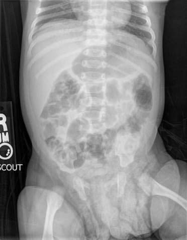
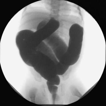
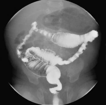
♦ Plain abdominal film reveals a distal bowel obstruction with dilated large bowel (Fig. 20-1). Barium enema reveals a narrow distal segment with dilated proximal bowel (Fig. 20-2). A postevacuation film reveals retained contrast material (Fig. 20-3).
Infants and Children
♦ Physical examination reveals a distended abdomen, normally placed anus, a rectal examination of normal caliber, a collapsed rectum, and production of explosive stool with removal of the digit. Barium enema reveals a collapsed distal colon and a dilated proximal colon. Postevacuation films reveal retained contrast material.
Management
♦ Most infants can be treated with colonic irrigations once or twice per day before scheduling a definitive procedure. Timing for a pull-through procedure is surgeon dependent, with some performing the procedure in the first several weeks of life and others waiting until infants are 2 to 3 months of age and weigh at least 10 pounds.
♦ If colonic irrigations do not sufficiently decompress the bowel and the infant is unable to tolerate feedings and gain weight, consideration should be made for the placement of a temporary colostomy.
♦ Infants with long-segment Hirschsprung disease or total colonic Hirschsprung disease are less likely to thrive with a colonic irrigation management scheme and will need a temporary colostomy placed in the colon or small bowel, where normal ganglion cells are present. A Kimura patch should be considered in infants with total colon Hirschsprung disease as a two-step procedure preceding pull-through.
Buy Membership for Pediatrics Category to continue reading. Learn more here