24 Hip and Thigh Fractures
Anatomy of the Hip and Thigh
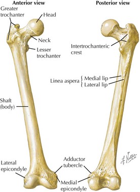
Femur
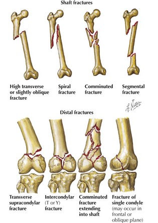
• Parts and landmarks: head; fovea (for round ligament); neck; greater trochanter; lesser trochanter; intertrochanteric line, crest, and fossa; pectineal line; gluteal tuberosity; linea aspera; shaft (body); popliteal surface; adductor tubercle; medial epicondyle; lateral epicondyle; medial condyle; lateral condyle; intercondylar fossa; patellar surface
Coxal (Hip) Bones
• Ilium, ischium, and pubis are fused in adults. (See Chapter 17, Pelvic Fractures, for more bone information.)


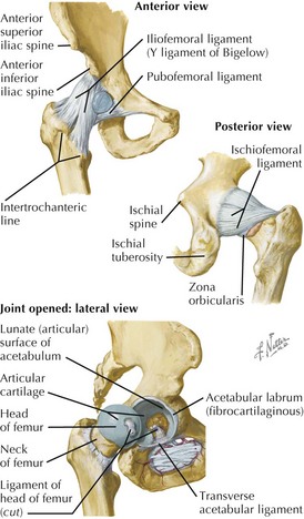
Hip Joint


• (Collateral) ligaments: spiraling thickenings of fibrous joint capsule, passing from acetabular rim to intertrochanteric line or trochanters


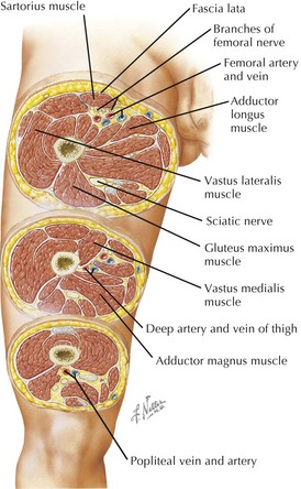
Compartments of the Thigh


• Fascia lata: investing deep fascia of thigh


• Gluteal compartment


• Anterior compartment


Vessels and Nerves
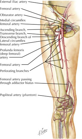
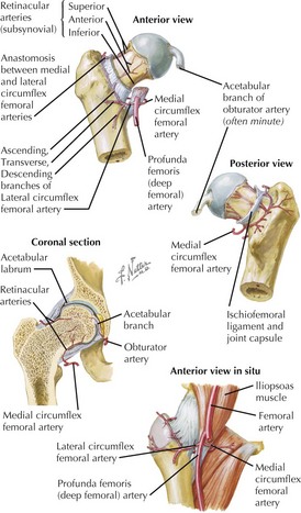
Arterial Supply to the Thigh and Hip Joint


• Hip joint is supplied by anastomotic branches of medial and lateral femoral circumflex and artery to head of femur (from obturator artery).
• Artery to head of femur runs along ligament of head; artery might contribute little blood to joint after adulthood.
• Immediate blood supply to hip joint provided by retinacular arteries, branches of circumflex vessels
• Retinacular arteries from medial circumflex usually provide more blood and pass beneath unattached posterior border of joint capsule.
Veins of the Hip and Thigh
• Run parallel to femoral artery and its major branches: valved; arterial counterpulsation effect pumps blood heartward


Clinical Correlates
Compartment Syndromes
• Relatively rare because large volume is required to cause pathological increase in tissue pressure
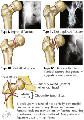
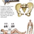
Hip Fractures
Intracapsular Fractures
• Compression-type fractures typically occur along inferior neck, more commonly in elderly persons with osteoporosis.