Hiccups
Hiccups (hiccoughs) are the characteristic sounds produced by the involuntary contractions of the diaphragm terminated by sudden closure of the glottis. In the majority, it is a self-limiting condition. The most common cause of hiccups is thought to be gastric distension after rapid ingestion of food, fluid or air. The causes of intractable hiccups are listed below.
History
The causes of hiccups predominantly originate in the respiratory, abdominal and nervous systems, therefore the history is structured similarly.
Respiratory history
The presence of cough with purulent sputum production implies respiratory tract infection. However, patients with pneumonia tend to be more unwell and may complain of rigors with high temperatures. Following pneumonia or thoracic surgery, chronic pyrexia and pleuritic chest pains with failure of resolution of symptoms of the original infection may herald the onset of an empyema. Hiccups may (rarely) complicate routine thoracic surgery. When a chronic cough is associated with haemoptysis and weight loss in a smoker, bronchial tumour is the most likely diagnosis.
Abdominal history
Symptoms of rapid progressive painless dysphagia (p. 109) may be due to oesophageal carcinoma. At birth, a diaphragmatic hernia may present with respiratory distress; adults with diaphragmatic hernias may, however, be completely asymptomatic, the hernia being detected incidentally on a chest X-ray. Unremitting pyrexia and malaise in patients after abdominal surgery, or following localised inflammatory conditions such as appendicitis and cholecystitis, may have developed an intra-abdominal abscess. The subphrenic region is the most common site of a collection of pus. This is associated with intractable hiccups and pain referred to the shoulder, scapula or neck. A pleural effusion may be an accompanying feature.
Neurological history
Pyrexia, headache, photophobia and neck stiffness are associated with meningitis. Patients with encephalitis tend to present with confusion, alterations in consciousness and seizures. The brainstem is a very anatomically complex structure, therefore symptoms of infarction or tumour may be very variable, depending on distribution of involvement of the lower cranial nerves. The presenting features may be diplopia, difficulty swallowing, dysarthria and facial sensory loss. Cortical lesions arising from infarction or haemorrhage may present with aphasia, visual field defects and hemiplegia. The principal differentiating feature between infarction and tumour is the speed of onset; in general, stroke presents suddenly and tumours progressively.
Others
Amelioration of hiccups by sleep, bizarre accompanying symptoms and inconsistencies in the history are suggestive of hysterical hiccups. Uraemia may be responsible for hiccups.
Examination
A substantial number of causes of intractable hiccups are associated with pyrexia. High swinging fevers are suggestive of collections of pus, such as empyema and subphrenic abscesses.
An abdominal examination should be performed. Areas of tenderness may suggest appendicitis or cholecystitis as a predisposing cause for a subphrenic abscess. More specifically, an abdominal abscess may point to the skin surface, or present with tenderness over the lower chest wall.
Respiratory examination may reveal tracheal deviation with a large empyema or pleural effusion from carcinoma. Areas of percussion dullness correspond to consolidation, effusion and empyema. A large diaphragmatic hernia may elevate the diaphragm sufficiently to produce clinical signs suggestive of an effusion. Localised coarse crepitations may be auscultated, corresponding to areas of consolidation by pneumonia. Vocal resonance is increased with consolidation but attenuated with effusion and empyema.
A neurological examination is performed to identify the presence of and define the anatomical territory of any deficit. Specialised tests should be performed for meningitis. This will include Kernig’s sign (pain on extension of the knee from a flexed hip and knee position) and Brudzinski’s sign (flexion of the neck produces flexion of the hip and knee).
General Investigations
In the absence of features in the history to guide specific investigations, the following general investigations should be performed.
■ FBC
WCC ↑ with infection, malignancy.
■ U&Es
↑ urea, ↑ creatinine with renal failure.
■ CXR
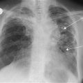
Peripheral shadow or cavitation, hilar mass, collapse of a lobe may suggest carcinoma. Consolidation is seen with pneumonia. Fluid with a meniscus may be due to empyema, effusion from malignancy or as a result of a subphrenic abscess. Elevation of a hemidiaphragm may be due to phrenic nerve palsy from carcinomatous infiltration or apparent elevation due to the presence of a diaphragmatic hernia.
Specific Investigations
■ Upper GI endoscopy and biopsy
Oesophageal carcinoma.
■ Bronchoscopy and mediastinoscopy
Biopsy for bronchial carcinoma.
■ CT thorax
Biopsy for bronchial carcinoma, definition and identification of loculations with empyema.
■ US abdomen
Subphrenic abscess; aspiration and drainage may also be performed.
■ CT/MRI head
Infarction and tumour may be appreciated as low-density areas. Mass effects may be produced by tumour or bleeding causing midline shifts. Intracranial bleeding may be identified as a high-density area. Cortical swelling may be seen with encephalitis. Raised intracranial pressure can be identified and is a contraindication to lumbar puncture.
■ EEG
Encephalitis – periodic complex formation and slowing of the background rhythm.
■ Lumbar puncture
Meningitis. Encephalitis – high lymphocyte count as the majority are due to viral aetiology.


