Mallavarapu, KM, et al. Multiple myeloma presenting with hepatosplenomegaly. J Assoc Physicians India. 2011; 59:456–458.
Patel, M, et al. Characterization of computed tomography scan abnormalities in patients with biopsy-proven hepatic metastases from uveal melanoma. Arch Ophthalmol. 2011; 129(12):1576–1582.
Valayannopoulos, V, et al. Successful treatment of severe cardiomyopathy in glycogen storage disease type III With D,L-3-hydroxybutyrate, ketogenic and high-protein diet. Pediatr Res. 2011; 70(6):638–641.
Weitz, N, et al. Education and imaging. Gastrointestinal: metastatic ocular melanoma causing massive hepatomegaly. J Gastroenterol Hepatol. 2011; 26(10):1577.
Wilson, S, et al. Hepatosplenomegaly associated with chronic malaria exposure: evidence for a pro-inflammatory mechanism exacerbated by schistosomiasis. Parasite Immunol. 2009; 31(2):64–71.
Wilson, S, et al. Hepatosplenomegaly in Kenyan schoolchildren: exacerbation by concurrent chronic exposure to malaria and Schistosoma mansoni infection. Trop Med Int Health. 2007; 12(12):1442–1449.
Oliva, MR, et al. Computed tomography features of nonalcoholic steatohepatitis with histopathologic correlation. J Comput Assist Tomogr. 2006; 30(1):37–43.
van Hoek, B, Non-alcoholic fatty liver disease: a brief review. Scand J Gastroenterol Suppl. 2004;(241):56–59.
Grazioli, L, et al. Liver adenomatosis: clinical, histopathologic, and imaging findings in 15 patients. Radiology. 2000; 216(2):395–402.


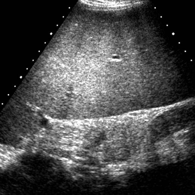
 , extending caudally below the level of the kidney
  .
.
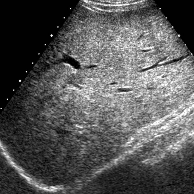
 extending below the level of the right kidney
  as well as echogenicity greater than that of the kidney due to diffuse steatosis.
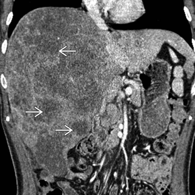
 .
.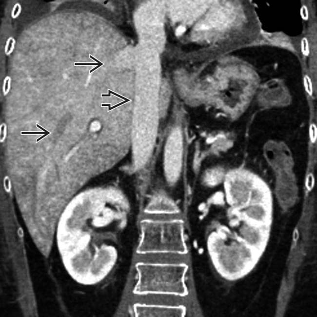
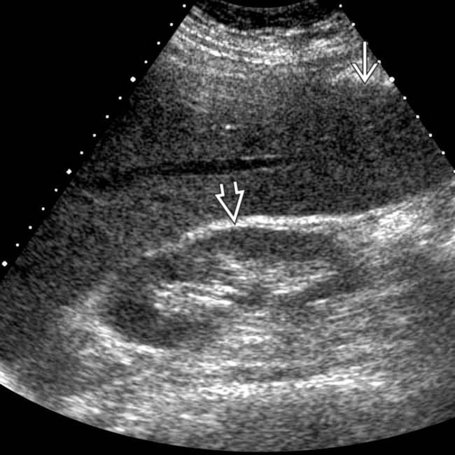
 and IVC
  due to passive congestion of the liver. Drug toxicity accounted for the cardiac dysfunction.