Hepatic Adenoma

Published on 19/07/2015 by admin

Filed under Radiology

Last modified 22/04/2025

Print this page

rate 1 star rate 2 star rate 3 star rate 4 star rate 5 star
Your rating: none, Average: 0 (0 votes)

This article have been viewed 2367 times

 Almost all in young women with high estrogen environment, including steatosis and oral contraceptive use

image Anabolic steroids, diabetes, and glycogen storage disease are other causes
• Key features (not always present): Hypervascularity, fat content, hemorrhage, encapsulation
• MR shows some elements better than CT (lipid and hemorrhage)
• Gadoxetate-enhanced MR (Eovist; Primovist)

image Adenoma shows no substantial uptake or retention
image Key distinction from FNH
• T1WI: Mass: Heterogeneous signal intensity

image Increased signal intensity (due to fat or recent hemorrhage)
image Decreased signal intensity (necrosis, calcification, old hemorrhage)
• Heterogeneous, hypervascular mass with foci of fat or hemorrhage in a young woman

TOP DIFFERENTIAL DIAGNOSES

• Hepatocellular carcinoma (HCC)

image HCC typically occurs in older, cirrhotic men
• Fibrolamellar HCC
• Focal nodular hyperplasia

image Homogeneously enhances; retains gadoxetate
• Hypervascular metastases

PATHOLOGY

• Hepatic steatosis, pregnancy, anabolic steroids,  and oral contraceptives increase number and growth rate of adenomas

CLINICAL ISSUES

• Risk factors for HCC

image Large adenoma, male sex, glycogen storage disease, anabolic steroid use, CTNNB1 -mutated subtype of HA
image
(Left) Graphic shows a hypervascular mass image 
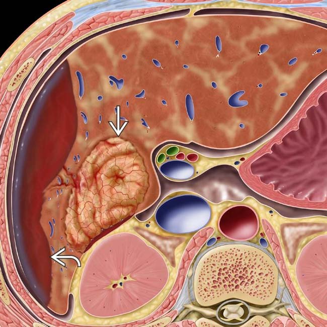
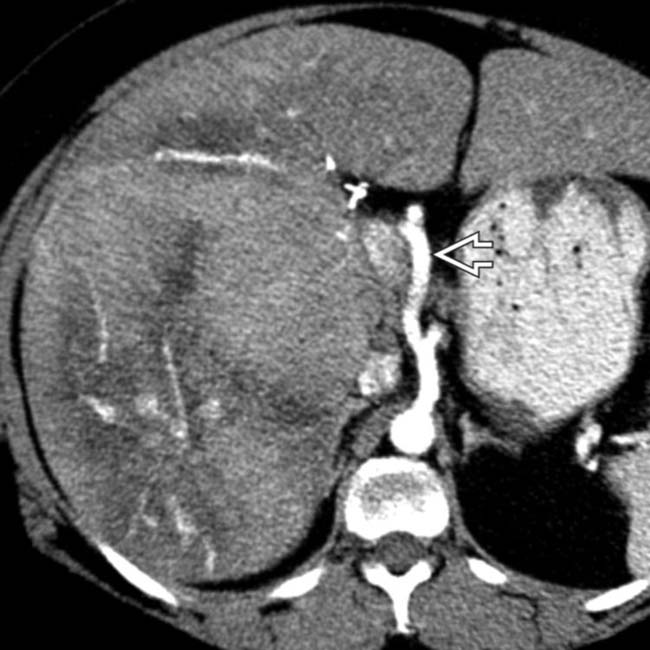
in the right lobe and spontaneous subcapsular bleeding image.

image
(Right) Axial CECT of a 40-year-old woman with sudden RUQ pain and syncope shows an intensely enhancing mass image 
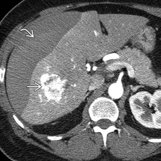
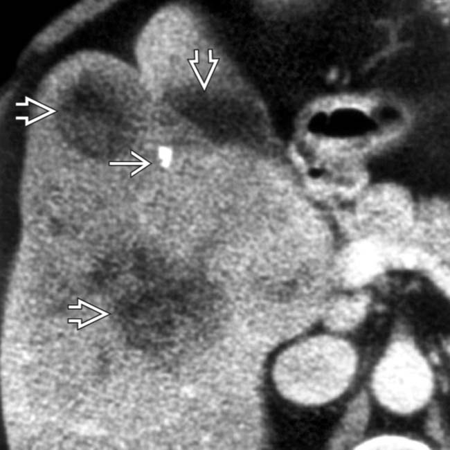
in the right lobe of the liver. A lentiform heterogeneous collection of fluid indents the surface of the liver, and within this collection is a focus of higher density image likely representing a sentinel clot. A ruptured inflammatory hepatic adenoma was resected.
image
(Left) Photograph of a resected specimen shows a large adenoma image 
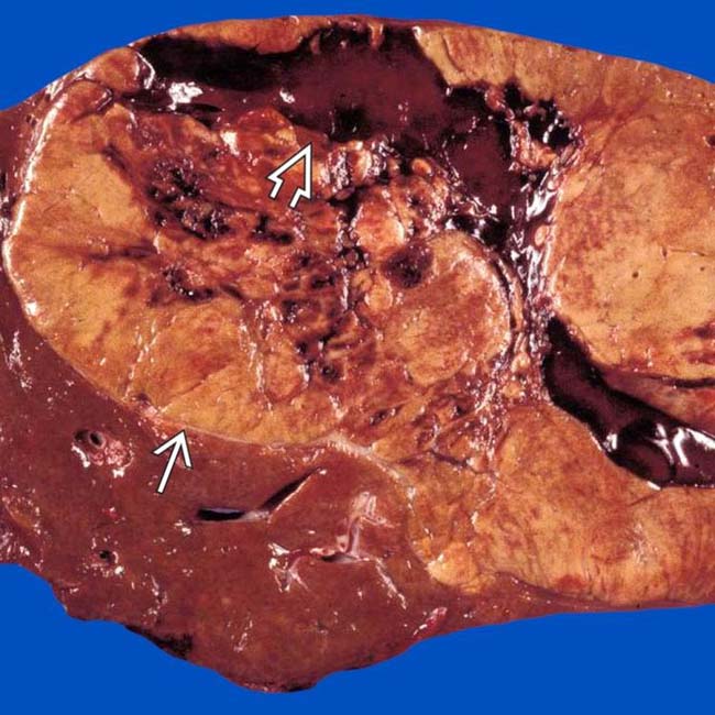
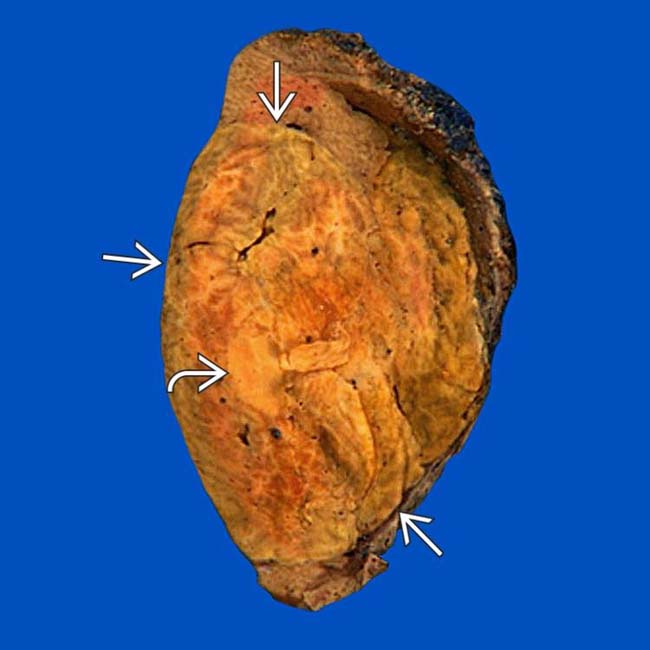
with central areas of rupture and hemorrhage image. (Courtesy M. Yeh, MD, PhD.)

image
(Right) Photomicrograph of a hepatic adenoma features a thin-walled unpaired vessel image 
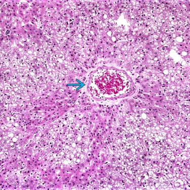
surrounded by neoplastic hepatocytes with abundant steatosis. Imaging often reveals these features, directly or indirectly. (Courtesy M. Yeh, MD, PhD.)

TERMINOLOGY

Abbreviations

• Hepatic adenoma (HA)

Synonyms

• Hepatocellular adenoma, liver cell adenoma

Definitions

• Heterogeneous group of benign hepatocellular neoplasms with distinctive genetic, pathologic, and clinical features

IMAGING

General Features

• Best diagnostic clue

image Heterogeneous, hypervascular mass with foci of fat or hemorrhage in a young woman
• Location

image Subcapsular region of right lobe of liver (75%)
image Intraparenchymal or pedunculated (10%)
• Size

image Varies from 6-30 cm
• Key concepts

image Very uncommon relative to focal nodular hyperplasia (FNH) and hepatocellular carcinoma (HCC)
image 3 distinct subtypes with different genetics, pathology, clinical features

CT Findings

• Depending on HA subtype

image Encapsulation seen in ∼ 20%, best on delayed phase CECT
image Hemorrhage within tumor, best seen on NECT as hyperdense foci
image Intratumoral lipid, best seen on NECT as hypodense foci
image Hypervascularity

– Most intense and persistent in inflammatory subtype of HA
image Calcification: Focal, present in ∼ 5%

MR Findings

• T1WI

image Mass: Heterogeneous signal intensity

– Increased signal intensity (due to fat and recent hemorrhage), more evident on MR than CT
– Decreased signal intensity (necrosis, calcification, old hemorrhage)
image Rim (fibrous pseudocapsule): Hypointense
• T2WI

image Mass: Heterogeneous signal intensity

– Increased signal intensity (old hemorrhage, necrosis)
– Decreased signal intensity (fat, recent hemorrhage)
image Rim (fibrous pseudocapsule): Hypointense
• T1WI C+

image Gadolinium, arterial phase

– Heterogeneous hypervascular enhancement (especially in inflammatory subtype)
image Delayed phase

– Pseudocapsule: Hyperintense to liver and adenoma
• Gadoxetate-enhanced MR (Eovist, Primovist)

image Hepatocellular-specific contrast agent
image Adenoma shows no substantial uptake or retention on delayed imaging

– Key distinction from FNH

Ultrasonographic Findings

• Grayscale ultrasound

image Complex, hyper-/hypoechoic, heterogeneous mass with anechoic areas

– Due to fat, hemorrhage, necrosis, and calcification
– Capsule may be seen
• Color Doppler

image Hypervascular tumor
image Large peripheral arteries and veins
image Intratumoral veins present

– Absent in FNH; useful distinction for adenoma

Angiographic Findings

• Conventional

image Hypervascular mass with centripetal flow
image Enlarged hepatic artery with feeders at tumor periphery (50%)
image Hypovascular; avascular regions

– Due to hemorrhage and necrosis

Nuclear Medicine Findings

• Technetium sulfur colloid (TcSC)

image Usually “cold” (photopenic) (80%)
image Uncommonly “warm” (20%)

– Due to uptake in sparse Kupffer cells
• HIDA scan

image Increased activity in some
• Gallium scan

image No uptake

Imaging Recommendations

• Best imaging tool

image Gadoxetate-enhanced MR, including multiphasic and delayed imaging
image In- and opposed-phase GRE

DIFFERENTIAL DIAGNOSIS

Hepatocellular Carcinoma

• May be hard to distinguish on imaging or pathology
• Biliary, vascular, nodal invasion and metastases = malignancy
• HCC typically occurs in older, cirrhotic men

image Adenoma occurs in young, healthy women

Fibrolamellar HCC

• Large, lobulated mass with scar and septa
• Vascular, biliary invasion and metastases common

Focal Nodular Hyperplasia

• Arterial phase: Homogeneously enhancing mass
• All other phases: Isodense to normal liver
• T2WI: Scar is typically hyperintense
• Uniformly retains gadoxetate on delayed phase MR

Hypervascular Metastases

• Usually multiple; look for primary tumors

image Breast, thyroid, kidney, and endocrine
• Arterial phase: Heterogeneous enhancement
• Portal and delayed phases: Iso-/hypodense washout

PATHOLOGY

General Features

• Etiology

image ↑ risk in oral contraceptive and anabolic steroid users
image Pregnancy increases growth rate and risk of rupture
image Hepatic steatosis ↑ growth and number of adenomas
image Diabetes mellitus
image von Gierke type I glycogen storage disease

– Multiple adenomas in 60% of affected patients

Staging, Grading, & Classification

• Proposed new classification based on genetics, pathology, and tumor biology
• 3 distinct subtypes

image Inflammatory HA

– Most common subtype (40-50%)
– Includes those previously called “telangiectatic HA”
– Occur in young women on oral contraceptives and obese women
– 60% have mutation of IL6ST gene with altered glycoprotein metabolism
– MR: No excessive fat or lipid within masses; persistent hypervascularity through arterial and venous phases due to sinusoidal dilation, peliotic areas, and abnormal vessels

image Bright on T2WI
image Likely to show MR (and clinical) evidence of hemorrhage (up to 30%)
image 10% estimated likelihood of malignant degeneration to HCC
image HNF1A -mutated HA

– 2nd most common subtype (30-35% of HAs)
– Association with diabetes and familial hepatic adenomatosis
– Exclusively in women; 90% have history of oral contraceptive use
– Mutated HNF1A  gene promotes lipogenesis and hepatocellular proliferation
– MR: Diffuse lipid deposition within HAs

image Most evident as signal dropout on opposed-phase GRE T1WI
image Macroscopic fat deposits are less common
image Only moderate enhancement on arterial phase; no persistent enhancement on venous and delayed
– Least aggressive subtype

image HAs of this subtype < 5 cm rarely bleed and have minimal risk of HCC
image CTNNB1 -mutated HA

– Least common subtype (10-15% of HAs)
– Subtype most likely to occur in men, those taking androgenic steroids, and in patients with glycogen storage disease

image Also associated with metabolic syndrome
– Mutation of CTNNB1 disrupts hepatocyte proliferation, growth, adhesion, etc.
– This subtype carries highest risk of malignant transformation (> 10%)
– MR features: No distinctive pattern established

image Usually hypervascular with evidence of hemorrhage or necrosis within tumor
image Unclassified HA subtype

– Does not fit other profiles of HA subtypes

Gross Pathologic & Surgical Features

• Well-circumscribed mass within noncirrhotic liver

image Soft, pale, or yellowish tan
image Frequently bile-stained nodules
image Foci of fat, hemorrhage, infarction
image Pseudocapsule and occasional pseudopods
• Adenomatosis

image No strict definition (generally > 10 adenomas)
image Associated with glycogen storage disease, steatosis, and diabetes
image May number > 100 adenomas

– May cause hepatic dysfunction, hemorrhage

Microscopic Features

• Sheets or cords of hepatocytes
• Absence of portal and central veins and bile ducts
• Increased amounts of glycogen and lipid
• Scattered, thin-walled, unpaired, vascular channels
• Few scattered Kupffer cells

CLINICAL ISSUES

Presentation

• Most common signs/symptoms

image RUQ pain (40%): Due to hemorrhage
image Asymptomatic (20%)

– Especially those with HNF1A  type
image No elevation of serum α-fetoprotein
• Diagnosis: Biopsy and histology

Demographics

• Age

image Young women in childbearing age group
image Predominantly in 3rd and 4th decades
• Gender

image 98% seen in females

– Not seen in males unless on anabolic steroids or with glycogen storage disease

Natural History & Prognosis

• Complications

image Hemorrhage: Intrahepatic or intraperitoneal (40%)
image Rupture: Increased risk in pregnancy
image May regress on withdrawal of oral contraceptives
• Risk factors for degeneration into HCC

image Male gender (10x more frequent than in women on per case basis)
image Concomitant glycogen storage disease
image Anabolic steroid use
image CTNNB1 -mutated subtype of HA
image HA mass size > 5 cm
• Prognosis

image Usually good

– After discontinuation of oral contraceptives
– After surgical resection of large or symptomatic HAs
image Poor

– Intraperitoneal rupture
– Rupture during pregnancy
– Malignant transformation
– Adenomatosis (> 10 adenomas)

image May hemorrhage, impair hepatic function

Treatment

• Adenoma < 5 cm

image Observation and discontinuation of oral contraceptives or other steroids
• Adenoma > 5 cm and near surface

image Surgical resection
image Consider transcatheter embolization
• Pregnancy should be avoided due to ↑ risk of rupture
• Transplantation for some cases of adenomatosis

DIAGNOSTIC CHECKLIST

Consider

• Rule out other benign and malignant liver tumors, which have similar imaging features

image Particularly HCC or FNH
• Check for history of oral contraceptives and glycogen storage disease (in case of multiple adenomas)

Image Interpretation Pearls

• Spherical, well-defined, hypervascular, and heterogeneous mass due to hemorrhage and fat; most evident on MR
• Gadoxetate-enhanced MR is most specific imaging test
image
(Left) Venous phase CECT of an 18-year-old girl injured in a vehicle crash shows a subcapsular, encapsulated mass image that is not very vascular, a typical (but not diagnostic) feature of the HNF1A-mutated subtype of hepatic adenoma.

image
(Right) An axial fat-suppressed T2WI in the same patient shows the mass as almost isointense to background liver with a thin hyperintense capsule image
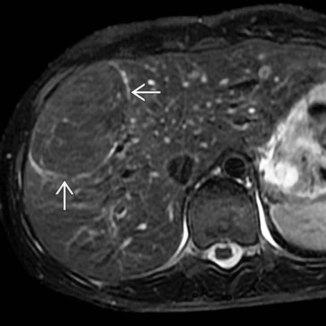
, typical features of an HNF1A-mutated subtype of hepatic adenoma.
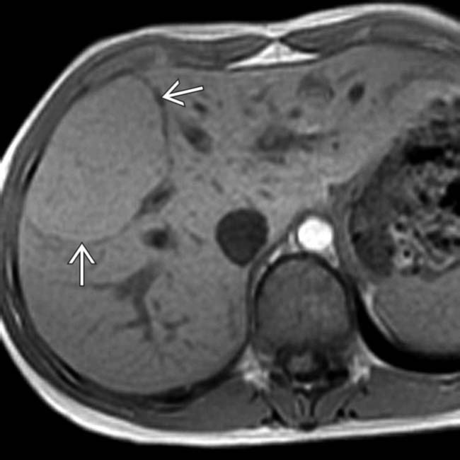
image
(Left) T1-weighted in-phase MR in the same case shows a uniform iso- to slightly hyperintense peripheral liver lesion with a thin low-intensity capsule image.

image
(Right) An opposed-phase GRE T1WI in the same case shows marked signal loss from the mass image, which indicates the presence of diffuse lipid, another typical feature of this subtype of hepatic adenoma.
image
(Left) On this delayed phase image in the same case, obtained after IV administration of gadoxetate (Eovist, Primovist), the normal liver enhances brightly whereas the mass shows no retained contrast, which indicates that it lacks functioning bile ductules and differentiates it from a focal nodular hyperplasia (FNH).

image
(Right) Photograph of the resected specimen, an HNF1A-mutated subtype of hepatic adenoma, shows an encapsulated, pale, tan mass image 
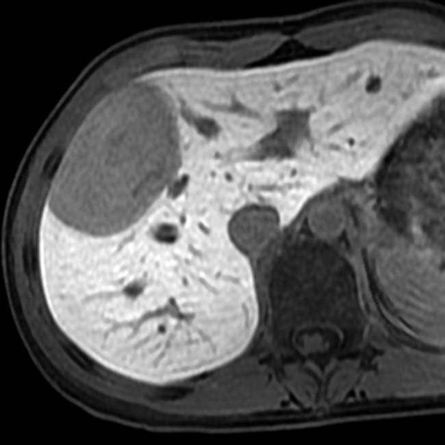
with diffuse and focal lipid image.
image
(Left) Axial T1WI GRE in-phase MR in a 54-year-old woman who had a liver mass detected on CT shows the mass image as slightly hyperintense to a normal-appearing liver. On opposed-phase GRE MR (not shown), there was no signal dropout from the lesion, indicating no lipid content.

image
(Right) On T2WI MR in the same case, the mass image 
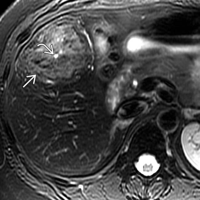
is slightly hyperintense to the liver, and a central scar image is quite hyperintense.
image
(Left) On MR sections in the same case taken after bolus injection of gadoxetate (Eovist) the mass is hypervascular image 
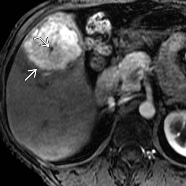
whereas the central scar is hypointense image. The mass and central scar were nearly isointense on the venous phase images (not shown).

image
(Right) On a 20-minute delayed image in the same case the mass image 
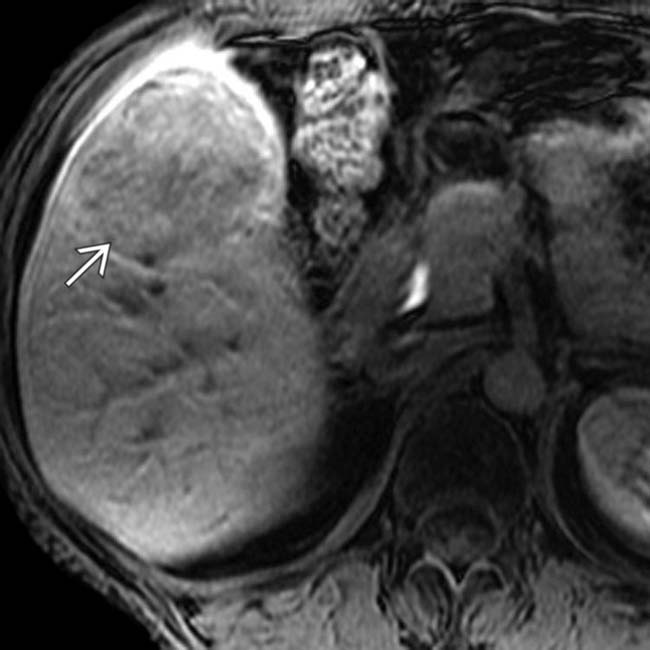
retains less contrast material than normal liver. Because there was concern that the lesion was not a typical FNH, it was resected.
image
(Left) Photo of the gross pathology specimen shows an encapsulated mass with blood pooling and foci of hemorrhage.

image
(Right) Histologic specimen in the same case shows disorganized hepatocytes image 
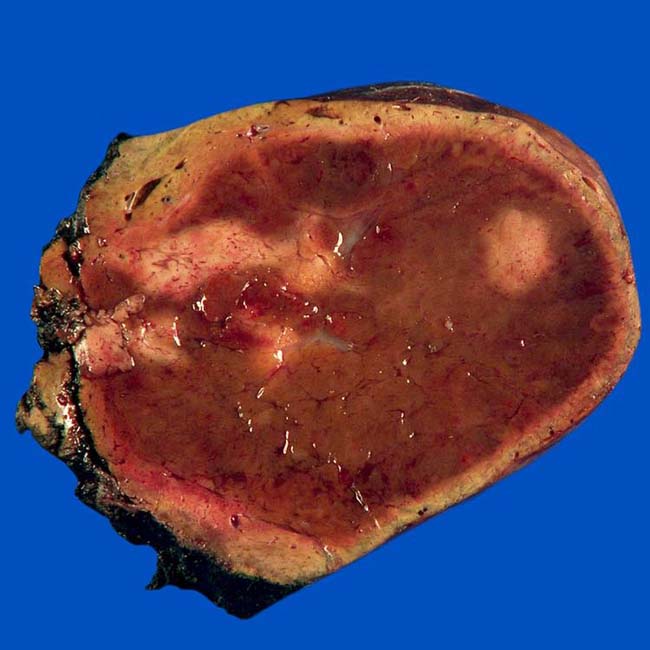
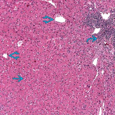
with abundant eosinophilic nuclei on H&E staining, unpaired blood vessels image distributed haphazardly throughout the lesion, and foci of chronic inflammation image. The final diagnosis was hepatic adenoma of the inflammatory subtype. (Courtesy T. Longacre, MD.)
image
(Left) Axial T1WI of a 35-year-old woman with RUQ pain shows a hepatic mass image 
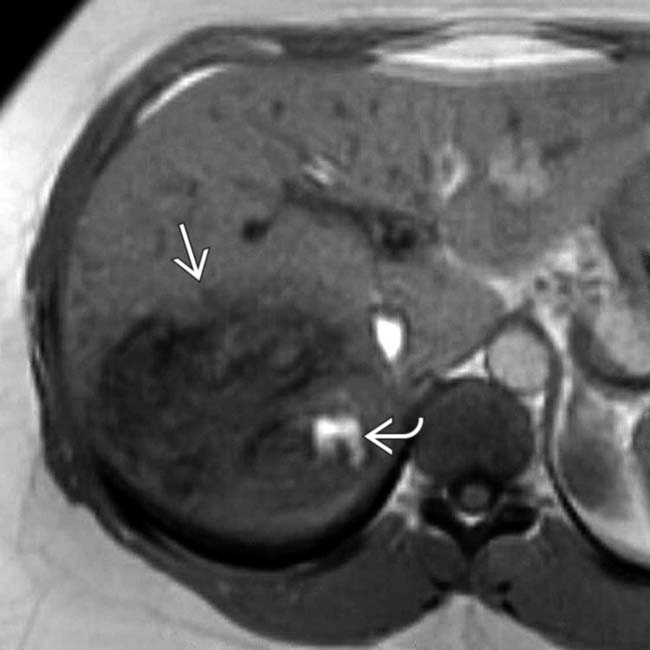
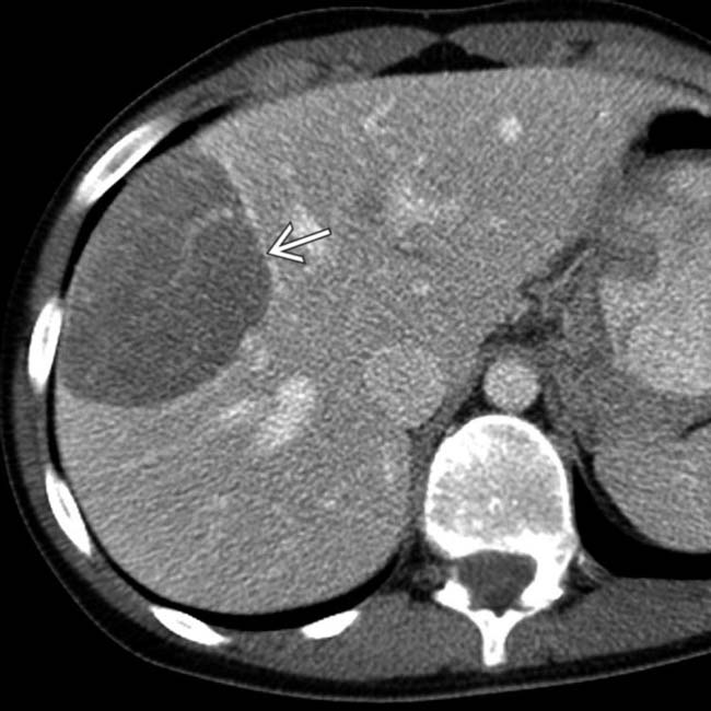
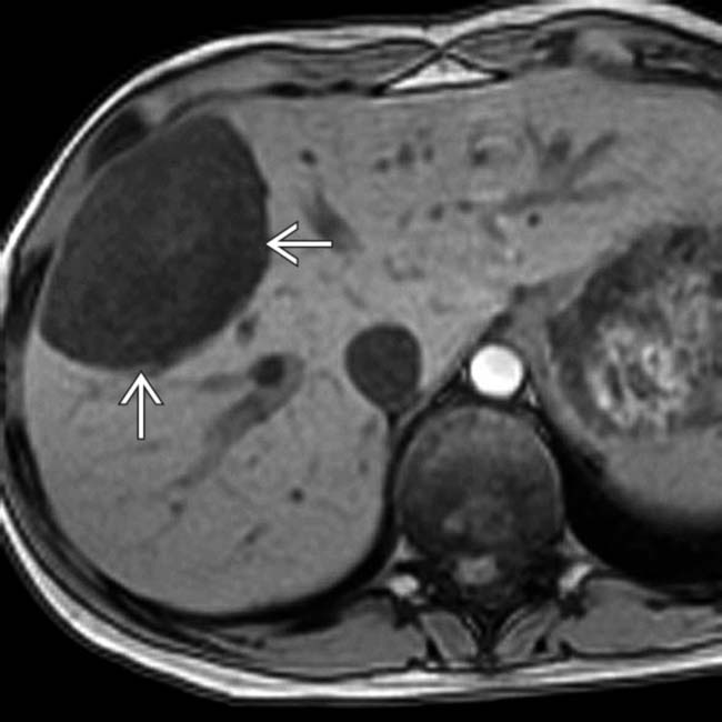
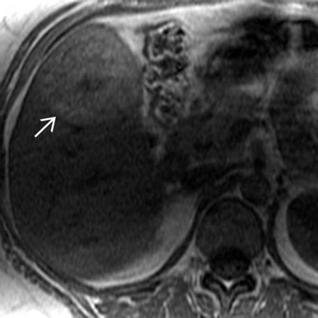
containing several hyperintense foci image that represent hemorrhage.

image
(Right) Axial T1WI shows more hyperintense foci image 
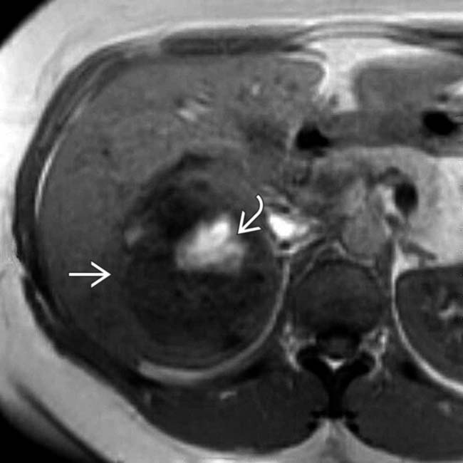
of hemorrhage within the hypointense mass image.
image
(Left) Axial T2WI in the same case shows a heterogeneous mass with some foci of slight hyperintensity image that represent lipid.

image
(Right) Axial portal venous phase T1WI in the same case shows persistent hypervascular enhancement image 
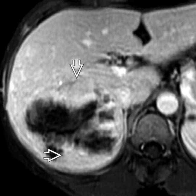
in the nonnecrotic and nonhemorrhagic portions of the mass, typical features of the inflammatory subtype of hepatic adenoma, which was confirmed on resection.
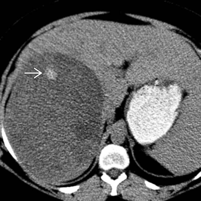
image
(Left) Axial NECT shows a very large, sharply demarcated, mostly homogeneous mass with a small focus of hemorrhage image.

image
(Right) Axial arterial phase CECT in the same patient shows hypervascularity with enlarged vessels within and on the surface of the tumor. Note the large hepatic artery image. Hepatic adenomas of the inflammatory subtype are usually heterogeneously hypervascular and often encapsulated with focal hemorrhage, as in this patient.
image
(Left) Axial opposed-phase GRE T1WI MR shows an encapsulated mass image 
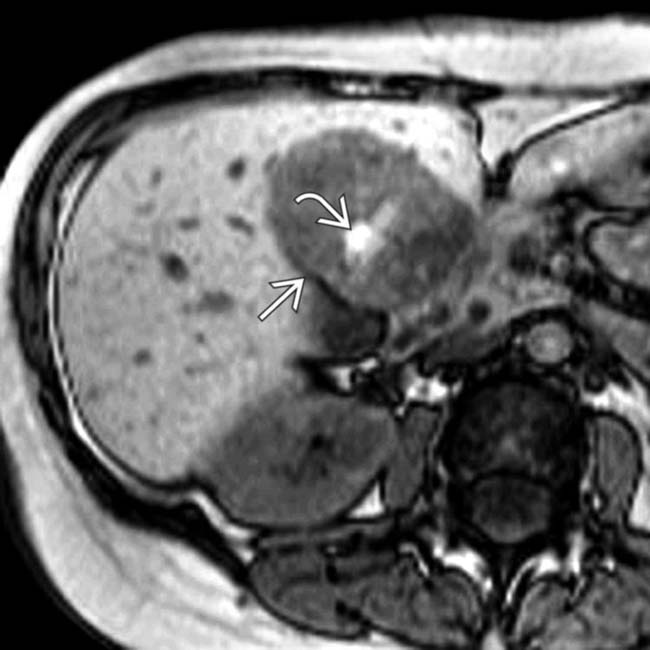
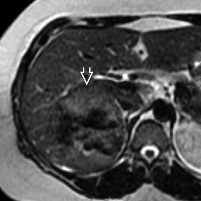
with hyperintense foci image representing hemorrhage or fat. The in-phase images showed increased signal within the mass.

image
(Right) Axial fat-suppressed T2WI MR in the same patient shows that the mass is nearly isointense to the liver with a central focus of hyperintensity image 
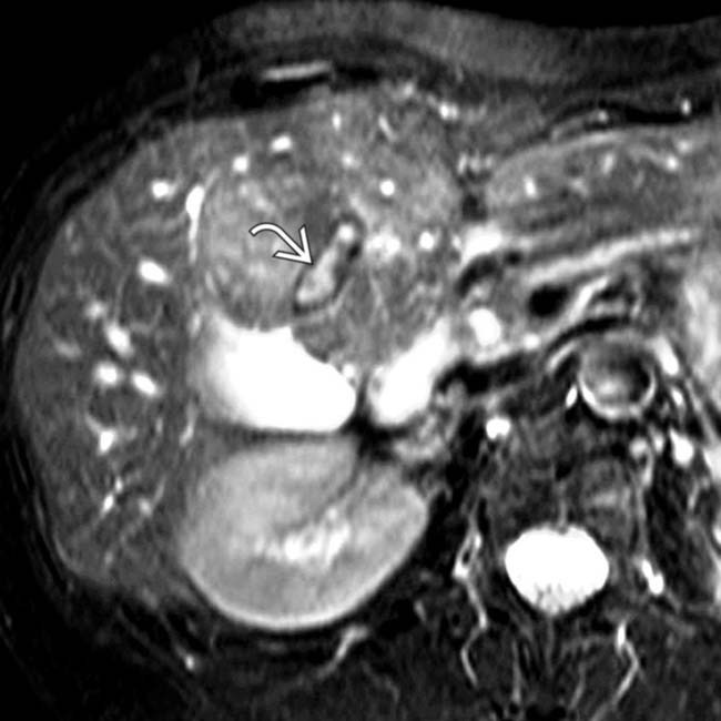
(hemorrhage). Fat content would have shown signal dropout on this sequence. A capsule or pseudocapsule is seen in ∼ 20% of adenomas.
image
(Left) Axial CT during the arterial phase of enhancement shows a brightly enhancing mass image 
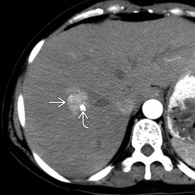
with a focal calcification image, the latter being a less common feature of adenomas.
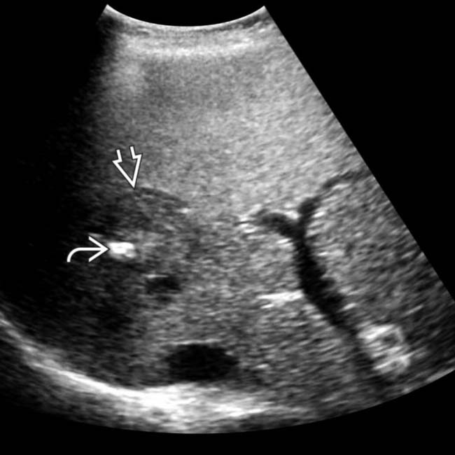
image
(Right) Ultrasound in the same case confirms the solid mass with calcification image and shows a capsule image around the mass. The capsule is a common feature and the focal calcification a less common feature of adenomas, as was proven in this case.
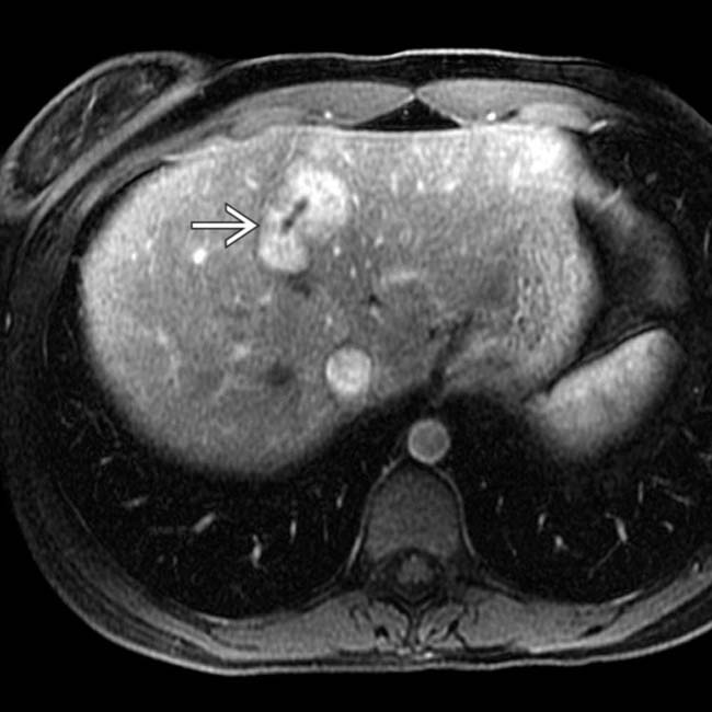
image
(Left) Arterial phase T1WI contrast-enhanced MR of a 32-year-old woman who had been using oral contraceptives for 14 years shows 1 of about 10 encapsulated, enhancing focal masses image.

image
(Right) More adenomas image are seen on this section. In the absence of chronic liver disease or a known malignancy, these findings were considered diagnostic of adenomas. Following cessation of oral contraceptive use, these adenomas decreased in size and number within 4 months.
image
(Left) Axial NECT of a 37-year-old man with type I glycogen storage disease and multiple adenomas shows hepatomegaly and low-attenuation masses image 
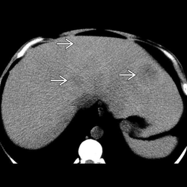
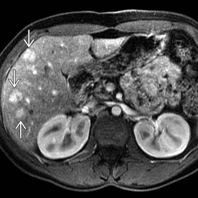
within the liver. The low attenuation is due to intratumoral lipid. These are foci of the CTNNB1-mutated subtype of hepatic adenoma.

image
(Right) A hepatic arterial phase image in the same patient shows that some lesions are hypervascular image 
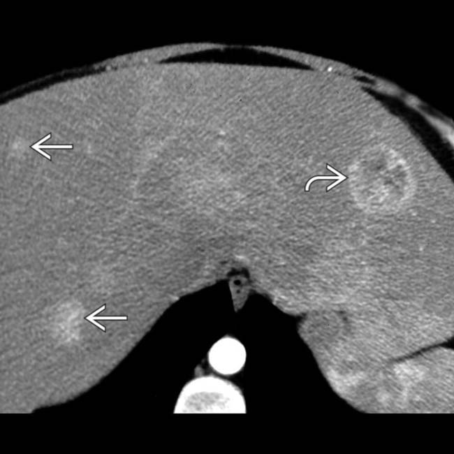
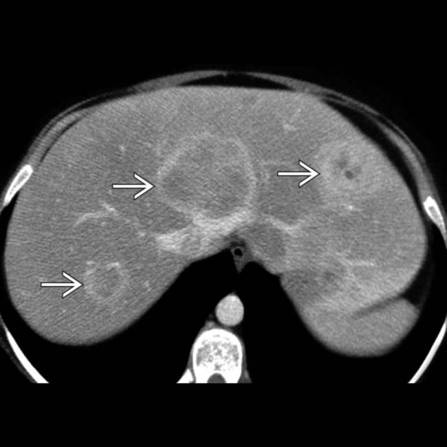
whereas others show heterogeneous enhancement image.
image
(Left) Portal venous phase image in the same case shows capsular enhancement or compressed hepatic parenchyma around most of the adenomas image. Adenomas are usually multiple in the setting of type I or III glycogen storage disease and carry a high risk of malignant degeneration, especially those > 5 cm in diameter and those in men.

image
(Right) Photograph of the resected specimens in a case of hepatic adenomatosis shows multiple adenomas with hemorrhage and necrosis. (Courtesy M. Yeh, MD, PhD.)
image
(Left) Axial CECT shows a large heterogeneous hepatic mass with several foci image 
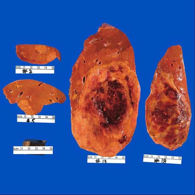
measuring less than water attenuation, indicating fat content. Also noted are foci of calcification image. Serial CT scans had shown slow growth of the mass over 7 years.
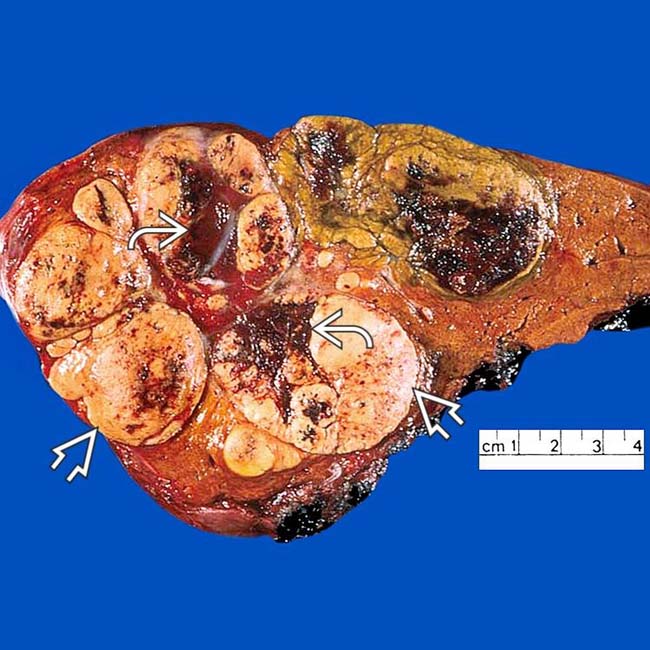
image
(Right) Gross pathology from the same case shows the mass with foci of fat image and hemorrhage image. Histology revealed foci of HCC within this adenoma, which had the CTNNB1 mutation, as did the foci of HCC.

SELECTED REFERENCES

1. Cogley, JR, et al. MR imaging of benign focal liver lesions. Radiol Clin North Am. 2014; 52(4):657–682.

2. De Kock, I, et al. Hepatic adenomatosis: MR imaging features. JBR-BTR. 2014; 97(2):105–108.

3. Grazioli, L, et al. Hepatocellular adenoma and focal nodular hyperplasia: value of gadoxetic acid-enhanced MR imaging in differential diagnosis. Radiology. 2012; 262(2):520–529.

Katabathina, VS, et al. Genetics and imaging of hepatocellular adenomas: 2011 update. Radiographics. 2011; 31(6):1529–1543.

Mounajjed, T, et al. Telangiectatic variant of hepatic adenoma: clinicopathologic features and correlation between liver needle biopsy and resection. Am J Surg Pathol. 2011; 35(9):1356–1363.

Ji, HF, et al. Reduced-size liver transplantation for glycogen storage disease. Hepatobiliary Pancreat Dis Int. 2009; 8(1):106–108.

Kobayashi, S, et al. Radiological and histopathological manifestations of hepatocellular nodular lesions concomitant with various congenital and acquired hepatic hemodynamic abnormalities. Jpn J Radiol. 2009; 27(2):53–68.

Santambrogio, R, et al. Liver transplantation for spontaneous intrapartum rupture of a hepatic adenoma. Obstet Gynecol. 2009; 113(2 Pt 2):508–510.

Furlan, A, et al. Multiple hepatic adenomas associated with liver steatosis at CT and MRI: a case-control study. AJR Am J Roentgenol. 2008; 191(5):1430–1435.

Prasad, SR, et al. Fat-containing lesions of the liver: radiologic-pathologic correlation. Radiographics. 2005; 25(2):321–331.

Grazioli, L, et al. Hepatic adenomas: imaging and pathologic findings. Radiographics. 2001; 21(4):877–892. [discussion 892-4].

Grazioli, L, et al. Liver adenomatosis: clinical, histopathologic, and imaging findings in 15 patients. Radiology. 2000; 216(2):395–402.

Ichikawa, T, et al. Hepatocellular adenoma: multiphasic CT and histopathologic findings in 25 patients. Radiology. 2000; 214(3):861–868.