Chapter 23 HEMATURIA, MICROSCOPIC
Asymptomatic isolated microscopic hematuria is common in children. Many experts recommend observation of these children if the examination is normal, with further evaluation only if proteinuria, hypertension, or gross hematuria is present. In the child with asymptomatic isolated microscopic hematuria, the early morning urinalysis should be repeated weekly for 2 weeks with no exercise before the collection of the urine sample. If the hematuria persists, urine culture should be obtained and, if positive, the patient should be treated with antibiotics. If the urine culture is negative, the patient should be followed up every 3 months with a history, physical examination, blood pressure measurement, and urinalysis. If the hematuria persists for 1 year, measurement of the urine calcium-creatinine ratio should be obtained, and the parents and siblings should be tested for hematuria. Hemoglobin electrophoresis should be performed if sickle cell trait is a consideration. Figure 23-1 provides an algorithm for the evaluation of a child with asymptomatic microscopic hematuria.
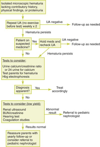
Figure 23-1 Evaluation of a child with asymptomatic microscopic hematuria.
(From Patel HP, Bissler JJ. Hematuria in children. Pediatr Clin North Am 2001;48:1519–1537, with permission.)
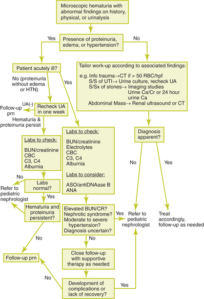
Asymptomatic microscopic hematuria with proteinuria is associated with a higher risk for significant renal disease. In this case, urinary protein excretion should be quantified. If protein excretion is greater than 4 mg/m2 per hour or the urine protein-to-creatinine ratio is greater than 0.2 mg protein per milligram of creatinine in children older than 2 years or greater than 0.5 mg of protein per milligram of creatinine in younger children, the patient should be referred to a pediatric nephrologist. If urinary protein excretion is less than these values, the patient should be reevaluated in a few weeks. If the hematuria and proteinuria have resolved, no further evaluation is indicated. If there is only asymptomatic microscopic hematuria, the patient may be monitored as for asymptomatic isolated microscopic hematuria. If the hematuria and proteinuria persist, the patient should be referred to a pediatric nephrologist. Additional testing is outlined below. Figure 23-2 provides an algorithm for the evaluation of a child with microscopic hematuria associated with proteinuria, symptoms, or abnormalities in history or physics examination.
Causes of Hematuria
• Acute poststreptococcal glomerulonephritis
• Idiopathic hypercalciuria without urolithiasis
• Membranoproliferative glomerulonephritis
• Mesangial proliferative glomerulonephritis
• Microangiopathic polyarteritis nodosa
• Rapidly progressive glomerulonephritis
• Systemic lupus erythematosus
• Thin basement membrane disease (benign familial hematuria)
Key Historical Features


Key Physical Findings


Suggested General Work-up of Microscopic Hematuria
| Test for proteinuria | To determine whether significant proteinuria is present |
| Microscopic examination of the urine for RBCs and RBC casts | To confirm the presence of hematuria and evaluate for underlying glomerulonephritis |
| Urine culture | To evaluate for (UTI) |
| Urine calcium-to-creatinine ratio | To evaluate for hypercalciuria if hematuria persists for 1 year (urine calcium-creatinine ratio less than 0.2 is normal) |
| Renal ultrasound | Should be considered to evaluate for stones, tumors, hydronephrosis, structural anomalies, renal parenchymal dysplasia, medical renal disease, inflammation of the bladder, bladder polyps, and posterior urethral valves |
| Hearing test | If there is any reason to suspect familial renal disease |
Suggested Work-up of Isolated Asymptomatic Microscopic Hematuria
| Urine culture | To evaluate for (UTI) |
| Serum creatinine | To evaluate for renal insufficiency |
| Renal ultrasound | Should be considered to evaluate for stones, tumors, hydronephrosis, structural anomalies, renal parenchymal dysplasia, medical renal disease, inflammation of the bladder, bladder polyps, and posterior urethral valves |
| Urine calcium-to-creatinine ratio | To evaluate for hypercalciuria if hematuria persists for 1 year (urine calcium/creatinine ratio less than 0.2 is normal) |
Additional Work-up of Isolated Asymptomatic Microscopic Hematuria
| 24-hour urine calcium excretion, serum electrolytes, calcium, phosphorus, magnesium | If hypercalciuria is identified |
| Coagulation studies | If an underlying coagulopathy is considered |
Suggested Work-up of Asymptomatic Microscopic Hematuria with Proteinuria
| Quantification of urinary protein excretion with 24-hour collection or spot urine protein/creatinine ratio | To quantify urinary protein excretion |
| Serum creatinine and BUN, CBC, serum albumin, antistreptolysin O (ASO) titers streptozyme test, serum albumin, serum complement C3 and C4 | To evaluate for glomerulonephritis |
| Antinuclear antibodies (ANA) | If lupus is suspected |
Suggested Work-up of Symptomatic Microscopic Hematuria
| Serum creatinine and BUN, CBC, serum albumin, ASO titers, streptozyme test, serum albumin, serum complement C3 and C4 | To evaluate for glomerulonephritis |
| ANA | To evaluate for lupus |
| Renal ultrasound or computed tomography (CT) scan | To evaluate for stones, tumors, hydronephrosis, structural anomalies, renal parenchymal dysplasia, medical renal disease, inflammation of the bladder, bladder polyps, and posterior urethral valves |
1. Gordon C., Stapleton F.B. Hematuria in adolescents. Adolesc Med. 2005;16:229–239.
2. Mahan J.D., Turman M.A., Mentser M.I. Evaluation of hematuria, proteinuria, and hypertension in adolescents. Pediatr Clin North Am. 1997;44:1573–1589.
3. Meyers K.E.C. Evaluation of hematuria in children. Urol Clin North Am. 2004;31:559–573.
4. Pan C.G. Evaluation of gross hematuria. Pediatr Clin North Am. 2006;53:401–412.
5. Patel H.P., Bissler J.J. Hematuria in children. Pediatr Clin North Am. 2001;48:1519–1537.