[level-membership-for-hematology-oncology-and-palliative-medicine-category]
2 HEMATOPOIESIS
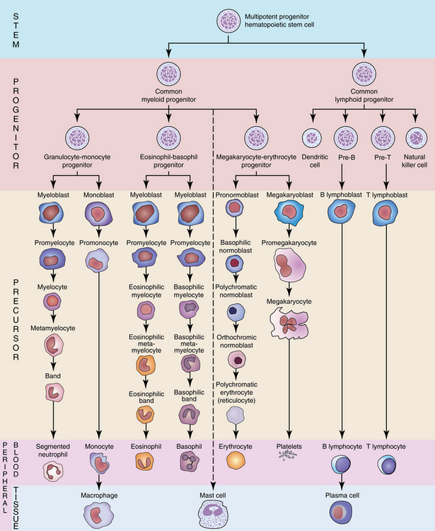
Hematopoiesis is a vigorous process of blood cell production and maturation that in the adult occurs primarily in the bone marrow. The process begins with the pluripotential hematopoietic stem cell (multipotent progenitor), which is capable of proliferation, replication, and differentiation. In response to cytokines (growth factors), the pluripotential stem cell will differentiate into a common myeloid or common lymphoid progenitor. Both the myeloid and lymphoid progenitors maintain their pluripotential capacity. The lymphoid progenitor proliferates and differentiates into T, B, and natural killer cells. The myeloid progenitor proliferates and differentiates into granulocyte, monocyte, erythrocyte, and megakaryocyte lineages. To this point in maturation, none of these stem cells can be morphologically identified, although it is postulated that they appear similar to a small resting lymphocyte. The blue shaded area in Figure 2-1 highlights the stem cell populations. Each lineage and maturation stage will be presented in detail in the following chapters.
Hematopoiesis is a dynamic continuum, that is, cells gradually mature from one stage to the next and may be between stages when viewed through the microscope. In general, the cell is then identified as the more mature stage. General morphological changes in blood cell maturation are demonstrated in Figure 2-2.
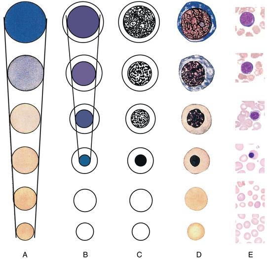
• An exception to the diameter decreasing is that in the granulocytic series, the promyelocyte may be larger than its precursor, the myeloblast (see Chapter 5).
• In the erythroid series, hemoglobin development in the cytoplasm imparts a pink/salmon color.
C, Nuclear chromatin becomes coarser, clumped, and condensed.
• In the granulocytic series, the nuclear shapes changes and the nucleus becomes segmented. Granules appear in cytoplasm (see Chapter 5).
• In the erythroid series, the nucleus becomes fully condensed and is ejected.
D, Composite of changes during maturation process. E, Representative cells from the erythroid series, demonstrating maturation changes.
(Modified from Diggs LW, Sturm D, Bell A: The morphology of human blood cells, ed 5, Abbott Park, Ill, 1985, Abbott Laboratories. Reproduction of The Morphology of Human Blood Cells has been granted with approval of Abbott Laboratories, all rights reserved by Abbott Laboratories.)
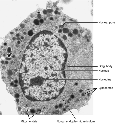
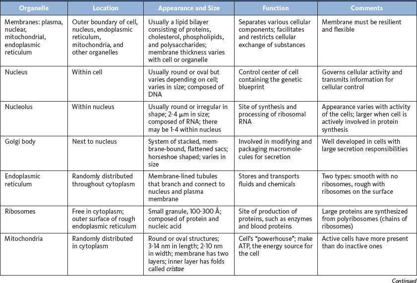
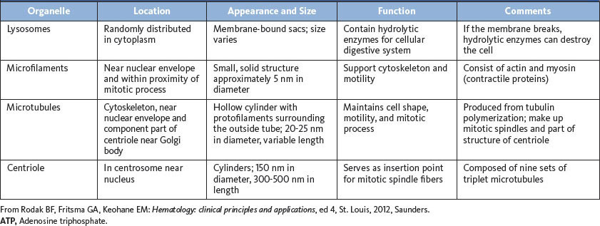
Figures 2-3 and 2-4 illustrate cell ultrastructure. A review of organelles will facilitate correlation of morphological maturation with cell function. This topic is explored in depth in hematology textbooks. Table 2-1 delineates the location, appearance, and function of individual organelles.
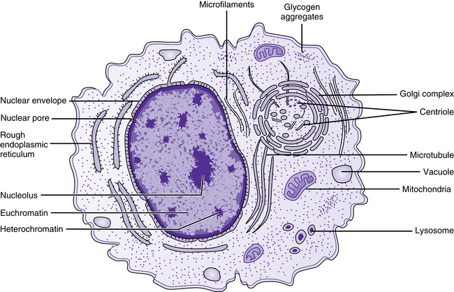
FIGURE 2–3 Schematic of electron micrograph. (From Rodak BF, Fritsma GA, Keohane EM: Hematology: clinical principles and applications, ed 4, St. Louis, 2012, Saunders.)
[/level-membership-for-hematology-oncology-and-palliative-medicine-category][not-level-membership-for-hematology-oncology-and-palliative-medicine-category]
2 HEMATOPOIESIS
Hematopoiesis is a vigorous process of blood cell production and maturation that in the adult occurs primarily in the bone marrow. The process begins with the pluripotential hematopoietic stem cell (multipotent progenitor), which is capable of proliferation, replication, and differentiation. In response to cytokines (growth factors), the pluripotential stem cell will differentiate into a common myeloid or common lymphoid progenitor. Both the myeloid and lymphoid progenitors maintain their pluripotential capacity. The lymphoid progenitor proliferates and differentiates into T, B, and natural killer cells. The myeloid progenitor proliferates and differentiates into granulocyte, monocyte, erythrocyte, and megakaryocyte lineages. To this point in maturation, none of these stem cells can be morphologically identified, although it is postulated that they appear similar to a small resting lymphocyte. The blue shaded area in Figure 2-1 highlights the stem cell populations. Each lineage and maturation stage will be presented in detail in the following chapters.
[/not-level-membership-for-hematology-oncology-and-palliative-medicine-category]
