140
Hemangiomas of infancy
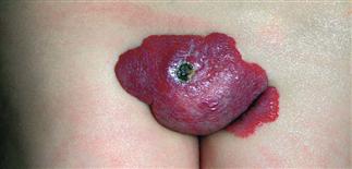
Ulceration is one of the most common complications of hemangiomas of infancy. Midline lumbar and sacral lesions can be associated with underlying vertebral and spinal cord anomalies.
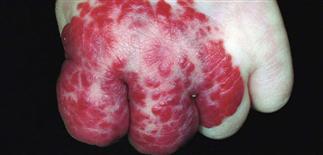
This early lesion on the dorsal aspect of the hand and fingers involuted without treatment and without functional loss of the hand.
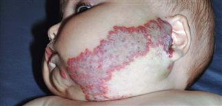
The head and neck regions are the most common locations for hemangiomas of infancy. This large parotid hemangioma required oral steroids, intralesional steroids, and surgical excision.
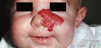
This plaque hemangioma involving the medial cheek and nasal dorsum resolved completely within 5 years. The patient had a 2-month course of oral corticosteroids.
DESCRIPTION
Benign red, purple, or blue vascular neoplasms due to endothelial hyperplasia occurring within the first year of life.
HISTORY
• Most common vascular tumor of infancy (1–3% of newborns and 10% of 1-year-olds); 30% noticeable at birth, most detected within the first 3 weeks of life. Deeper lesions noted within first month of life; predictable pattern of growth, stabilization, and involution. • Half of all hemangiomas resolve by age 5. • Lesion size, depth, location do not affect rate of involution. • Most infants have single lesion; multiple lesions in 15–20%. • Visceral involvement possible. • More common in girls, premature infants, and head and neck region; can damage function of eyes, ears, mouth when located near these organs. • Mandible (beard distribution) hemangiomas of infancy can be associated with glottic hemangiomas. Large segmental facial hemangiomas can be associated with malformations of other organs (PHACES syndrome: posterior fossa malformations, hemangioma, arterial anomalies, coarctation of the aorta and cardiac defects, eye anomalies, and sternal defects). Midline hemangiomas of infancy can be associated with underlying bony, soft tissue abnormalities. • Other complications: pain, ulceration, infection.
PHYSICAL FINDINGS
• Nascent (early) hemangiomas of infancy appear flat and pale white, with a few telangiectasias and large dilated blood vessels. Growing hemangiomas appear bright red (superficial) or blue (deep), feel firm and rubbery. • The surface color of deeper lesions can be very subtle. • Involuting hemangiomas become slate-gray and begin to soften.
TREATMENT
• Follow closely for complications. • Avoid scarring procedures, except when medically necessary to prevent permanent deformity. • Ulcerations: gently cleanse with mild soap, apply thin layer of a topical antibiotic such as mupirocin 2% ointment (Bactroban), metronidazole gel (MetroGel), or bacitracin ointment; cover with air-permeable barrier dressing such as polyurethane film dressing, Tegaderm, and OpSite. Barrier creams such as zinc oxide 20% ointment, Desitin ointment, A&D ointment can be used for the perineum and other sites not amenable to topical dressings. Regranex (platelet-derived growth factor) speeds healing and decreases pain. • Pulsed dye laser for ulcerations can be useful but can worsen ulceration in a small percentage. • Systemic prednisone or prednisolone 2–3 mg/kg orally, given as a single morning dose, is a widely used therapy for complicated lesions. • Propranolol given at a dose of 2 mg/kg/day divided b.i.d. or t.i.d. • Other medical therapies include intralesional and topical corticosteroids, and topical beta-blockers such as timolol maleate. • Embolization, surgical resection, and radiation are sometimes used for complicated hemangiomas.


