Heart Failure

Published on 06/06/2015 by admin

Filed under Pediatrics

Last modified 06/06/2015

Print this page

rate 1 star rate 2 star rate 3 star rate 4 star rate 5 star
Your rating: none, Average: 0 (0 votes)

This article have been viewed 1447 times

46 Heart Failure

A healthy child’s heart may beat more than 200,000 times and transport more than 3 tons of blood each day. The heart and vascular system are primarily responsible for delivering blood to the tissues while providing oxygen and nutrients and withdrawing waste products. Each is involved in the regulation of a perfusing blood pressure, including those homeostatic alterations necessitated with changes in posture. They play a primary role in the circulation of hormones. Heart failure is the inability of the heart to meet the metabolic demands of the body.

The failure of the heart and vascular system to keep up with metabolic requirements may arise from a number of causes, including cellular deficiencies, arrhythmias, metabolic insufficiencies, and congenital defects. Although most frequently a child presents in heart failure because of decompensation from congenital anomalies, the more prevalent of these are discussed elsewhere within this text, and only the principles common to heart failure will be repeated here. It is important to remember “heart failure” as a clinical picture may present in many of these anomalies when the physiology markedly worsens. However, to illustrate the fundamental concepts of pediatric heart failure, the remainder of this chapter focuses on that which may arise in children with normal anatomy. This state of “heart failure” in children may present indolently or very suddenly; in the latter, less-specific symptoms may be present for longer.

Regardless of the source, the goals of treatment are always (1) discovering and addressing the cause of failure along with (2) decreasing myocardial work load and oxygen consumption and (3) augmenting and supporting function and systemic oxygenation. With such interventions, the hope is that the heart will remodel, if not recover. Because of the danger of cardiovascular collapse, which may be very rapid, prompt consultation with pediatric cardiologists and intensivists is important, and early recognition requires diagnostic vigilance. Through a careful history and examination and with tests such as electrocardiography (ECG), echocardiography, and cardiac catheterization, the cause may often be learned and treatment instituted. Invasive arterial or venous monitoring is helpful in some cases if the patient is very ill.

Etiology And Presentation

The heart is a pump. It may fail as a pump either from the inability to generate enough contractile force or from structural anomalies that prevent or inappropriately direct blood to the tissues. Examples of the former include myocarditis, an infection of heart muscle cells (myocytes), or a cardiomyopathy, a primary structural or metabolic abnormality of the microscopic elements of a myocyte. Examples of structural anomalies that might lead to heart failure include left-to-right shunt lesions such as ventricular septal defect; patent ductus arteriosus, and, rarely, atrial septal defect, or valvar regurgitation because of the increased volume load on the heart; and severe valvar stenosis because of the increased pressure load. When the heart is no longer able to compensate, the state of heart failure exists.

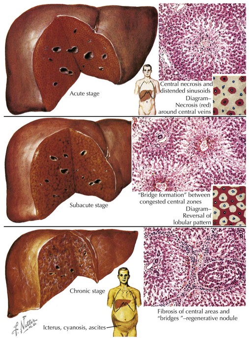
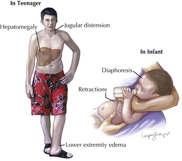
When the heart fails to adequately pump blood forward for any reason, venous congestion occurs. On the right side, the liver in particular becomes distended and engorged with blood. This leads to abdominal discomfort or vomiting initially, but in certain prolonged cases, it may lead to liver dysfunction and distension of venous collaterals, some of which may be visible under the skin (Figure 46-1). The signs of peripheral edema and jugular venous distension (Figure 46-2, left), classically seen in adults are not typically seen in infants or young children. On the left side, pulmonary venous congestion leads to fluid extravasation within the lungs, typically interstitial in infants and intraalveolar in older children and adults (Figure 46-2, right). This leads to shortness of breath, tachypnea, and dyspnea and can present on chest radiography similar to pneumonia. Indeed, in some cases, a secondary infection may develop superimposed on these wet and stiff lungs.

As the heart begins to fail in the volume of blood it can pump, the sympathetic nervous system seeks to compensate by increasing the heart rate. In these cases, an ECG may be useful in differentiating a sinus tachycardia in response to a failing heart (P-wave axis between 0 and 90 with a one-to-one relationship to the QRS complexes) versus a primary arrhythmia. If prolonged and untreated, some nonsinus heart rhythms can be the cause of heart failure. Finally, upregulation of the sympathetic nervous system may also cause vasoconstriction and diaphoresis, and the child may present as cold, pale, and diaphoretic.

Buy Membership for Pediatrics Category to continue reading. Learn more here