Heart Failure

Published on 06/06/2015 by admin

Filed under Pediatrics

Last modified 22/04/2025

Print this page

rate 1 star rate 2 star rate 3 star rate 4 star rate 5 star
Your rating: none, Average: 0 (0 votes)

This article have been viewed 1682 times

46 Heart Failure

A healthy child’s heart may beat more than 200,000 times and transport more than 3 tons of blood each day. The heart and vascular system are primarily responsible for delivering blood to the tissues while providing oxygen and nutrients and withdrawing waste products. Each is involved in the regulation of a perfusing blood pressure, including those homeostatic alterations necessitated with changes in posture. They play a primary role in the circulation of hormones. Heart failure is the inability of the heart to meet the metabolic demands of the body.

The failure of the heart and vascular system to keep up with metabolic requirements may arise from a number of causes, including cellular deficiencies, arrhythmias, metabolic insufficiencies, and congenital defects. Although most frequently a child presents in heart failure because of decompensation from congenital anomalies, the more prevalent of these are discussed elsewhere within this text, and only the principles common to heart failure will be repeated here. It is important to remember “heart failure” as a clinical picture may present in many of these anomalies when the physiology markedly worsens. However, to illustrate the fundamental concepts of pediatric heart failure, the remainder of this chapter focuses on that which may arise in children with normal anatomy. This state of “heart failure” in children may present indolently or very suddenly; in the latter, less-specific symptoms may be present for longer.

Regardless of the source, the goals of treatment are always (1) discovering and addressing the cause of failure along with (2) decreasing myocardial work load and oxygen consumption and (3) augmenting and supporting function and systemic oxygenation. With such interventions, the hope is that the heart will remodel, if not recover. Because of the danger of cardiovascular collapse, which may be very rapid, prompt consultation with pediatric cardiologists and intensivists is important, and early recognition requires diagnostic vigilance. Through a careful history and examination and with tests such as electrocardiography (ECG), echocardiography, and cardiac catheterization, the cause may often be learned and treatment instituted. Invasive arterial or venous monitoring is helpful in some cases if the patient is very ill.

Etiology And Presentation

The heart is a pump. It may fail as a pump either from the inability to generate enough contractile force or from structural anomalies that prevent or inappropriately direct blood to the tissues. Examples of the former include myocarditis, an infection of heart muscle cells (myocytes), or a cardiomyopathy, a primary structural or metabolic abnormality of the microscopic elements of a myocyte. Examples of structural anomalies that might lead to heart failure include left-to-right shunt lesions such as ventricular septal defect; patent ductus arteriosus, and, rarely, atrial septal defect, or valvar regurgitation because of the increased volume load on the heart; and severe valvar stenosis because of the increased pressure load. When the heart is no longer able to compensate, the state of heart failure exists.

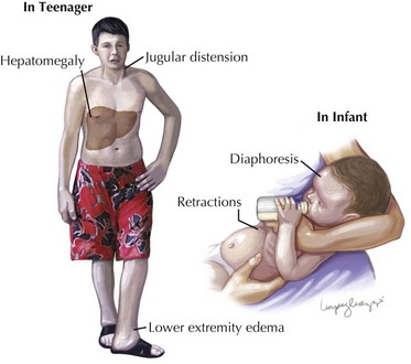
When the heart fails to adequately pump blood forward for any reason, venous congestion occurs. On the right side, the liver in particular becomes distended and engorged with blood. This leads to abdominal discomfort or vomiting initially, but in certain prolonged cases, it may lead to liver dysfunction and distension of venous collaterals, some of which may be visible under the skin (Figure 46-1). The signs of peripheral edema and jugular venous distension (Figure 46-2, left), classically seen in adults are not typically seen in infants or young children. On the left side, pulmonary venous congestion leads to fluid extravasation within the lungs, typically interstitial in infants and intraalveolar in older children and adults (Figure 46-2, right). This leads to shortness of breath, tachypnea, and dyspnea and can present on chest radiography similar to pneumonia. Indeed, in some cases, a secondary infection may develop superimposed on these wet and stiff lungs.

As the heart begins to fail in the volume of blood it can pump, the sympathetic nervous system seeks to compensate by increasing the heart rate. In these cases, an ECG may be useful in differentiating a sinus tachycardia in response to a failing heart (P-wave axis between 0 and 90 with a one-to-one relationship to the QRS complexes) versus a primary arrhythmia. If prolonged and untreated, some nonsinus heart rhythms can be the cause of heart failure. Finally, upregulation of the sympathetic nervous system may also cause vasoconstriction and diaphoresis, and the child may present as cold, pale, and diaphoretic.

The apex beat may be displaced downward and laterally and may be full and bounding or weak depending on both the cause of the heart failure and how successful the child is compensating. In newborns, anything more than a slight pulsation felt with two fingers placed beneath the xiphisternum is concerning for cardiac involvement. Auscultation may reveal a number of clues as to the cause. Difficult-to-hear heart sounds may signify a pericardial effusion with fluid between the heart and the pericardium that does not permit adequate filling of blood into the heart but more often is caused by poor contraction from a primary cardiomyopathy. A gallop is frequently heard in failing hearts and refers to an extra (third or fourth) heart sound. Third heart sounds may be normal in some children and are thought to be caused by rapid diastolic filling. A fourth heart sound is never normal and is indicative of a poorly compliant ventricle reflecting a high-pressure wave back toward the atrium.

Murmurs may be heard in the setting of heart failure caused by a primary structural abnormality (discussed elsewhere). Some murmurs represent turbulence across an incompetent valve caused by severe left ventricular dilatation seen in some failing hearts.

Evaluation and Management

Because of the high risk of cardiovascular collapse, concerns of heart failure should be managed in close consultation with pediatric intensivists and cardiologists. A careful, directed history is important to frame the presentation and learn its time course. Understanding the clinical associations listed above are important in evaluating the vital signs. Resting vital signs, including heart rate, systolic and diastolic blood pressures, and respiratory rate, are of primary importance. High heart rates with borderline or low blood pressures are concerning findings that the cardiovascular system is involved, whether from primary cardiac disease or secondary to another condition such as sepsis. A posteroanterior chest radiograph may be useful in evaluating both the lungs and size of the cardiac silhouette. A child’s cardiac shadow should be 50% or less than the diameter of the chest; a larger shadow may indicate a large, overloaded, and potentially failing heart. Evidence of pulmonary infiltration may be of a primary lung cause or secondary to heart failure. An ECG is important to differentiate sinus rhythm from an arrhythmia. An echocardiogram may be necessary in suspicious cases to evaluate both heart function and structure.

If signs of heart failure are present (tachycardia, borderline or low blood pressure, tachypnea, hepatomegaly), judicious use of fluid replacement along with early support with pressor medications such as milrinone and dopamine and intubation, sedation, and paralysis may be necessary to reduce metabolic demand in the most serious cases. In cases of complete cardiovascular collapse (shock) when the heart function and blood pressure are unable to maintain adequate perfusion despite maximal pharmacotherapeutic support, extracorporeal membrane oxygenation (ECMO) is able to support the child. These large external devices are able to maintain adequate cardiac output to support the patient for a number of weeks. If longer periods are necessary, implantable ventricular assist devices are available and intended to support the child until a heart transplant is available.

Volume status is critical in the treatment of heart failure because fluid overload and increased myocardial wall stress exert extra strain on a failing heart. A history of recent weight gain, peripheral or (if an infant) periorbital edema, signs of hepatic distension, and electrolyte abnormalities (including hyponatremia) may indicate a picture consistent with heart failure. Increasing central venous pressure along with diminished arterial mean pressure common in this setting will diminish renal perfusion and impair fluid excretion. Inpatient intravenous (IV) diuresis to the patient’s estimated dry weight along with the addition of middle-dose milrinone or low-dose dopamine may be necessary. Carefully following orthostatic blood pressures is important and, if possible, measurement of central venous pressure and mixed venous oxygen saturation through a central catheter is often additionally helpful in estimating cardiac output and systemic vascular resistance. Brain-type natriuretic peptide is often used in trending fluid overload and heart failure.

Failing hearts are more prone to arrhythmias, and 24-hour telemetry as an inpatient or 24-hour surveillance Holter monitors should play a routine role in the evaluation of children in heart failure. Finally, hearts that are markedly dilated and hypocontractile are prone to thrombus formation, and such children are at risk for thromboembolism and stroke. Medications to reduce in situ coagulation and platelet function are important in this population.

Because of natural changes in vascular physiology that start at birth, an important clue in the evaluation of a child in heart failure is the patient’s age. Infants who present at birth with heart failure most likely have sustained an in utero insult such as asphyxiation, maternal sepsis, intrauterine myocarditis, prolonged fetal arrhythmia, perinatal hemorrhage, or (rarely) primary valvar regurgitation (because shunt lesions are hemodynamically inconsequential before pulmonary resistance falls postnatally). An infant who is presented around 1 week of age typically has structural congenital cardiac anomalies depending on the flow through a patent ductus arteriosus because this structure typically completes closure between 4 and 10 days of age. Examples of ductal-dependent lesions include hypoplastic left heart syndrome, critical aortic or pulmonary valve stenosis or atresia, severe coarctation of the aorta, and interruption of the aortic arch. Administration of prostaglandin E1 may temporarily reverse the closure of the ductus. Finally, children who present from several weeks to a few months of age in heart failure often have shunt defects because this is the timeframe in which pulmonary vascular resistance decreases, leading to preferential shunting of oxygenated blood back to the lungs. At all of these times or anytime thereafter, genetic, metabolic, or infectious causes of primary myocardial causes of heart failure may present.

Case Example

Myocarditis occurs when there is inflammation of the myocardium with cell damage or death leading to decreased ventricular function and cardiac output. In the United States and Europe, most cases are of viral origin. Although the suspected causative agents have changed over recent decades, today when a successful diagnosis is made, adenovirus and enterovirus are the most frequently isolated viral species. Myocarditis may however arise from a host of other agents, including bacteria, protozoa, pharmaceuticals, and even rheumatologic and autoimmune processes.

There are wildly differing estimations as to the incidence of this disease, but it is likely that many cases remain subclinical and are never discovered. Catheter-based biopsy-proven diagnosis in this disease is often fraught with difficulty. Taking a heart muscle biopsy from an acutely ill child has its dangers, and the result of such a biopsy may not lead to any changes in the acute course of treatment. When biopsy is possible, samples consistent with viral myocarditis will show a lymphocytic infiltrate and cell necrosis. Magnetic resonance imaging may come to play an important role in future diagnoses with tissue characterization suggestive of inflammation, edema, and possibly myocardial scarring.

The decrease in myocardial function caused by myocarditis may present either suddenly or indolently. Paradoxically, patients who present most suddenly (and often in cardiogenic shock, or “fulminant myocarditis”) tend to recover most readily if the initial course is supported; more than 90% of these patients survive transplant free 11 years later. But patients in whom function limps along with little recovery rarely regain full function and either develop chronic although stable dilated cardiomyopathy or end-stage heart failure. Of these, fewer than half of patients are alive and transplant free after 11 years. Myocarditis may present at any age.

Infants and children present in varying degrees of heart failure as detailed above, and primary complaints may revolve around chest pain (pericarditis), respiratory distress, or abdominal distress and decreased appetite from increasing venous stasis. There may be a fever or a history thereof from the primary infection; a history of a flulike illness may precede the presentation by 1 to 4 weeks. A resting tachycardia with or without a gallop will be present along with tachypnea or cough. More severe cases may have a signs of cardiogenic shock (hypotension, cool and pale extremities). There may be a systolic murmur of mitral regurgitation, a finding seen when significant left ventricular dilation has occurred. Atrial and ventricular fibrillation along with atrioventricular nodal block are sometimes seen and may lead to sudden death.

The ECG tends to show low-voltage and often markedly widened QRS complexes. There may be inversion of the T waves along with Q waves indicative of myocardial infarction. Cardiomegaly may be present on chest radiography, but its absence does not rule out the disease, particularly in the very beginning of fulminant myocarditis. Echocardiography, the most sensitive of these tests, demonstrates left ventricular dilatation and decreased ventricular function. Global and uniform hypokinesis is the norm, although segmental wall motion abnormality (dyskinesia) may also be present. With increasing left ventricular volume and worsening valve leaflet apposition, color-Doppler ultrasonography will demonstrate mitral regurgitation. Cardiac troponin T will be elevated in patients with myocarditis and is often a useful test in distinguishing myocarditis from cardiomyopathy, in which it is not elevated.

Because of the rapidity with which cardiovascular collapse may ensue, prompt consultation is recommended. Inotropic support with milrinone or dopamine (or both), arterial monitoring, 24-hour telemetry to monitor for ectopy, and ECMO are often needed in the acute fulminant phase of this disease. Lidocaine infusions and magnesium may be necessary if ventricular ectopy develops. Treatment is therefore primarily supportive. Immunosuppressive regimens are controversial, with many studies showing no improvement with combinations of steroids and medications, including cyclosporine and azathioprine. Some evidence has been emerging that IV immune globulin with the possible addition of vitamin C may have a beneficial effect on some forms of myocarditis.