Hands and feet

Published on 16/03/2015 by admin

Filed under Dermatology

Last modified 22/04/2025

Print this page

rate 1 star rate 2 star rate 3 star rate 4 star rate 5 star
Your rating: none, Average: 0 (0 votes)

This article have been viewed 1358 times

22 Hands and feet

Soft tissue augmentation of the hands

Calcium hydroxylapatite

Calcium hydroxylapatite (CaHA) has become the most commonly recommended filler for rejuvenation of the aging hand. First used for medical purposes in areas such as vocal fold and maxillofacial augmentation, CaHA (Radiesse®, Merz Aesthetics) was approved by the US Food and Drug Administration (FDA) in 2006 both for volume restoration in patients with HIV-related lipoatrophy and for cosmetic correction of moderate to severe facial wrinkles and folds. Specific approval for hand rejuvenation was obtained in Canada in 2010, but remains an off-label usage in the USA.

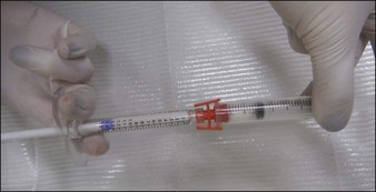
As an opaque, white substance, CaHA has the advantage of acting as both a filler and camouflage for underlying anatomical architecture. However, its use for hand rejuvenation was initially limited by the product’s high viscosity and pain upon injection. In 2007, Busso & Applebaum reported a novel approach that overcame both issues. Utilizing a Luer-to-Luer lock connector (Fig. 22.1A), the authors mixed 0.1 mL 2% lidocaine without epinephrine with 1.3 mL of CaHA (the standard syringe volume at that time). After tenting the skin of the central dorsal hand to separate it from underlying large vessels and tendons, they injected a single bolus of 0.5–1.4 mL of the lidocaine–CaHA mixture into the space created between the subcutaneous layer and the superficial fascia. The bolus was then massaged and molded into a cosmetically smooth appearance. The addition of lidocaine reduced both the viscosity and pain.

Since this initial report, physicians have modified the technique by increasing the volume of lidocaine used for dilution (e.g. the 2009 study by Edelson). In 2010, Marmur et al published the first organized institutional review board-approved study of CaHA filler for hand rejuvenation. They mixed 2.0 mL of 2% lidocaine without epinephrine per 1.3 mL syringe of CaHA. A total of 0.3–1.0 mL of the product was injected interdigitally using a 25-gauge 1.5-inch (3.75 cm) needle at three to five insertion points, as opposed to a single bolus injection. They reported high patient satisfaction rates in five patients who each received a single treatment with follow-up extending to 24 weeks postoperatively. Although localized edema initially resolved in all patients by 1 week, one patient had recurrent edema at day 10 that persisted for 3 additional weeks and required oral corticosteroids.

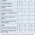
Several larger studies by Busso et al, Bank, and Marmur et al have recently been published supporting the safety and efficacy of CaHA as an anti-aging hand filler. Busso et al published a multicenter, randomized, controlled trial of 101 patients followed over 6 months and established a new hand volume severity scale 3 to assess results (Table 22.1). Following administration of a lidocaine bolus preoperatively, CaHA was administered as a bolus into the dorsal hand using a 27-gauge 0.75-inch (20 mm) needle. Aside from reporting statistically significant improvement in the treatment group, the study also found that adverse events, including bruising, itching, pain, redness, and swelling (Fig. 22.2), were frequent yet short in duration and did not affect overall patient function significantly. Similarly, Sadick noted only brief and minor side effects in 10 patients following CaHA treatment with a 25-gauge 0.5-inch (12 mm) needle to the dorsal hands and residual cosmetic correction lasting up to 1 year postoperatively.

Table 22.1 Busso hand volumizing severity scale

Severity Characteristics of hand
4 All three central tendons are fully exposed when hand is at rest
3 All three central tendons are partially exposed, with one or two tendons fully exposed when hand is at rest
2 All three central tendons are partially exposed when hand is at rest
1 One or two central tendons are slightly exposed when hand is at rest
0 No tendon is exposed when hand is at rest

Adapted from Busso M, Moers-Carpi M, Storck R, et al 2010 Multicenter, randomized trial assessing the effectiveness and safety of calcium hydroxylapatite for hand rejuvenation. Dermatologic Surgery 36(s1):790-797.

Most recently, the availability of blunt-tipped small-gauge cannulas has revolutionized the procedure by reducing the number of puncture sites required, patient discomfort, and postoperative bruising. The authors recommend delivering boluses or linear threads with one entry point at the dorsal wrist (Fig. 22.3). The product is massaged until smooth. Patients are instructed to massage several times daily and use topical ice packs for the first 48 hours. A small latex-free examination glove can be applied immediately with an ice pack (Fig. 22.4). Despite these interventions, some patients complain of severe pain, which limits range of motion. The authors’ clinical experience validates the mean volume of 3.1 mL for optimal improvement of both hands reported by Busso et al. We have found that dividing the treatment into two planned sessions at 2–4-week intervals has reduced the incidence of significant pain and made the treatment viable for patients who work with their hands.

Poly-L-lactic acid

Poly-l-lactic acid (PLLA) was initially considered to be a good filler for hand rejuvenation but its use has gone out of favor for this purpose. PLLA (Sculptra®) was approved by the FDA in 2004 for the treatment of HIV-associated lipoatrophy and was largely used off-label cosmetically in the USA until cosmetic facial approval in 2009. Vleggaar in 2006 reported soft tissue augmentation of the hands with PLLA, lasting up to 2 years. Redaelli published a study of 27 patients treated in Italy in 2004–2005 with 150 mg of PLLA diluted with 0.5 mL of 3% mepivacaine in water to a final volume between 5 and 8 mL. The patients were treated with 1.5–2 mL of PLLA mixture over several sessions, with multiple intermetacarpal injections using 25–27-gauge needles for the first three sessions at monthly intervals and evaluations for treatment every 3 months thereafter. Despite postoperative massage, both of these European articles noted nodule formation as late as 1 year after the last injection as a potential complication of therapy.

Sadick et al in 2008 described the experience of three clinical practices that treated the hands of 26 patients with PLLA diluted with 5–6 mL sterile water and 2–5 mL 1% lidocaine. Therapy was performed with a 25-gauge 1.5-inch needle at 1-month intervals over for a total of two or three treatments and resulted in a high rate of patient satisfaction. Though 46% of patients reported adverse events such as bruising / swelling (30.8%), pain (15.4%), itching (3.8%), and arterial spasm (3.8%), they were minor and resolved within a few days of treatment. No nodule formation postoperatively was noted, although an ‘uneven result’ was reported with a low incidence of 3.8%. As with facial use of PLLA, higher dilutions of 8–10 mL of sterile water plus lidocaine (as opposed to the 5 mL sterile water dilution written in the package insert) have been recommended by Rendon et al to try to reduce the risk of nodule formation. However, unlike in the face, injection depth is limited for the dorsal hands so that any nodules that develop will be noticeable, and this has limited its usefulness for this area.

Fat transplantation

Autologous fat grafting was been utilized as early as the 1980s by Coleman for soft tissue augmentation of the dorsal hands. Though considerable difference still exists regarding harvesting and transplant, many experts report positive results with this modality. The ‘Coleman technique’ involves infiltration of 0.5% lidocaine with 1 : 200 000 epinephrine in lactated Ringer’s solution into the donor fat site (i.e. lower abdomen) through a blunt Lamis infiltrator following a small incision at a ratio of 1 mL of solution per cubic centimeter of fat graft to be harvested. Through the same incision site a blunt tip harvesting cannula 3 mm in diameter and 23 cm in length is inserted; a Luer-Lok® syringe is then connected to the cannula to aspirate the fat as the cannula is advanced and retracted. After removal of the syringe, a Luer-Lok® plug is attached to the bottom and it is centrifuged at 3000 rpm for 3 minutes to separate the material into three layers: the upper oily layer, the middle fat tissue, and the lower layer of blood, water, and lidocaine. The upper layer should be decanted and the lower layer drained. The hand is subsequently prepared through preoperative regional blocks, and six or seven 1–2 mm incisions at the periphery of the hand near the metacarpophalangeal joints are made in the direction of wrinkle lines. After transferring the fat to a 1 mL Luer-Lok® syringe and attaching an infiltration cannula, between 0.02 and 1 mL of fat should be injected as the cannula is passed through the incision sites proximally toward the phalanges. Coleman reported high patient satisfaction, with bruising and prolonged swelling as potential adverse events.

Variations in the procedure include utilizing frozen fat as opposed to fresh samples, and Butterwick et al reported equivalent to improved results in a 2006 study with frozen autologous fat as opposed to fresh. Some sources claim longevity of a year or beyond although others, such as Haq et al, claim that multiple treatment sessions may be needed and that duration is not easy to predict; a recent publication by Giunta et al estimated 69% residual injection volume at 6 months postoperatively following hand rejuvenation with autologous fat transplant. Despite these promising results there is still a lack of rigorous studies confirming the safety and efficacy of this therapy, and the American Society of Plastic Surgeons Fat Graft Task Force in 2009 (reported by Gutowski) was unable to make recommendations on the application of fat grafting for hand rejuvenation. Because it is a labor-intensive treatment, in clinical practice, fat transfer for hand augmentation may be best suited to patients seeking fat reduction at donor areas and those for whom fat transfer will also be used for facial augmentation.

Hyaluronic acid

Although hyaluronic acid (HA) fillers are the most commonly used category of dermal filling agents worldwide, there are fewer supporting studies and reports of their use for hand augmentation than for the therapies described above. The only FDA-approved HAs are Belotero®, Juvéderm® Ultra, Juvéderm® Ultra Plus, Restylane®, and Perlane®. Man et al in 2008 published a study comparing 10 patients randomized to receive either 1.4 cm3 HA or 2.0 cm3 human collagen for rejuvenation of the dorsal hands; HA was superior in terms of decreasing signs of intrinsic aging. In a case report, Hartmann et al in 2009 found that both Macrolane®, a volumizing HA used in Europe for breast augmentation but not available in the USA, and Restylane® were efficacious in improving appearance. The patient treated with Restylane® received a dermal injection with a 30-gauge needle via tunneling and tenting technique followed by gentle massage. The patient treated with Macrolane® received a subcutaneous injection with a blunt 18-gauge needle through a small incision with a scalpel following preoperative 1% prilocaine injection. The patient then received gentle massage and the incision was closed by a Steri-Strip®.

Despite safety and efficacy, hand augmentation with HA products is limited by the Tyndal effect, the blue hue that results when a clear gel filler is placed superficially or beneath thin skin. Lax, aged, and photoaged dorsal hand skin tends to be particularly thin and transparent and therefore more prone to this effect. A blue discoloration in this area can simulate or aggravate the appearance of solar purpura.

Soft tissue augmentation of the feet

Feet mirror the aging process happening throughout the body. Soft tissue augmentation of the foot is most frequently cited in the podiatric literature as a means of redistributing plantar pressure in those with atrophied pedal fat pads, such as diabetics. The pioneer of this technique, Sol Balkin, utilized silicone as a tissue-volumizing agent beneath the plantar prominence in over 1600 patients. In one of his studies, 41 pedal ulcers associated with diabetic neuropathy were injected with silicone in doses of 0.10–025 mL at the site of ulceration at 1–4-week intervals to a mean total of 1.65 mL. All ulcers healed initially and 73% remained healed at a mean follow-up of 6.3 years.

CaHA and HAs have also been used in the plantar foot for issues that bridge the medical and cosmetic. About 1.0 mL of CaHA can be injected into the bursa under the metatarsal head to create an extra cushion at the balls of the foot. In this case, one should be careful to remain superficial to the tendons of the deep foot. This procedure, coined ‘pillows for feet’, is advertised to target women seeking relief from the metatarsalgia secondary to the chronic use of high-heeled shoes (described by Beck). Unlike the hands, there is no means to massage the pedal injection area post-administration to adjust filler placement; however, X-rays taken at the 2-week follow-up visit can help the clinician to visualize CaHA location.

Although the dorsal feet undergo many of the same cosmetic changes as the back of hands, there are only a few anecdotal reports of foot rejuvenation done under similar technical guidelines as that of hand augmentation. Intradigital CaHA injections can relieve the pressure associated with a bunion impinging on an adjacent toe. In this case about 0.3 mL of CaHA is injected between the toes after a pretibial nerve block is conducted with 1.5 mL of lidocaine. Werschler & Busso improved the surface appearance by injecting CaHA prepared in the same manner as used for the hands. Tight-fitting footwear and high-pressure pedal-intensive exercises like running or bicycling are to be avoided for up to 4 weeks after injection. Intradigital or subpedal padding can be custom fabricated and placed in loose-fitting shoes to be worn for at least 2 weeks (Ribotsky, personal communication, 2011). Those patients who have compromised circulation, diabetes, active infection, or any other type of medical condition that could impair healing and / or predispose to infection, ulceration, or skin compromise are not recommended for this procedure.

Further reading

Balkin SW, Kaplan L. Silicone injection management of diabetic foot ulcers: a possible model for prevention of pressure ulcers. Decubitus. 1991;4(4):38–40.

Bank DE. A novel approach to treatment of the aging hand with Radiesse. Journal of Drugs in Dermatology. 2009;8(12):1122–1126.

Beck M. Toe the line: doctors fight cosmetic foot surgery. Health Journal. 2010. 27 July. Online. Available http://online.wsj.com/article/.

Busso M, Applebaum D. Hand augmentation with Radiesse (calcium hydroxylapatite). Dermatologic Therapy. 2007;20(6):385–387.

Busso M, Moers-Carpi M, Storck R, et al. Multicenter, randomized trial assessing the effectiveness and safety of calcium hydroxylapatite for hand rejuvenation. Dermatologic Surgery. 2010;36(s1):790–797.

Butterwick KJ. Rejuvenation of the aging hand. Dermatologic Clinics. 2005;23:515–527. vii

Butterwick KJ, Bevin AA, Iyer S. Fat transplantation using fresh versus frozen fat: a side-by-side two-hand comparison pilot study. Dermatologic Surgery. 2006;32(5):640–644.

Coleman SR. Hand rejuvenation with structural fat grafting. Plastic and Reconstructive Surgery. 2002;110(7):1731–1744. discussion 1745-1747

Edelson KL. Hand recontouring with calcium hydroxylapatite (Radiesse). Journal of Cosmetic Dermatology. 2009;8(1):44–51.

Giunta RE, Eder M, Machens HG, et al. Structural fat grafting for rejuvenation of the dorsum of the hand. Handchirurgie, Mikrochirurgie, plastische Chirurgie. 2010;42(2):143–147.

Gutowski KA. Current applications and safety of autologous fat grafts: a report of the ASPS fat graft task force. Plastic and Reconstructive Surgery. 2009;124(1):272–280.

Haq S, Storck R, Martine B, et al. Multinational, multipatient study of calcium hydroxylapatite for treatment of the aging hand: European cosmetic physician group on hand augmentation. Dermatologic Surgery. 2010;36(s1):782–789.

Hartmann V, Bachmann F, Plaschke M, et al. Hand augmentation with stabilized hyaluronic acid (Macrolane VRF20 and Restylane Vital, Restylane Vital Light). Journal der Deutschen Dermatologischen Gesellschaft. 2010;8(1):41–44.

Man J, Rao J, Goldman M. A double-blind, comparative study of nonanimal-stabilized hyaluronic acid versus human collagen for tissue augmentation of the dorsal hands. Dermatologic Surgery. 2008;34(8):1026–1031.

Marmur ES, Al Quran H, De Sa Earp AP, et al. A five-patient satisfaction pilot study of calcium hydroxylapatite injection for treatment of aging hands. Dermatologic Surgery. 2009;35(12):1978–1984.

Pu LL, Coleman SR, Cui X, et al. Autologous fat grafts harvested and refined by the Coleman technique: a comparative study. Plastic and Reconstructive Surgery. 2008;122(3):932–937.

Redaelli A. Cosmetic use of polylactic acid for hand rejuvenation: report on 27 patients. Journal of Cosmetic Dermatology. 2006;5(3):233–238.

Rendon MI, Cardona LM, Pinzon-Plazas M. Treatment of the aged hand with injectable poly-l-lactic acid. Journal of Cosmetic and Laser Therapy. 2010;12(6):284–287.

Sadick NS. A 52-week study of safety and efficacy of calcium hydroxylapatite for rejuvenation of the aging hand. Journal of Drugs in Dermatology. 2011;10(1):47–51.

Sadick NS, Anderson D, Werschler WP. Addressing volume loss in hand rejuvenation: a report of clinical experience. Journal of Cosmetic and Laser Therapy. 2008;10(4):237–241.

Vleggaar D. Soft-tissue augmentation and the role of poly-L-lactic acid. Plastic and Reconstructive Surgery. 2006;3 suppl:S46–S54.

Werschler PBM, Busso M. Prepackaged injectable soft-tissue rejuvenation of the hand and other nonfacial area. Body Rejuvenation. 2010;7:221–225.