66
Hand, foot, and
mouth disease
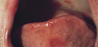
Oral vesicles of hand, foot, and mouth. There are painful, white, aphthae-like erosions on the tongue, lips, and hard palate. Eating can be painful.
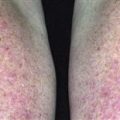
Palmar and plantar vesicles appear cloudy with a small red halo. These lesions may be tender or asymptomatic. They may be sparse or numerous.
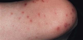
Typical vesicles of hand, foot, and mouth disease on the heel. Pain may impede walking, although generally this is a mild illness.
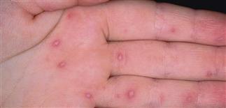
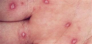
Multiple discrete cloudy vesicles with inflammatory halos on the palm are characteristic of hand, foot, and mouth disease.
DESCRIPTION
Highly contagious viral infection that causes aphthae-like oral erosions and a vesicular eruption on hands and feet. The classic benign, self-limited form of this disease is associated with coxsackievirus A16. Enterovirus 71 is a picornavirus genetically related to coxsackie A16. It can cause oral ulcers and similar skin exanthem, but epidemic outbreaks have associated potentially serious neurologic and cardiopulmonary complications, particularly in children under 4 years.
HISTORY
• Incubation period 4–6 days. Epidemics in summer and autumn but may appear any time. • Usually presents with acute stomatitis and mild fever. Mild sore throat and malaise or abdominal pain for 1–2 days may occur. • About 20% of patients develop submandibular lymphadenopathy, cervical lymphadenopathy, or both. • Children younger than 5 years most commonly affected; rate of infection among close household contacts is high. • Enterovirus 71 outbreaks commonly include fever, oral ulcers and/or extremity rash, vomiting, cough.
PHYSICAL FINDINGS
• Number of oral aphthae-like erosions (3–6 mm) varies from few to 10 or more. They are irregularly distributed anywhere in oral cavity. More painful in younger children. Each erosion lasts 3–5 days. • Cutaneous lesions in coxsackie-induced hand, foot, and mouth disease occur in two-thirds of patients and appear less than 24 h after oral lesions. • Skin lesions begin as 3- to 7-mm red macules that rapidly become pale, white, oval vesicles with red areolae. Vesicles have unique rhomboidal shape of ‘square blisters’. May be a few or dozens. • Vesicles occur on palms, soles, dorsal aspects of fingers and toes, and occasionally on face, buttocks, legs. • Heal in approximately 7 days, usually without crusting or scarring. • Diagnosis usually made clinically; laboratory tests unnecessary for the benign presentation of the disease.
TREATMENT
• Hand, foot, and mouth disease caused by coxsackie A16 usually mild and self-limited. Resolves without treatment in about 10 days. • Oral ulcerations painful in infants and interfere with feeding. • Recent emergence of epidemics of hand, foot, and mouth disease caused by enterovirus 71 are associated with varied neurologic syndromes, including aseptic meningitis, Guillain–Barré syndrome, polio-like paralysis, acute transverse myelitis, acute cerebellar ataxia, intracranial hypertension, febrile convulsions. Prodrome is 1–7 days before neurologic disease and has fever, coryza, malaise, headache, diarrhea. Two-thirds of those affected may have a rash, often truncal; herpangina-type lesions may occur in mouth.