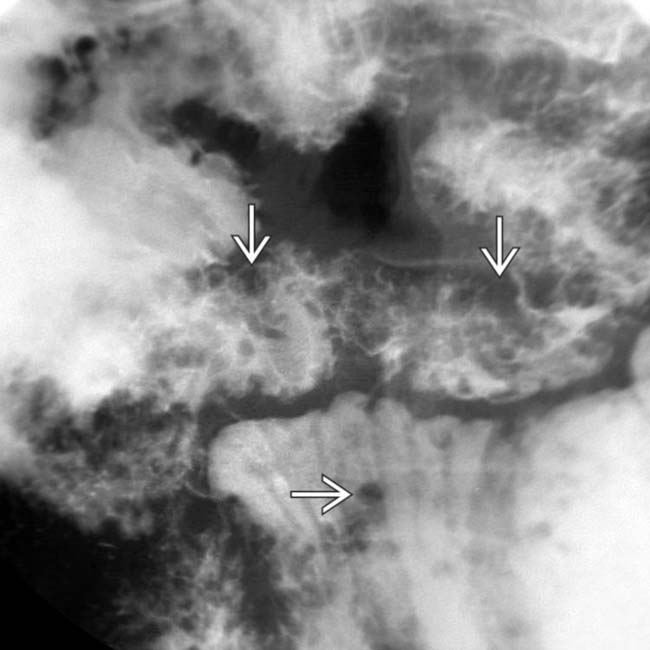
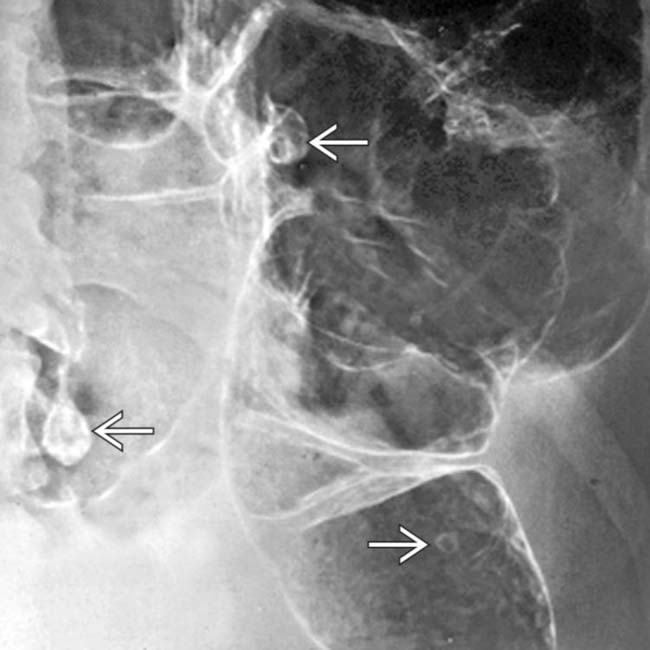
Hamartomatous Polyposis Syndromes

Published on 09/08/2015 by admin

Filed under Radiology

Last modified 22/04/2025

Print this page

rate 1 star rate 2 star rate 3 star rate 4 star rate 5 star
Your rating: none, Average: 3 (3 votes)

This article have been viewed 2900 times

 Cluster of small filling defects in small bowel with intussusception

• Peutz-Jeghers syndrome (PJS)

image Jejunum and ileum > duodenum > colon > stomach

TOP DIFFERENTIAL DIAGNOSES

• Familial adenomatous polyposis and related syndromes
• Brunner gland hyperplasia (hamartoma)
• Lymphoid follicles (hyperplasia)
• Metastases and lymphoma (GI tract)

PATHOLOGY

• Sessile/pedunculated; carpet-like, clustered, or scattered polyps

CLINICAL ISSUES

• Most common signs/symptoms

image PJS: Pain, mucocutaneous pigmentation, melena
• PJS complications

image Intussusception, SB obstruction, malignant neoplasms

– Bowel > breast > pancreas > reproductive tract
– Prognosis: 40% risk of cancer by age 40
• Intestinal polyposis syndromes encompass a wide spectrum of diseases with considerable overlap

image Gene mutations and phenotypes
image Polyp histology; disease severity; extraintestinal manifestations
image Blurs distinction between polyposis syndromes
image
(Left) This 28-year-old man has known Peutz-Jeghers syndrome (PJS). A film from a small bowel follow-through shows some of the hundreds of small polyps image, presumably hamartomas, throughout his bowel.

image
(Right) Axial CT section in the same patient shows a large mass image 
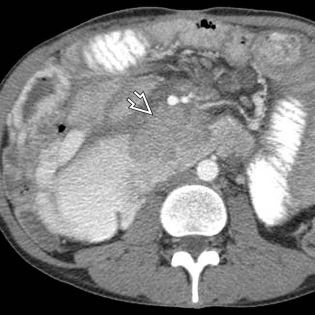
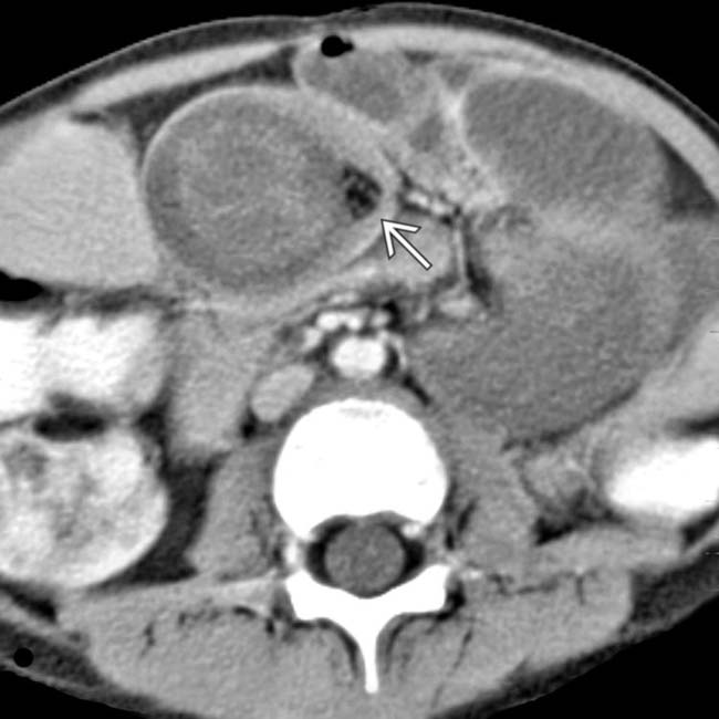
causing partial obstruction of the duodenum. The mass proved to be a metastasis from a testicular nonseminomatous germ cell tumor.
image
(Left) Another CT section from the same patient shows 2 segmental, nonobstructing intussusceptions image
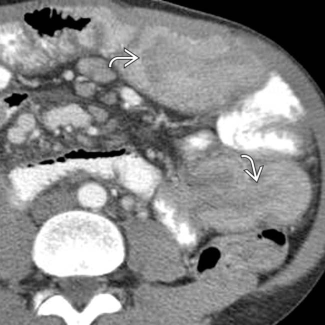
, probably due to the hamartomatous polyps.

image
(Right) Another CT section in the same patient shows some of the innumerable small bowel polyps image
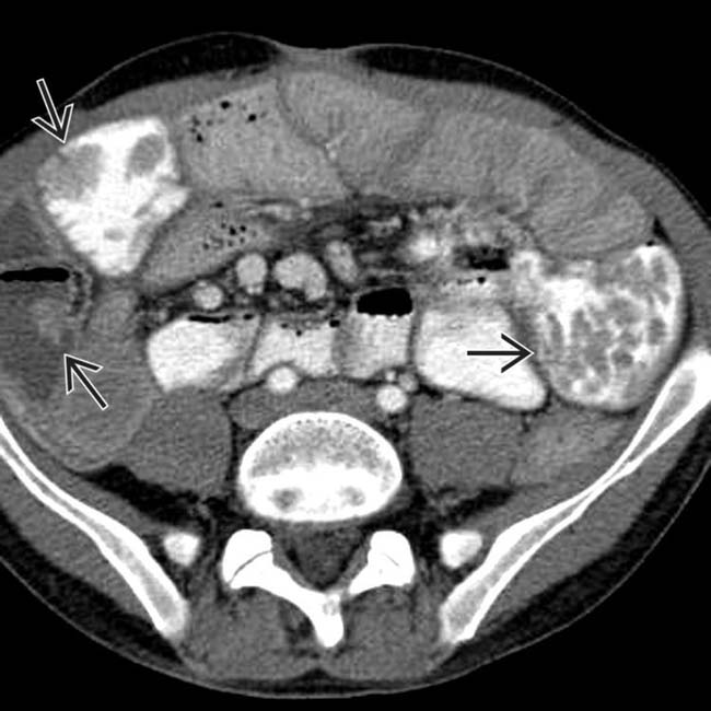
. As illustrated by this case, some patients with PJS may develop malignant tumors, not just of the bowel, but also of the breast, pancreas, or reproductive tract.

TERMINOLOGY

Abbreviations

• Peutz-Jeghers syndrome (PJS)
• Multiple hamartoma (Cowden) syndrome (MHS)
• Juvenile polyposis (JP)
• Cronkhite-Canada syndrome (CCS)

Synonyms

Definitions

• Spectrum of hereditary and nonhereditary polyposis syndromes characterized by gastrointestinal (GI) tract polyps and other associated lesions

IMAGING

General Features

• Best diagnostic clue

image PJS: Cluster of small filling defects in small bowel (SB) with intussusception
• Location

image PJS: Jejunum and ileum > duodenum > colon > stomach
image MHS + JP: Most polyps in rectosigmoid colon
image CCS: Stomach (100%), colon (100%), SB (50%)
• Size

image Varied
• Morphology

image Sessile or pedunculated polypoid lesions
image Pattern: Carpet-like, clustered, or scattered

Radiographic Findings

• Fluoroscopic-guided double-contrast studies

image Multiple, variably sized radiolucent filling defects
• Polyps in PJS occur from stomach to rectum; mouth and esophagus spared

image SB involved in 95%

– Usually multiple, broad-based polyps
– Clustered appearance more than carpeting bowel
– Large polyps have characteristic lobulated surface
image Colorectal involvement 30%: Multiple, scattered, no carpeting
image Stomach/duodenal involvement 25%: Diffuse involvement

Imaging Recommendations

• Best imaging tool

image Double-contrast barium studies (multiple views)
image CT enterography

DIFFERENTIAL DIAGNOSIS

Familial Adenomatous Polyposis and Related Syndromes

• Hundreds or thousands of polyps carpeting colonic mucosa
• Tubular or tubulovillous; colorectal cancer risk 100%

Brunner Gland Hyperplasia (Hamartoma)

• Duodenal bulb and descending duodenum
• Hyperplasia: Multiple nodules (“Swiss cheese” pattern)
• Hamartomas simulate hamartomatous polyps
• Differentiated by associated thickened, irregular folds

Lymphoid Follicles (Hyperplasia)

• Innumerable tiny/small radiolucent nodules
• Usually generalized (duodenum, SB, colon)
• Distinguished by clinical history and generalized pattern

Metastases and Lymphoma (GI Tract)

• Rarely as numerous as in polyposis syndromes
• Metastases: May be polypoid, mimicking polyps
• Lymphoma: Small/bulky polypoid, mimicking polyps

image Thickened bowel wall and folds; adenopathy seen

PATHOLOGY

General Features

• Etiology

image Hereditary autosomal dominant (AD): PJS, MHS, only 25% of juvenile polyposis cases
image Nonhereditary: CCS, 75% of juvenile polyposis cases
• Genetics

image Spontaneous mutation of gene on chromosome 19 (PJS) and 10 (MHS)
image PTEN is a tumor suppressor gene
image Loss or mutation associated with variety of rare syndromes known collectively as PTEN hamartoma tumor syndromes

– Includes Cowden (MHS), and Bannayan-Riley-Ruvalcaba syndromes

Staging, Grading, & Classification

• Classification: Hamartomatous polyposis syndromes

image PJS: Autosomal dominant (AD)

– Hamartomatous GI tract polyps, mucocutaneous pigmentation of lips, oral mucosa, palms, and soles
– Risk for cancer (10%) of stomach, duodenum, colon
– Extra-GI tract cancers: Pancreas, breast, reproductive organs
image MHS (Cowden): AD genodermatosis

– Mucocutaneous: Facial papules, oral papillomas, keratosis
– Breast: Fibrocystic (50%), ductal-type cancer (30%)
– Thyroid (65%): Adenomas, goiter, follicular cancer
– Pancreatic ductal and intraductal papillary mucinous (IPMN) tumors
– Clinically: Bird-like face, high-arched palate
image Bannayan-Riley-Ruvalcaba syndrome

– AD inheritance
– GI tract hamartomas
– subcutaneous lipomas, macrocephaly, etc.
– Less prone to cancer
image Juvenile polyposis: 2types

– Isolated juvenile polyps of childhood (nonhereditary)
– JP of colon or entire GI tract (autosomal dominant)
image Cronkite-Canada:  Inflammatory polyps with ectodermal defects

Gross Pathologic & Surgical Features

• Sessile/pedunculated; carpet-like, clustered, or scattered polyps

Microscopic Features

• Extensive smooth muscle arborization of polyps

CLINICAL ISSUES

Presentation

• Most common signs/symptoms

image PJS: Pain, mucocutaneous pigmentation, melena
• Lab data: Hypochromic anemia, positive stool guaiac test

Demographics

• Age

image PJS (10-30), MHS (30-40), CCS (> 60) years
• Gender

image PJS (M = F), MHS (M < F), JP (M > F), CCS (M < F)
• Epidemiology

image PJS  = 1:10,000

Natural History & Prognosis

• PJS complications: Intussusception, SB obstruction, malignant neoplasms

image Bowel > breast > pancreas > reproductive tract
• Prognosis: 40% risk of cancer by age 40
• Intestinal polyposis syndromes encompass wide spectrum of diseases with considerable overlap

image Gene mutations and phenotypes
image Polyp histology; disease severity; extraintestinal manifestations
image Blurs distinction between polyposis syndromes

Treatment

• Follow-up and surveillance; surgery for malignant neoplasms

DIAGNOSTIC CHECKLIST

Image Interpretation Pearls

• PJS: Small bowel polyps, mucocutaneous pigmentation
image
Axial CECT in a 4-year-old boy shows small bowel intussusception image proven at surgery to be due to hamartomatous polyps.

image
Axial CECT shows markedly dilated small bowel due to intussusception.
image
Air-contrast barium enema shows numerous polyps image 
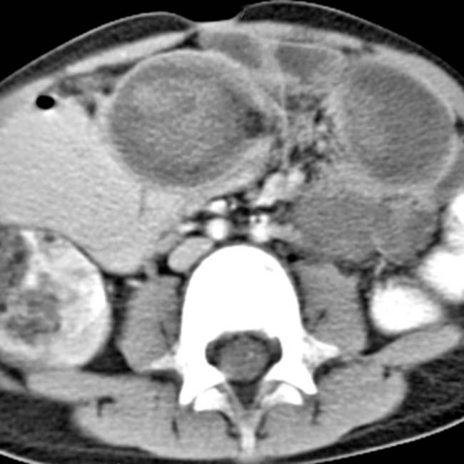
in a patient with Peutz-Jeghers syndrome.
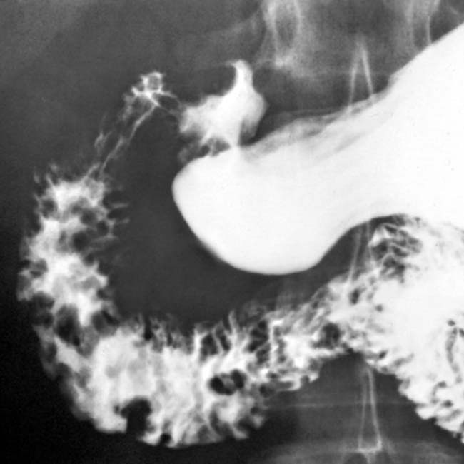
image
Upper GI series shows multiple polyps in the duodenum of a patient with Peutz-Jeghers syndrome.

SELECTED REFERENCES

1. Kobayashi, Y, et al. A tumor of the uterine cervix with a complex histology in a Peutz-Jeghers syndrome patient with genomic deletion of the STK11 exon 1 region. Future Oncol. 2014; 10(2):171–177.

Tsai, HL, et al. Rectal carcinoma in a young female patient with Peutz-Jeghers syndrome: a case report. Med Princ Pract. 2014; 23(1):89–91.

Lucci-Cordisco, E, et al. The growing complexity of the intestinal polyposis syndromes. Am J Med Genet A. 2013; 161A(11):2777–2787.

Gammon, A, et al. Hamartomatous polyposis syndromes. Best Pract Res Clin Gastroenterol. 2009; 23(2):219–231.

Hokama, A, et al. Telescope sign of intussusception in Peutz-Jeghers syndrome. Dig Liver Dis. 2009. [Epub ahead of print].

Huang, SC, et al. Pediatric juvenile polyposis syndromes: an update. Curr Gastroenterol Rep. 2009; 11(3):211–219.

Itaba, S, et al. Education and imaging. Gastrointestinal: Solitary Peutz-Jeghers-type hamartoma of the colon. J Gastroenterol Hepatol. 2009; 24(3):498.

Joshi, V. Peutz-Jeghers Syndrome: A Rare Cause of Gastric Outlet Obstruction. Clin Gastroenterol Hepatol. 2009. [Epub ahead of print].

Yano, T, et al. Vascular, polypoid, and other lesions of the small bowel. Best Pract Res Clin Gastroenterol. 2009; 23(1):61–74.

Cho, GJ, et al. Peutz-Jeghers syndrome and the hamartomatous polyposis syndromes: radiologic-pathologic correlation. Radiographics. 1997; 17(3):785–791.

Harned, RK, et al. The hamartomatous polyposis syndromes: clinical and radiologic features. AJR Am J Roentgenol. 1995; 164(3):565–571.

Buck, JL, et al. Peutz-Jeghers syndrome. Radiographics. 1992; 12(2):365–378.