Haematuria in a 60-year-old man

Published on 10/04/2015 by admin

Filed under Surgery

Last modified 22/04/2025

Print this page

rate 1 star rate 2 star rate 3 star rate 4 star rate 5 star
Your rating: none, Average: 0 (0 votes)

This article have been viewed 894 times

Problem 20 Haematuria in a 60-year-old man

The patient is obese, with a pulse of 65/min and a blood pressure 130/80 mmHg. There is a large mass palpable in his left abdomen below his costal margin.

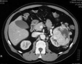
His urine microscopy shows >1000 red blood cells, and culture had no growth. Haemoglobin is 132 g/L, creatinine 128, and liver function tests are normal. A representative slice from the contrast-enhanced CT scan is shown in Figure 20.1.

For tumour staging the CT also assesses for invasion of adjacent structures (renal vein, perinephric fat, adrenal gland, pancreas) and metastases (lungs, bone, liver, adrenal). None of these is identified.

Tumour stage is the most important prognostic factor (Table 20.1).

Table 20.1 Prognosis according to tumour stage

Renal Tumour 5-Year Survival
Confined to kidney 75–90%
Invades perinephric fat, adrenal, renal vein, IVC 50–70%
Lymph node involvement 30%
Distant metastases 5%

The histopathology confirms a clear cell RCC which is invading perinephric fat and into the collecting system. There is no local lymph node involvement and the surgical margins are free of tumour. The patient makes an uneventful recovery.

Six months later the patient is reviewed. He has no urinary symptoms, has a good appetite, stable weight and enjoys being back at work. His creatinine is 108, and a CT chest, abdomen and pelvis is performed. This shows enlarged subcarinal nodes. The solitary right kidney appears normal and there are no pulmonary lesions (Figure 20.2).

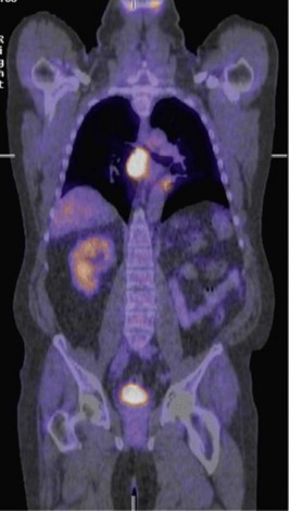
This location is very difficult to sample due to its deep, central location, and a percutaneous biopsy is not possible. A transtracheal needle aspirate via a bronchoscopy is performed and only yields inflammatory material. A PET/CT scan is arranged and shows the following (Figure 20.3).

A further attempt at biopsy of this enlarged lymph node is made by transoesophageal aspirate via endoscopy with ultrasound guidance. Cytology confirms malignant clear cells consistent with metastatic RCC.

The patient proceeds with treatment using a tyrosine kinase inhibitor and his lymphadenopathy halves in size over 4 months. One year later he continues with the tyrosine kinase inhibitor, and the lymphadenopathy is still present. He remains active and is asymptomatic.

Answers

A.1 Haematuria can arise from upper or lower urinary tract causes.

Lower Tract

Trauma and coagulation disorders must also be excluded.

A.2 Details of the haematuria may help to identify the site of bleeding. Renal bleeding may have associated flank pain if clots are being passed (clot colic). These clots can be elongated due to passage down the ureters. Prostatic bleeding usually occurs at initiation or termination of the urinary stream, and may occur independent to voiding. This often has associated bothersome lower urinary tract symptoms, such as hesitancy, frequency, nocturia and terminal dribbling.

A history of smoking (TCC of kidney or bladder), radiation therapy (radiation cystitis), chemotherapy (haemorrhagic cystitis), occupational exposure to chemicals (TCC bladder), kidney stones, bleeding disorder and anticoagulant use should be elucidated.

The classical triad presentation of RCC with gross haematuria, flank pain and abdominal mass is now rarely seen and is usually indicative of advanced disease. The most common presentation occurs incidentally in asymptomatic patients undergoing radiological imaging for an unrelated condition. Occasionally patients present with systemic symptoms including fever, nausea, loss of appetite and loss of weight. Less commonly patients present with a paraneoplastic syndrome due to cytokine release by the tumour, such as hypercalcaemia, polycythaemia, anaemia, hypertension (HT), Stauffer’s syndrome (hepatic dysfunction), neuropathy.

A.3

CT urogram is performed before and after intravenous contrast. Contrast should not be given if significant renal impairment is present. This CT carefully assesses the urinary tract and identifies any renal stones, tumours and filling defects of the collecting system (often transitional cell cancers). If no cause is identified, then a cystoscopy is needed to further evaluate the lower urinary tract.

A.4 This is a CT scan of the abdomen after administration of intravenous contrast. There is an 8 cm diameter solid tumour of the left kidney and a normal right kidney. No lymphadenopathy is seen.

A.5 An urgent specialist opinion is required – with a view to left radical nephrectomy.

Radical nephrectomy remains the gold standard for treatment of RCC. This involves excision of the kidney within its enveloping perinephric fat and fascia, and any regional lymph nodes. It can be performed via an open incision or laparoscopic approach. Nephron-sparing surgery with partial nephrectomy is considered in patients with smaller exophytic tumours, or in patients with renal impairment, a solitary kidney, renal stones or von Hippel–Lindau syndrome. This involves excising the tumour along with overlying fat and leaving the remaining kidney in situ.

A.6 The patient almost certainly has metastatic disease. Clear cell RCCs usually metastasize via haematogenous spread to the lung and lymph nodes. The metastatic behaviour is often unpredictable and may occur early or very late, with deposits often being found in unusual sites. PET scanning may have a role. If the tumour proves to be FDG-avid, this imaging tool may be helpful in subsequent management.

A tissue biopsy will differentiate metastatic RCC from an unrelated malignancy or benign lymphadenopathy. This information is important in deciding on further management.

A.7 This image confirms that the enlarged subcarinal lymph node is metabolically active and the most likely cause is metastatic disease. The radioactive tracer is also taken up by the solitary right kidney and is present within the bladder.

A.8 Metastatic RCC is considered incurable. It is insensitive to chemotherapy and radiotherapy has limited use. Treatment with immune modulators (interferon alpha, interleukin-2) is only available to patients with good performance status, and has a partial response rate of 10–20%. The newest agents available are tyrosine kinase inhibitors (TKIs) such as sunitinib. These block multiple receptor tyrosine kinases which are implicated in tumour growth and angiogenesis. TKIs show an improved partial response rate (20–40%) and longer period of disease stabilization. Referral to a palliative care team may be required in the near future if his disease progresses.