[level-membership-for-internal-medicine-category]
Haematemesis
Haematemesis is the vomiting of blood. This may be frank blood or blood altered by the action of gastric acid and digestive enzymes, i.e. ‘coffee grounds’. Haematemesis is usually caused by lesions proximal to the duodenojejunal junction.
Causes
Oesophagus
Stomach
Duodenum
History
Swallowed blood
Check for a recent history of epistaxis or haemoptysis.
Oesophagus
There may be a history of excess alcohol consumption or a history of other liver disease to suggest oesophageal varices. A history of retrosternal burning pain and heartburn would suggest oesophagitis. The bleeding associated with varices is often torrential; that associated with oesophagitis is minor. Check for a history of dysphagia, which can result from carcinoma.
Stomach
History of epigastric pain to suggest peptic ulceration. There may be a history of steroid or NSAID medication. Mallory–Weiss syndrome usually occurs in the younger patient who has had a large meal with a large quantity of alcohol, followed by a forceful vomit. The first vomit contains food, the second contains blood. Acute gastric erosions may occur with stressful illnesses, e.g. major surgery, acute pancreatitis, burns (Curling’s ulcer) or head injuries (Cushing’s ulcer). It is unusual to get a large bleed with a carcinoma. Anaemia is a common presentation, although there may be ‘coffee grounds’ vomit. Leiomyoma causes a moderate haematemesis. There is usually no preceding history. There will also be no preceding history with vascular malformations. Hereditary haemorrhagic telangiectasia is rare. The patient may present with a history of the condition, or it may be apparent from the telangiectasia around the lips and oral cavity.
Duodenum
Melaena tends to be a more common symptom than haematemesis with duodenal lesions. There may be a history of chronic duodenal ulceration, although often presentation may be acute with little background history. Bleeding from invasive pancreatic tumours is rare. The patient will present with malaise, lethargy, weight loss and vomiting. Haemobilia is rare. Aortoduodenal fistula is rare and usually follows repair of an aneurysm, with subsequent infection of the graft. There is massive haematemesis and melaena.
Bleeding disorders
The patient may present with a history of a bleeding disorder, e.g. haemophilia. There may be a history of spontaneous bruising or bleeding from other orifices.
Drugs
There may be a history of anticoagulant, steroid or NSAID therapy. Always question the patient carefully to see whether he or she has bought any proprietary, ‘over-the-counter’ preparations that may contain aspirin or NSAIDs.
Others
Uraemia may cause bleeding. Other symptoms such as dyspnoea, nausea, malaise, peripheral oedema or coma may be present. Rarely, gastrointestinal bleeding may be a presenting symptom of a connective tissue disease.
Examination
Depending upon the severity of the bleed, there may be shock. The patient will be cold, clammy, with peripheral vasoconstriction; there will be a tachycardia and hypotension.
Swallowed blood
Check for blood around the nose. Examine the chest for a possible cause of haemoptysis.
Oesophagus
There may be little to find on examination, except clinical signs of anaemia and weight loss, unless the cause is oesophageal varices. In the latter case, there may be jaundice, abdominal distension due to ascites, spider naevi, liver palms, clubbing, gynaecomastia, testicular atrophy, caput medusae, splenomegaly or hepatomegaly.
Stomach
There may be little to find on examination. There may be an epigastric mass with a carcinoma or a palpable left supraclavicular node (Virchow’s node). There may be epigastric tenderness. With hereditary haemorrhagic telangiectasia, there may be telangiectasia on the lips and mucous membrane of the mouth.
Duodenum
Again, there may be little to find on examination other than epigastric tenderness. In the rare case where duodenal bleeding comes from an invasive pancreatic carcinoma, there may be a palpable mass in the region of the pancreas.
Bleeding disorders
There may be signs of bruising or bleeding from other orifices.
Drugs
The signs will depend on the severity of the bleed and the site of the lesion produced.
Others
There may be signs of uraemia, e.g. pallor, bronze colour of the skin, pulmonary oedema, peripheral oedema, pericarditis, cardiac tamponade, hypertension, retinopathy. Rarely, haematemesis may be associated with connective tissue diseases, e.g. polyarteritis nodosa. Other manifestations of polyarteritis nodosa may be apparent, e.g. neuropathies, cardiac disease or skin lesions.
General Investigations
■ FBC, ESR
Hb ↓ anaemia due to chronic bleeding, e.g. from carcinoma. ESR ↑ connective tissue disease.
■ U&Es
Urea and creatinine will be raised in uraemia. Urea may be raised due to absorption of blood from the bowel.
■ LFTs
Liver failure, oesophageal varices, haemobilia.
■ Clotting screen
Liver disease. Bleeding diatheses. Anticoagulants.
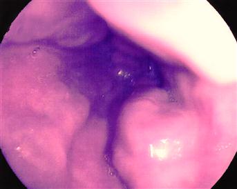
■ OGD
Will demonstrate most lesions, e.g. varices, oesophagitis, peptic ulcer, gastric erosions, Mallory–Weiss tear, carcinoma and rarer causes of bleeding. Biopsy may be carried out where necessary.
Specific Investigations
[/level-membership-for-internal-medicine-category][not-level-membership-for-internal-medicine-category]
Haematemesis
Haematemesis is the vomiting of blood. This may be frank blood or blood altered by the action of gastric acid and digestive enzymes, i.e. ‘coffee grounds’. Haematemesis is usually caused by lesions proximal to the duodenojejunal junction.
Causes
Oesophagus
Stomach
Duodenum
History
Swallowed blood
Check for a recent history of epistaxis or haemoptysis.
Oesophagus
[/not-level-membership-for-internal-medicine-category]
