H
Haab’s pupillometer See pupillometer.
haemangioma, cavernous A benign orbital tumour in adults, mostly females. The tumour is composed of large blood-filled spaces possibly due to dilatation and thickening of the capillary loops. The most common site is the muscle cone behind the globe causing proptosis, hyperopia and choroidal folds. Visual acuity may be reduced. Treatment is surgical in most cases.
haemangioma, retinal A benign tumour affecting the retinal capillaries and sometimes the optic nerve. It is often associated with systemic lesions, such as von Hippel–Lindau disease. The retinal lesion grows from a small red nodule to a larger yellowish mass accompanied by dilatation and tortuosity of the supplying artery and draining vein, and perhaps hard exudates, macular oedema, epiretinal membrane and retinal detachment. Treatment includes laser photocoagulation, cryotherapy and radiotherapy.
haematoma A swelling containing blood. It may result from injury (e.g. black eye) or from some blood disease, such as leukaemia. Note: also spelt hematoma.
haematoma, ocular A swelling due to a large haemorrhage into the tissues of the eye.
haemophthalmia An effusion of blood into the eye.
haemorrhage The escape of blood from any part of the vascular system. Note: also spelt hemorrhage.
blot h. A form of intraretinal haemorrhage often noted in background (nonproliferative) diabetic retinopathy, branch retinal vein occlusion, carotid occlusive disease and child abuse. The haemorrhage is located within the inner retina and is limited by the orientation of the inner nuclear and plexiform layers. A small blot haemorrhage is often referred to as a ‘dot’ haemorrhage.
flame h. See haemorrhage, preretinal.
preretinal h. Haemorrhage occurring between the retina and the vitreous body. It is usually large and often shaped like a D with the straight edge at the top. Syn. subhyaloid haemorrhage. Others are flame shaped and occur at the level of the nerve fibre layer and tend to parallel the course of the nerve fibres (flame haemorrhage). Retinal haemorrhages are usually round and originate in the deep capillaries of the retina. Retinal and preretinal haemorrhages usually absorb after a period of time (except those that break into the vitreous), but subarachnoid haemorrhage (which is usually due to a rupture of an aneurysm in an artery of the circle of Willis) must be suspected as they often accompany it.
See retinopathy, proliferative.
subconjunctival h. A red patch of blood on the conjunctiva of the eye, due to the rupture of a small blood vessel beneath. The condition is nearly always unilateral and the haemorrhage absorbs spontaneously although it frequently alarms the subject. It may be associated with hypertension, especially in people over 50 years of age.
See disease, sickle-cell.
subarachnoid h.; subhyaloid h. See haemorrhage, preretinal.
Haidinger’s brushes An entoptic phenomenon observed when viewing a large diffusely illuminated blue field through a polarizer. It appears as a pair of yellow, brush-like shapes, which seem to radiate from the point of fixation. The brushes are believed to be due to double refraction by the radially oriented fibres of Henle around the fovea. This phenomenon is used in detecting and treating eccentric fixation.
Halberg clip See clip, Halberg.
half-eyes See spectacles, half-eye.
Haller’s layer See layer, Haller’s.
hallucinations, visual Visual perception not evoked by a light stimulus. They may be provoked by some pathological process anywhere along the visual pathway, or as a result of an organic brain disease.
See syndrome, Charles Bonnet.
halo A coloured ring of light seen around a light source as a result of aberrations, internal reflections, diffraction or scattering. It also appears when the eye is diseased and the cornea is oedematous, as in glaucoma.
haplopia Single normal vision, as distinguished from diplopia.
haploscope Instrument used mainly in the laboratory to study various aspects of binocular vision. It presents separate fields of view to the two eyes while allowing changes in convergence or accommodation of one or both eyes, as well as providing for controls of colour, intensity or size of target and field.
See amblyoscope, Worth; dichoptic; masking, dichoptic.
haptic 1. See scleral zone. 2. Pertaining to the sense of touch.
See lens, scleral contact.
Harada’s disease See disease, Harada’s.
hard resin See CR-39 material.
harmonious ARC See retinal correspondence, abnormal.
Hartman–Shack principle See aberration, wavefront.
Hasner’s valve See valve of Hasner.
Hassall–Henle bodies See cornea guttata.
head posture, abnormal A deviation in position of the head, aimed at mitigating the effects of diplopia. It may be due to a field restriction or shyness, but the most frequent reason is an incomitant strabismus. Patients usually adjust their heads to permit fusion. If the deviation is too large to achieve fusion, patients may adjust their heads so as to increase the separation between the diplopic images and thereby making the diplopia less troublesome. Examples: if the right medial rectus or the left lateral rectus is affected, the face may be turned to the left, and vice versa if the other horizontal muscles are affected; if the left superior oblique is affected, the face may be turned to the right, the chin may be depressed and the head may be tilted to the right. If the cause of abnormal head posture remains untreated it may produce torticollis.
See paralysis of the fourth nerve; paralysis of the sixth nerve; strabismus, paralytic.
head tilt test See test, Bielschowsky’s head tilt.
headache, ocular (HA) A headache believed to result from excessive use of the eyes, uncorrected refractive error, especially hyperopia and low grades of astigmatism, binocular vision anomaly or eye diseases. This headache typically occurs in the brow region but also in the occipital or neck regions.
See accommodative insufficiency; asthenopia.
headlamp 1. Lighting device fitted to a vehicle and used to provide illumination on the road. 2. A lamp strapped to the forehead of a surgeon or miner, enabling light to be directed where required leaving both hands free.
heliophobia Neurotic fear of exposure to sunlight.
Helmholtz’s law of magnification See law, Lagrange’s.
Helmholtz’s theory of accommodation See theory, Helmholtz’s of accommodation.
Helmholtz’s theory of colour vision See theory, Young–Helmholtz.
HEMA Transparent hydrophilic plastic used in the manufacture of soft contact lenses. It stands for 2-hydroxyethyl methacrylate.
See index of refraction; lens, contact.
hematoma See haematoma.
hemeralopia Term used to mean either night blindness in which there is a partial or total inability to see in the dark associated with a loss of rod function or vitamin A deficiency; or day blindness in which there is reduced vision in daylight while vision is normal in the dark. Syn. nyctalopia (this term is only synonymous with night blindness); night sight (this term is only synonymous with day blindness).
See atrophy, girate; blindness, congenital stationary night; choroideremia; disease, Oguchi’s; retinitis pigmentosa.
hemiachromatopsia Colour blindness in one half of the visual field of one or both eyes.
See achromatopsia.
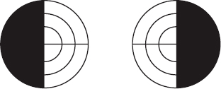
hemianopia Loss of vision in one half of the visual field of one eye (unilateral hemianopia) or of both eyes (bilateral hemianopia) (Fig. H1). Syn. hemianopsia.
See quadrantanopia; reflex, hemianopic pupillary.
absolute h. Hemianopia in which the affected part of the retina is totally blind to light, form and colour.
altitudinal h. Hemianopia in either the upper or lower half of the visual field. A common cause is anterior ischaemic optic neuropathy.
binasal h. Hemianopia in the nasal halves of the visual fields of both eyes.
bitemporal h. Hemianopia in the temporal halves of the visual fields of both eyes.
congruous h. Hemianopia in which the defects in the two visual fields are identical. A common cause is a lesion in the posterior optic radiations.
heteronymous h. A loss of vision in either both nasal halves (binasal hemianopia) or both temporal halves of the visual field (bitemporal hemianopia). A common cause of the latter is a lesion in the optic chiasma.
homonymous h. A loss of vision in the nasal half of the visual field of one eye and the temporal half of the visual field of the other eye. Left homonymous hemianopia is a loss of vision in the temporal half of the visual field of the left eye and the nasal half of the visual field of the right eye. Right homonymous hemianopia is a loss of vision in the temporal half of the visual field of the right eye and the nasal half of the visual field of the left eye. Common causes are occlusion of the posterior cerebral artery (stroke), trauma and tumours.
See macular sparing.
incongruous h. Hemianopia in which the defects in the two affected visual fields differ in one or more ways. A common cause is a lesion of the optic tract.
quadrantic h. See quadrantanopia.
relative h. Hemianopia involving a loss of form and colour but not of light.
h. spectacles See spectacles, hemianopic.
hemianopsia See hemianopia.
hemidecussation The rearrangement of the fibres of the optic nerves occurring in the optic chiasma in which about half of them from each optic nerve pass on to the contralateral optic tract. Thus each optic tract contains one half of the fibres of the ipsilateral optic nerve (representing the ipsilateral half visual field) and one half from the contralateral optic nerve (representing the contralateral half visual field). Syn. semidecussation.
See decussation.
hemidesmosome See desmosome.
hemifield One half of the visual field, usually divided vertically through the fovea into the left or the right visual field. It occurs following transection of the optic chiasma. Hemifield neglect sometimes occurs following trauma to the posterior lobe of one hemisphere.
See visual neglect.
hemorrhage See haemorrhage.
Henle, crypts; glands of See glands of Henle.
Henle, fibre layer of See layer of Henle, fibre.
hepatolenticular degeneration See disease, Wilson’s.
Herbert’s pits See trachoma.
hereditary Pertains to a condition that is genetically transmitted from parent to offspring.
See acquired; congenital; familial; inheritance.

van Herick, Shaffer and Schwartz method See method, van Herick, Shaffer and Schwartz.
Hering’s after-image; after-image test See under the nouns.
Hering’s law See law of equal innervation, Hering’s.
Hering’s theory of colour vision; visual illusion See under the nouns.
Hering–Hermann grid A grid consisting of perpendicularly crossed white stripes on a black background. The observer sees a dark shadow at the intersections of the white stripes. This phenomenon is due to lateral inhibition and does not occur for the fixated point (Fig. H2). Syn. Hermann’s grid; Hermann’s visual illusion.
Hering–Hillebrand deviation See deviation, Hering–Hillebrand.
herpes simplex of the cornea See herpesvirus; keratitis, herpes simplex.
herpes zoster A viral infection of the posterior root ganglia of the spinal cord due to a reactivation of the varicella-zoster virus (also called chickenpox virus) which had remained latent. It is characterized by a circumscribed vesicular eruption of the skin and neuralgic pain in the areas supplied by the sensory nerves. This is due to the migration of the virus from the affected ganglia to the sensory nerves. Ocular manifestations include iritis, keratitis, scleritis, uveitis, and retinal necrosis. Syn. shingles.
herpes zoster ophthalmicus An inflammation of that portion of the gasserian ganglion receiving fibres from the ophthalmic division of the trigeminal nerve, due to an infection by a latent varicella-zoster virus identical to that causing chickenpox. The disease that occurs most commonly in people over 50 years of age begins with a severe, unilateral, disabling neuralgia in the region of distribution of the nerve. It is followed by a vesicular eruption of the epithelium of the forehead, the nose, eyelids and sometimes the cornea. The vesicles rupture leaving haemorrhagic areas that heal in several weeks. Pain usually disappears in about two weeks but in a few cases neuralgia persists for a long time. Ocular complications occur in approximately 50% of all cases of herpes zoster ophthalmicus. Corneal involvement appears as acute epithelial keratitis, which is characterized by small fine dendritic or stellate lesions in the peripheral cornea in association with a conjunctivitis. This keratitis usually resolves within a week. As the disease progresses it may give rise to mucous plaque keratitis, which occurs usually between the third and the sixth month after the onset of the rash. It is characterized by the plaque lines on the surface of the cornea, which can be easily lifted, and stromal haze. Iridocyclitis also accompanies this keratitis in approximately 50% of cases.
herpesvirus Any virus belonging to a group of DNA-containing viruses, which have similar structures but few other properties in common. They are: herpes simplex virus 1 (HSV-1) (human herpesvirus 1); herpes simplex virus 2 (HSV-2) (human herpesvirus 2); varicella-zoster virus (VZV) (human herpesvirus 3); Epstein–Barr virus (EBV) (human herpesvirus 4); cytomegalovirus (CMV) (human herpesvirus 5); human herpesvirus 6 (HHV-6); human herpesvirus 7 (HHV-7); and human herpesvirus 8 (HHV-8). Herpesviruses may cause infections including blepharoconjunctivitis, herpes zoster ophthalmicus, iridocyclitis, keratitis, uveitis, retinal necrosis, retinitis and cytomegalovirus retinitis.
See antiviral agents; virus.
herpetic keratitis See keratitis, herpetic.
Herschel prism See prism, rotary.
Hess after-image See after-image.
Hertel exophthalmometer See exophthalmometer.
hertz A unit of frequency equal to one cycle per second. Symbol: Hz.
Hess–Lancaster test See test, Hess–Lancaster.
heterochromatic iridocyclitis See iridocyclitis, Fuchs’ heterochromic.
heterochromatic stimuli Visual stimuli that give rise to different colour sensations.
heterochromia Difference in colour of the two irides or of different parts of the same iris. It is usually congenital but some cases are associated with some eye diseases such as cataract, corneal precipitates, glaucoma, iridocyclitis, iris melanoma or as a result of siderosis. Syn. anisochromia.
See syndrome, Fuchs’; syndrome, Horner’s; syndrome, Marfan’s.
heterodeviation A form of ocular alignment that differs from the normal orthophoria. These include the general groups of heterophoria (e.g. esophoria, hyperphoria, etc.) and heterotropia (e.g. esotropia, exotropia, etc.).
heterometropia See anisometropia.
heteronymous diplopia; hemianopia See under the nouns.
heterophoria The tendency for the two visual axes of the eyes not to be directed towards the point of fixation, in the absence of an adequate stimulus to fusion. Thus, the active and passive positions do not coincide for that particular fixation distance. This tendency is characterized by a deviation that can take various forms according to its relative direction such as esophoria, exophoria, excyclophoria, incyclophoria, hyperphoria, hypophoria. Syn. phoria.
See anisophoria; dextrophoria; dissociation; kataphoria; laevophoria.
associated h. A term sometimes used to denote the prism power necessary to align the nonius markers of a fixation disparity test. It is not strictly speaking a heterophoria because only part of the visual field is dissociated while the rest of the field is fused (that fused area is often referred to as fusion lock or binocular lock). The dissociation of only part of the field is achieved by using either a method of cross-polarization (e.g. Mallet fixation disparity unit) or a septum (e.g. Turville infinity balance test). Syn. aligning prism; compensating prism.
See disparity, retinal; Disparometer; Mallett fixation disparity unit; test, Turville infinity balance.
compensated h. Any heterophoria that does not give rise to symptoms or to suppression.
decompensated d. See heterophoria, uncompensated.
dissociated h. Any heterophoria which is revealed by methods which produce complete dissociation such as the cover test, the Maddox rod test, the Thorington test, the von Graefe’s test, the diplopia test, etc.
uncompensated h. Any heterophoria which gives rise to symptoms or to suppression. The symptoms are associated with visual tasks, especially close work, but also occasionally, inadequate illumination. Resting the eyes will usually lessen the symptoms. Unbalanced spectacle correction, deterioration in the patient’s general health, worry and anxiety can also sometimes give rise to an uncompensated heterophoria. This type of heterophoria is presumed to manifest itself as fixation disparity. Syn. decompensated heterophoria.
See Disparometer; Mallett fixation disparity unit; prism, relieving.
heterophthalmia A difference in the appearance of the two eyes as in heterochromia.
heteropsia See anisometropia.
heterotopia maculae See macula, ectopia of the.
heterotropia See strabismus.
heterotropia, cyclic See strabismus, cyclic.
hinge joint of a spectacle frame A joint made up of plates, charniers and a pivot by means of which the sides hinge upon the front of the spectacles.
von Hippel’s disease See disease, von Hippel’s.
von Hippel–Lindau disease See disease, von Hippel–Lindau.
hippus Small rhythmic variations in the size of the pupils. They are present in everybody and increase slightly at high luminances. The frequency of these oscillations is about 1.4 Hz. Hippus may also be associated with systemic disorders such as multiple sclerosis, neurosyphilis and myasthenia gravis.
Hirschberg’s method See method, Hirschberg’s.
histoplasmosis An infection caused by inhalation of the fungus Histoplasma capsulatum found in soil dust. It manifests by a self-limited pneumonitis, which occasionally progresses and may affect the eye. It is then characterized by a disseminated choroiditis with small, yellowish scattered lesions called ‘histo spots’ which represent atrophic choroidal scars appearing as ‘punched-out’ spots. At a later stage the patient may present subretinal neovascularization with blurred vision. Many cases have been reported in parts of the world where the fungus is nonexistent. Thus the condition has been termed ‘presumed ocular histoplasmosis syndrome (POHS)’. The main treatment for neovascularization consists of laser photocoagulation and steroids for the maculopathy.
history, case A record of a patient’s chief complaint, ocular and general health and that of close relatives, and visual requirements. It is a very important part of the examination, which facilitates the diagnosis and treatment of the patient’s complaint.
HIV (human immunodeficiency virus) An RNA retrovirus of the genus Lentivirus that infects and destroys vital cells of the human immune system, such as helper T cells (CD4+ cells). It causes the acquired immunodeficiency syndrome (AIDS) and may lead to complications such as anterior uveitis, viral keratitis, cytomegalovirus retinitis, microvascular abnormalities of the conjunctiva and/or retina, etc.
See syndrome, acquired immunodeficiency (AIDS).
hole in the card test See test, hole in the card.
hole in the hand test See test, hole in the hand.
Hollenhorst’s plaques See plaques, Hollenhorst’s.
Holmes–Adie syndrome See syndrome, Adie’s.
Holmes–Wright lantern See test, lantern.
Holmgren’s test See test, wool.
hologram See holography.
holography A technique for obtaining a stereoscopic image of an object without the use of lenses. It consists of recording on a photographic plate the pattern of interference between coherent light reflected from the object and light that comes directly from the same source (or is reflected from a mirror). The coherent light is usually provided by a laser. The photographic recording on the plate (called a hologram) when illuminated with coherent light yields an image that is identical in amplitude and phase distribution with the original wave from the object. It thus provides a three-dimensional image of the object in the sense that the observer’s eyes must refocus to examine foreground and background and indeed ‘look around’ objects by simply moving the head laterally.
homatropine hydrobromide Alkaloid derived from atropine. It is an antimuscarinic drug used as a mydriatic and as a weak cycloplegic.
homocentric pencil of rays 1. One in which all rays converge or diverge from a single point. 2. One in which more than one beam of light share the same pathway. Example: in an ophthalmoscope the illumination and observation paths are shared.
homochromatic after-image See after-image, homochromatic.
homocystinuria An autosomal recessive inherited disorder caused by a cystathionine beta synthase deficiency, which leads to an accumulation of the amino acid methionine and homocysteine. The first signs are ocular; a dislocated lens which may cause diplopia or glaucoma, myopia and occasionally retinal detachment. Systemic signs are blond hair, intellectual impairment and some of the features of Marfan’s syndrome (e.g. tall, thin build).
homonymous diplopia; hemianopia See under the nouns.
hordeolum, external An acute suppurative infection (usually caused by staphylococci) of an eyelash follicle or of the sebaceous gland of Zeis or of the sweat gland of Moll (located in the region of an eyelash). The condition occurs most commonly in people who have a lower resistance to staphylococci as in debility, or who have a blepharitis. It has the appearance of a hyperaemic elevated area, indurated on the eyelid margin where it may rupture and discharge yellowish pus. The symptom is tenderness of the eyelid that may become marked as the suppuration progresses and the eyelid near the margin is red and swollen. Treatment usually consists of hot compresses and application of an antibiotic ointment and perhaps removal of the affected eyelash. Surgical incision is rare. Syn. stye.
hordeolum, internal An acute purulent infection (usually caused by staphylococci) of the meibomian glands. It usually causes more discomfort than an external hordeolum as it is located on the conjunctival side of the eyelid. Treatment is similar to that of external hordeolum but surgical incision is required more frequently. Syn. meibomian stye.
See chalazion; meibomianitis.
horizontal cell See cell, horizontal.
Horner’s muscle; syndrome See under the nouns.
Horner–Trantas’ dots See conjunctivitis, vernal.
horopter The locus of object points in space that stimulate corresponding retinal points of the two eyes when the eyes are fixating binocularly one of these object points. The horopter is a curve that passes through the fixation point and changes shape with fixation distance. Objects closer to the eyes than the horopter are seen double (crossed disparity) and objects further than the horopter are seen double (uncrossed disparity). There are various types of horopters depending upon the method of determination.
apparent frontoparallel plane h. The locus of object points in space which appear to the observer to lie on a plane through the fixation point, parallel to the plane of the face. Syn. frontal plane horopter.
See deviation, Hering–Hillebrand; plane, apparent frontoparallel.
empirical h. A horopter determined experimentally by having an observer judge a series of targets to be neither ‘nearer than’ nor ‘farther than’ the fixation point. Examples: the frontoparallel plane horopter; the nonius horopter.
longitudinal h. Horopter that is plotted by only considering the longitudinal section where the rods meet the plane of fixation. Thus this horopter is a curve located in that plane and not a surface.
nonius h. Horopter plotted by fixating binocularly a central vertical rod while the other rods located in the periphery have their upper halves seen by one eye only and their lower halves seen by the other eye only. Each rod is moved individually until the two halves are seen aligned. Syn. vernier horopter.
rectilinear h. The assemblage of all lines in space that stimulate corresponding retinal lines. It is a pencil of quadratric surfaces with the space horopter as their common curve of intersection.
space h. The horopter consisting of all object points in space which stimulate corresponding retinal points as distinguished from the two-dimensional cases such as the apparent frontoparallel plane, longitudinal or nonius horopters.
theoretical h. A horopter based on theoretical concepts. Example: the Vieth–Müller horopter.
vernier h. See horopter, nonius.
Vieth–Müller h. A theoretical horopter formed by a circle passing through the point of fixation and the anterior nodal points of the two eyes. Thus any point on this horopter forms an image in the two retinas, which is at equal distances from their respective foveas. Syn. Vieth–Müller circle.
See deviation, Hering–Hillebrand.
horror fusionis Avoidance of fusion resulting from two retinal images which are so different that they are impossible to fuse. This is often the case in strabismus. Syn. fusion aversion.
horseshoe tear A type of retinal tear in which a strip of tissue is torn from the retina. The tear commonly follows a vitreous detachment in which the vitreous adheres to the retina and pulls it from the point of adherence during or just after an abrupt eye movement. This type of retinal tear is particularly dangerous since it is often a precursor to a retinal detachment.
Howard–Dolman test See test, Howard–Dolman.
HRR test See plates, pseudoisochromatic.
Hudson–Stahli See line, Hudson–Stahli.
hue Attribute of colour sensation, such as blue, red, green, etc., which is ordinarily associated with a given wavelength of the light stimulating the retina, as distinguished from the attributes of brightness and saturation. See phenomenon, Bezold–Brücke; threshold, differential.
Hummelsheim’s procedure See transposition.
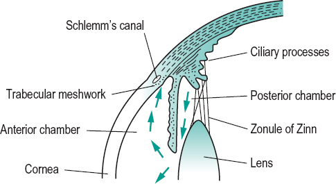
humour, aqueous Clear, colourless fluid that fills the anterior and posterior chambers of the eye. It is a carrier of nutrients for the lens and to a larger extent the cornea, especially of glucose and essential amino acids. It contributes to the maintenance of the intraocular pressure. It is formed in the ciliary processes, flows into the posterior chamber, then through the pupil into the anterior chamber and leaves the eye through the trabecular meshwork passing to the canal of Schlemm and then to veins in the intrascleral venous plexus (Fig. H3). A small amount (10% to 15%) also flows out of the eye via the uveoscleral pathway. The aqueous in the anterior chamber is a component of the optical system of the eye. It has an index of refraction of 1.336, slightly lower than that of the cornea, so that the cornea/aqueous surface acts as a diverging lens of low power. It is a fluid very similar to blood plasma but with a much lower concentration of protein and a higher concentration of ascorbate.
The rate of aqueous humour outflow varies between 2.0 μl/min and 3.0 μl/min via both the conventional (trabecular meshwork and Schlemm’s canal) and the unconventional (uveoscleral) pathways. This rate is normally equal to the rate of aqueous secretion. If the rate of outflow is lower than the rate of secretion intraocular pressure increases.
See flare, aqueous; ultrafiltration.
humour, vitreous A transparent, colourless, gelatinous mass of a consistency somewhat firmer than egg white which fills the space between the crystalline lens, the ciliary body and the retina, and constitutes four-fifths of the volume of the eye. The vitreous is about 99% water, the remaining 1% includes hyaluronic acid, organic salts and soluble and insoluble proteins especially collagen (mainly type II). It is firmly attached to the pars plana of the ciliary body near the ora serrata in an area known as the vitreous base and around the optic disc. In older people and in pathological conditions the vitreous is no longer in a gel state, tending to become fluid. Syn. vitreous body.
See artery, hyaloid; asteroid hyalosis; floaters; hyaloid remnant; synchisis scintillans; syndrome, Wagner’s; vitreous base; vitreous detachment.
Humphrey Vision Analyser See Analyser, Humphrey Vision.
Humphriss immediate contrast test; method See method, Humphriss.
Hutchinson’s pupil See pupil, Hutchinson’s.
Huygens’ eyepiece See eyepiece, Huygens’.
hyaline bodies See drusen.
hyaloid artery; canal; fossa; membrane See under the nouns.
hyaloid remnant A rare condition in which there remain some parts of the hyaloid artery. Posteriorly there may be a vascular loop or the thread of an obliterated vessel running forward from the optic disc and floating freely in the vitreous. Anteriorly there may be some fibrous remnants attached to the posterior lens capsule and others sometimes floating in the vitreous. The anterior attachment of the hyaloid artery to the lens may also remain throughout life as a black dot, called Mittendorf’s dot, and can be seen within the pupil by direct ophthalmoscopy (it appears as a white dot with the biomicroscope). There is rarely any visual interference although patients may sometimes report seeing muscae volitantes. Syn. persistent hyaloid artery.
See canal, hyaloid; papilla, Bergmeister’s; pseudopapilloedema; vitreous, persistent hyperplastic primary.
hydrocortisone acetate See antiinflammatory drug.
hydrogel Type of plastic material which contains water, and is commonly used in the manufacture of soft contact lenses, e.g. HEMA.
hydrogen peroxide See antiseptic.
hydrophilic lens See lens, contact.
hydrophthalmos See glaucoma, congenital.
hydrops, corneal Excessive accumulation of watery fluid in the stroma of the cornea as a result of rupture of the posterior layers of the cornea (Descemet’s membrane and the endothelium). It is often found in advanced keratoconus.
hydrops, acute A complication of advanced keratoconus in which Descemet’s membrane ruptures resulting in acute corneal oedema. It is due to stretching of Descemet’s membrane as the cornea steepens. The corneal oedema usually clears spontaneously, although the scar may interfere with vision. Hyperosmotic agents may be needed to reduce corneal oedema.
hydroxyethylcellulose; hydroxymethyl-cellulose, hydroxypropylcellulose See tears, artificial.
hydroxypropylmethylcellulose See hypromellose.
hyoscine hydrobromide An antimuscarinic (or parasympatholytic) drug with actions similar to those of atropine (i.e. mydriasis and cycloplegia) but of shorter duration. It is used in the measurement of refraction of children and in the treatment of keratitis, anterior uveitis and in cases of burns to the anterior segment of the eye to prevent posterior synechia, as well as to reduce the pain secondary to iridociliary spasm. Syn. scopolamine hydrobromide.
hyperaccommodation See accommodative excess.
hyperacuity The ability of the eye to detect the differences in the spatial locations of two or more stimuli. Hyperacuity thresholds are not based on resolution and are usually below about 15 seconds of arc. Hyperacuity tests include vernier acuity, stereoscopic acuity, orientation discrimination in which differences in the tilts of lines must be detected, the movement displacement threshold, the vertical alignment of a cluster of dots, or the ability to bisect two parallel lines. Hyperacuity is less affected by optical defocus or light scattering (as occurs for example in corneal leukoma, cataract, vitreous haemorrhage) than is Snellen acuity and can therefore be helpful in assessing macular function behind a cataract or other media opacity before surgery (Fig. H4).
See maxwellian view system, clinical.
hyperaemia Excessive accumulation of blood in a part of the body. Note: also spelt hyperemia. Syn. injection.
See injection, ciliary; injection, conjunctival.
hyperaesthesia, corneal Abnormally high corneal sensitivity (threshold less than 15 mg/0.013 mm2 near the limbus in the adult eye) as distinguished from hypoaesthesia (beyond 70 mg/0.013 mm2 near the limbus in the adult eye) which is abnormally low corneal sensitivity.
See aesthesiometer; corneal touch threshold; sensitivity, corneal.

hypercapnia The presence of a raised carbon dioxide content or tension in a milieu (e.g. blood, tears). Contact lens wear tends to give rise to this condition, especially lenses of low gas transmissibility.
See acidosis.
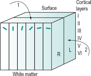
hypercolumn A complete set of orientation columns over a cycle of 180° and of right and left dominance columns in the visual cortex. A hypercolumn may be about 1 mm wide. A hypercolumn of orientation columns is perpendicular to a hypercolumn of ocular dominance columns (Fig. H5).
hypercomplex cell See cell, hypercomplex.
hyperfocal distance See distance, hyperfocal.
hyperlacrimation Overflow of tears due to excessive secretion by the lacrimal gland. It may be caused by drugs (e.g. pilocarpine), strong emotion; or as a reflex from trigeminal stimulation by an inflamed eye; or irritation of the cornea or conjunctiva by a chemical irritant in the air; cold wind; or a foreign body in the eye. The main symptoms are discomfort and blurring of vision and sometimes embarrassment. Management depends on the cause. Syn. hypersecretion. See epiphora.
hypermetria An abnormally large movement. Example: a saccade that overshoots its target.
See dysmetria, ocular; movement, saccadic eye.
hypermetrope A person who has hypermetropia.
hypermetropia (H) See hyperopia.
hyperope A person who has hyperopia.
hyperopia (H) Refractive condition of the eye in which distant objects are focused behind the retina when the accommodation is relaxed. Thus, vision is blurred. In hyperopia, the point conjugate with the retina, that is the far point of the eye, is located behind the eye (Fig. H6). At birth the mean refractive error is a hyperopia of about +2.00 D. As the child grows into adolescence the average refraction tends towards emmetropia. The percentage of people with hyperopia increases beyond the age of 40. Syn. far sight; long sight; hypermetropia.
See choroidal folds; cornea plana; glaucoma, angle-closure; headache, ocular; luxation of the lens; pseudopapilloedema; sclerocornea; test, plus 1.00 D blur.
absolute h. That hyperopia which cannot be compensated for by accommodation.
acquired h. Hyperopia resulting from changes in the refractive indices of the media due to either age or disease, or to surgery.
facultative h. That portion of hyperopia which can be compensated for by accommodation.
latent h. That portion of total hyperopia which is compensated for by the tonus of the ciliary muscle. It can be revealed wholly or partially by the use of a cycloplegic.
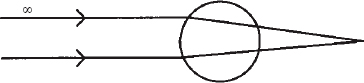
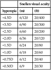
Table H1
Approximate relationship between uncorrected absolute hyperopia and visual acuity
| Snellen visual acuity | ||
| hyperopia | (m) | (ft) |
| +4.5D | 6/120 | 20/400 |
| +3.5D | 6/90 | 20/300 |
| +2.5D | 6/60 | 20/200 |
| +2.0D | 6/36 | 20/120 |
| +1.5D | 6/24 | 20/80 |
| +1.0D | 6/18 | 20/60 |
| +0.75D | 6/12 | 20/40 |
| +0.50D | 6/9 | 20/30 |
manifest h. That portion of total hyperopia which can be determined by the strongest convex lens in a subjective routine examination while retaining the best visual acuity.
simple h. Hyperopia uncomplicated by disease, trauma or astigmatism.
total h. The sum of the latent and manifest hyperopia.
hyperopic defocus See defocus, hyperopic.
hyperosmotic agent A drug that makes blood plasma hypertonic thus drawing fluid out of the eye and leading to a reduction in intraocular pressure. It is used in solution in the treatment of angle-closure glaucoma and sometimes before surgery to decrease the intraocular pressure. Common agents include glycerin (glycerol), isosorbide, mannitol and urea.
See solution, hypertonic.
hyperphoria The tendency for the line of sight of one eye to deviate upward relative to that of the other eye, in the absence of an adequate stimulus to fusion. If the deviation tends to be downward relative to the other eye or if the other eye in hyperphoria is used as a reference, the condition is called hypophoria.
See kataphoria.
left h. (L/R) Hyperphoria in which the line of sight of the left eye deviates upward relative to the other eye.
paretic h. Hyperphoria due to a paresis of one or several of the extraocular muscles.
right h. (R/L) Hyperphoria in which the line of sight of the right eye deviates upward relative to the other eye.
hyperplasia Any condition in which there is an increase in the number of cells in an organ or a tissue. It usually excludes tumour formation. Example: choroidal naevus.
hyperpolarization A change in the value of the resting membrane potential towards a more negative value. The inside of the cell becomes more negative than the outside. Hyperpolarization is inhibitory because the membrane potential moves away from the neuron’s threshold at which an action potential could occur. Example: the retinal photoreceptor potentials when stimulated by light.
Table H2
Common ocular and systemic diseases with hyperopia as an associated sign
cornea plana
microphthalmia
sclerocornea
angle-closure glaucoma
branch retinal vein occlusion (BRVO)
fragile X syndrome
growth hormone deficiency
diabetes type 1
hypertension
See depolarization; potential, receptor; potential, resting membrane; synapse.
hypersecretion See hyperlacrimation.
hypersensitivity An excessive reaction, local or systemic, or inappropriate immune response to an antigen. Four types of immune responses are usually described, but the main reaction involving the eyes is type 1. They are also called allergic reactions types 1–4.
type 1 hypersensitivity An immediate, abnormal reaction occurring when an antigen reacts with an antibody (e.g. immunoglobulin E (IgE)) attached to a mast cell or basophil. This leads to the release of specific chemical mediators of allergy (e.g. histamine) that react with target organs throughout the body. Systemic signs include: itching, lacrimation, skin rash and possibly haemodynamic collapse and shock. Allergic conjunctivitis is an example of this type of hypersensitivity. Type 2h. (cytotoxic h.) is caused by an interaction of antibody and antigens on cell surfaces. Examples: Graves’ disease, myasthenia gravis. Type 3h. (immunecomplex mediated h.) is mediated by a combination of antigen-antibody. Example: systemic lupus erythematosus. Type 4h. (T cell-mediated h.) is a delayed reaction (several days to develop) mediated by T lymphocytes. Example: rheumatoid arthritis.
See antihistamine; mast cell stabilizers.
hypertelorism, ocular A developmental, congenital anomaly in which the distance between the orbits is abnormally large resulting in a large distance between the eyes. This can be associated with mental deficiency, divergent strabismus, exophthalmos or optic atrophy.
See syndrome, Apert’s; syndrome, Crouzon’s.
hypertension Abnormally high blood pressure beyond 140–150 mmHg for systolic blood pressure or beyond 90–95 mmHg for diastolic blood pressure. These figures are higher for older people. Elevated blood pressure can give rise to hypertensive retinopathy. See paralysis of the sixth nerve; paralysis of the third nerve; retinal vein occlusion; sphygmomanometer.
hypertension, ocular A condition in which the intraocular pressure (IOP) is above normal (>21 mmHg) but in which there are neither visual field defects nor optic disc changes. Open-angle glaucoma may or may not develop later: risk factors include thin central corneal thickness, large cup/disc ratio, high IOP and lack of treatment to reduce IOP greater than 30 mmHg.
hypertensive retinopathy See retinopathy, hypertensive.
hyperthyroidism See disease, Graves’; ophthalmopathy, thyroid.
hypertonic solution See solution, hypertonic.
hypertropia Strabismus in which one eye is directed to the fixation point while the other is directed upward (right or left hypertropia). If one eye fixates while the other is directed downward, the condition is called hypotropia (right or left hypotropia). Syn. for hypertropia is sursumvergens strabismus and anoopsia; for hypotropia is deorsumvergens strabismus.
See test, three-step.
hypertropia, alternating A condition in which, on dissociation (e.g. by occlusion) of either eye, the eye behind the cover deviates upward but reverts to its fixating position when dissociation ceases. The condition can either occur as an isolated phenomenon or be associated with strabismus or latent strabismus. Syn. anaphoria; anatropia; dissociated vertical divergence; double hyperphoria. See phenomenon, Bielschowsky’s.
hyphaemia Haemorrhage into the anterior chamber of the eye. It usually occurs as a result of trauma to the eye, especially in sports, or as a result of intraocular surgery (e.g. trabeculectomy). It is advisable to have the patient admitted to hospital because of the possibility of recurrent haemorrhage and secondary glaucoma. Note: also spelt hyphema.
hypoaesthesia See hyperaesthesia, corneal.
hypoexophoria Combined hypophoria and exophoria.
hypolacrima See alacrima.
hypometria See dysmetria, ocular.
hypophoria See hyperphoria.
hypoplasia Any condition in which there is an underdevelopment, or a decrease in the number of cells, of an organ or tissue. Example: optic nerve hypoplasia in which there is a reduction of axons, which, in severe cases, leads to visual impairment.
See hyperplasia.
hypopyon The presence of pus in the anterior chamber of the eye associated with infectious diseases of the cornea (e.g. severe microbial keratitis, corneal ulcer), the iris or the ciliary body (e.g. severe anterior uveitis). The pus usually accumulates at the bottom of the chamber and may be seen through the cornea.
See keratitis, hypopyon; syndrome, Behçet’s.
hypothalamus A group of nuclei at the base of the brain located in the floor of the third ventricle. It consists of the optic chiasma, the paired mammillary bodies, the tuber cinereum, the infundibulum and the pars posterior of the pituitary gland.
hypothesis See significance.
hypotonic solution See solution, hypotonic.
hypotony, ocular Abnormally low intraocular pressure, usually less than 6 mmHg. It may result from trauma in which there is a perforating wound with loss of aqueous, concussion injury or following surgery for glaucoma. Persistent hypotony may result in visual impairment due to choroidal folds, choroidal detachment, retinal folds or retinal detachment. Syn. ocular hypotonia; ocular hypotonus.
See choroidal folds; cyclodialysis.
hypotropia See hypertropia.
hypoxia An inadequate supply of oxygen to tissues. It may occur in some pathological conditions. Examples: in long-standing cases of diabetes there is corneal hypoxia (with consequent high epithelial fragility and some neovascularization) and retinal hypoxia (with consequent neovascularization). Corneal hypoxia (with consequent oedema, loss of sensitivity, etc.) may also occur in contact lens wear.
See anoxia; microcysts, epithelial; mitosis; oedema; oxygen requirement, critical; retinopathy, proliferative; syndrome, corneal exhaustion; syndrome, overwear; tear pumping.
hypoxic stress See strain.
hypromellose A highly viscous, watersoluble, non-irritating compound used as a thickening, lubricating and clinging agent. It is used principally as artificial tears (as for example in the management of keratoconjunctivitis sicca) and sometimes as a wetting agent. Syn. hydroxypropylmethylcellulose. See alacrima; methylcellulose; staining, 3 and 9 o’clock.
hysteresis, accommodative A term used to indicate an incomplete and temporary relaxation of the accommodation of the eye after a period of fixation. The amount of relaxation varies according to the position of the fixation point relative to the position of the tonic accommodation, and to the ametropia of the eye. In general, a sustained near visual task leads to an increase in accommodation, while a sustained distant visual task leads to a decrease in accommodation.
See accommodation, resting state of.
hysterical amblyopia See amblyopia, hysterical.
hysteropia Visual disorder due to hysteria.


