Gynaecological examination

Published on 09/03/2015 by admin

Filed under Obstetrics & Gynecology

Last modified 22/04/2025

Print this page

rate 1 star rate 2 star rate 3 star rate 4 star rate 5 star
Your rating: none, Average: 0 (0 votes)

This article have been viewed 1408 times

2 Gynaecological examination

As with the examination of any organ system, a gynaecological examination starts with a general examination. One should assess from the end of the bed whether the patient appears healthy or not, obese, average or thin, anxious or at ease. One should specifically examine for anaemia, jaundice or lymphadenopathy. If the patient is a young fit woman, then further general examination is unnecessary; however, if the patient is elderly or unwell then a more detailed examination is required. The practice of routine breast examination is controversial, but if it is performed one should initially observe for tethering or peau d’orange. All four breast quadrants are palpated, followed by the axilla on each side with the patient’s arm resting on the examiner’s arm so that there is no tension in the axilla.

Pelvic examination

Pelvic examination may be both uncomfortable and embarrassing for the patient and one must be sensitive to ensure that the environment is as friendly as possible. It is important to ensure privacy and explain to the patient exactly what she needs to remove and what you plan to do. A chaperone is recommended in all situations and is mandatory when the examiner is male. If a Cusco’s speculum is being used, the speculum should be warmed and lubricating jelly placed on the blades (a very small amount if a cervical smear needs to be taken). The examination may be uncomfortable, but it should not be painful in the absence of pathology.

The vulva and perineum should be inspected and one should observe any areas of discoloration, lumps or areas where the skin may have broken down. One should look for any evidence of prolapse. The Cusco’s speculum allows inspection of the cervix so that a cervical smear or endocervical swabs may be easily taken. It may be difficult to observe the cervical walls with a traditional Cusco’s speculum, but the plastic variety is now available where one can observe the entire vaginal wall. The patient lies in the supine position with the heels together and knees apart. The speculum is inserted with the blades closed and parallel to the labia and the opening mechanism towards the patient’s right. The speculum is gently inserted and when it is halfway into the vagina it is rotated 90° (so the locking mechanism is anterior) and inserted as far as it will go. The blades are then opened slowly under direct vision and if there has been correct placement the cervix should come into view. The cervix should be closely observed to detect any abnormalities and then a cervical smear can be taken, plus or minus endocervical swabs depending upon the clinical presentation. A cervical smear is taken by inserting the spatula or brush into the endocervical canal and rotating it 360° in one direction and 360º in the other direction. The speculum is then withdrawn, watching carefully so that the blades do not clamp down on to the cervix. The vaginal walls are observed as the Cusco’s speculum is withdrawn.

A Sims’ speculum is used to assess women with uterovaginal prolapse or when the cervix is difficult to find. The patient should be placed in the Sims’ position or the left lateral position. The patient’s buttocks should be separated, with assistance if necessary, or by asking the woman to raise the upper leg, and the patient should be asked to cough in order to observe for obvious prolapse or stress incontinence. The blade of the Sims’ speculum should then be inserted along the posterior vaginal wall and retracted posteriorly to display the anterior vaginal wall. The patient should be asked to cough again, to assess any degree of prolapse of the anterior vaginal wall and to note any stress incontinence. The anterior vaginal wall is then supported with a sponge forceps and the patient is asked to cough again, while continuing to support the anterior vaginal wall with the sponge forceps. One can then observe for cervical descent. To assess cervical descent fully a sponge forceps can be placed on the anterior lip of the cervix and traction can be applied, but this can be uncomfortable for the patient and should only be done as a preoperative assessment to determine the route of hysterectomy. The patient is asked to bear down again and the speculum is then slowly removed while the anterior vaginal wall is supported. The top of the vaginal vault and the posterior vaginal wall can thus be visualized sequentially to detect the presence of an enterocele or rectocele, respectively.

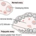
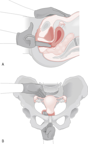
Both the Cusco’s and the Sims’ speculum examination should be followed by a bimanual examination. The patient lies in a similar position as for the Cusco’s speculum examination and the left hand parts the labia as two fingers are inserted into the vagina and then placed behind the cervix. The left hand is then placed on the abdomen above the symphysis pubis and pushes down onto the pelvis so that the organs are palpated between the left hand and the two fingers within the vagina. The uterus, which is situated centrally, should be assessed for size, consistency, mobility, regularity, anteversion, retroversion and tenderness. The adnexae are palpated on each side, particularly looking for tenderness and any suggestion of a mass. In very thin women normal-size ovaries may be felt (Fig. 2.1).

The area behind the cervix is palpated for any areas of tenderness or masses.

The pelvic examination occasionally needs to be concluded with a rectal examination if indicated.