[level-membership-for-anesthesiology-category]
59 Great Auricular Nerve Block
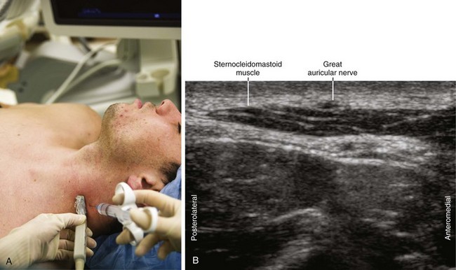
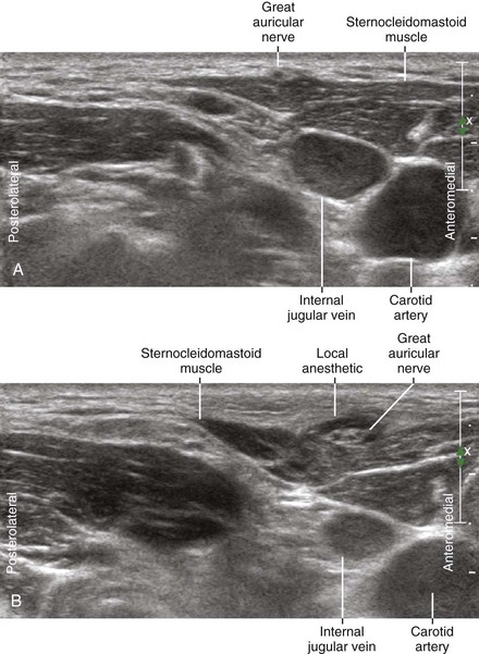
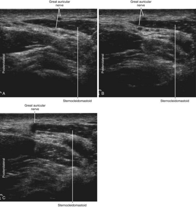
The great auricular nerve (GAN) is the largest branch of the superficial cervical plexus (see Chapter 60, Fig. 60-1). It provides cutaneous innervation to the periauricular region.1 The GAN wraps around the posterior border of the sternocleidomastoid muscle (SCM) and then courses superiorly and anteriorly, dividing into anterior and posterior branches.2 Because of its superficial location, the GAN can be damaged during surgical procedures in the neck.3 The nerves of the superficial cervical plexus lie deep to the platysma when first emerging from the plexus, but superficial to the prevertebral fascia.
Suggested Technique
Clinical Pearls
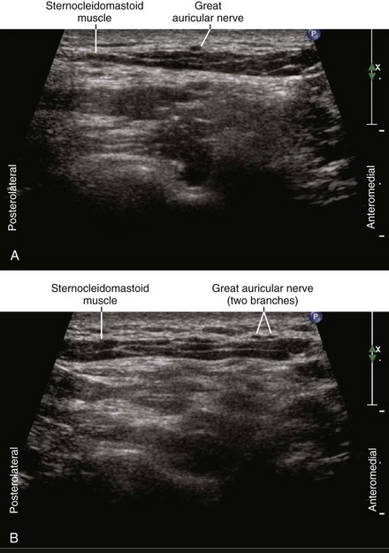
• The GAN consists of contributions from the second and third cervical nerves. The GAN divides into anterior and posterior branches.
• The GAN often can be seen both superficial and deep to the sternocleidomastoid muscle within one plane of imaging because the nerve loops around the posterolateral border of this muscle.
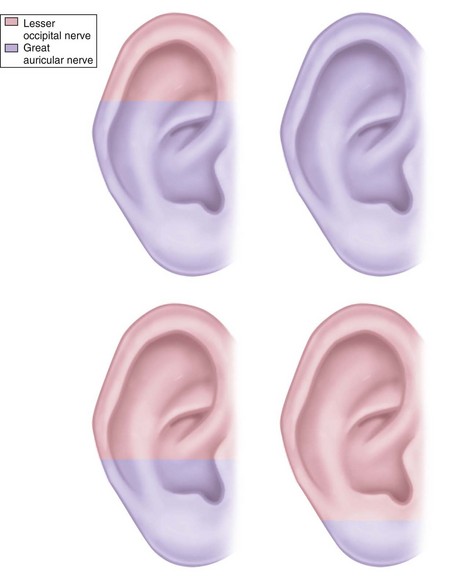
• Dominance patterns of cutaneous innervation of the external ear have been examined.4 Most patterns are either lesser occipital or great auricular dominant.
1 Thallaj A, Marhofer P, Moriggl B, et al. Great auricular nerve blockade using high resolution ultrasound: a volunteer study. Anaesthesia. 2010;65:836–840.
2 Ginsberg LE, Eicher SA. Great auricular nerve: anatomy and imaging in a case of perineural tumor spread. AJNR Am J Neuroradiol. 2000;21:568–571.
3 Nusair YM, Dickenson AJ. Great auricular causalgia: an unusual complication of excision of the submandibular gland. Br J Oral Maxillofac Surg. 2003;41:334–335.
4 Pantaloni M, Sullivan P. Relevance of the lesser occipital nerve in facial rejuvenation surgery. Plast Reconstr Surg. 2000;105:2594–2599.
[/level-membership-for-anesthesiology-category][not-level-membership-for-anesthesiology-category]
59 Great Auricular Nerve Block
The great auricular nerve (GAN) is the largest branch of the superficial cervical plexus (see Chapter 60, Fig. 60-1). It provides cutaneous innervation to the periauricular region.1 The GAN wraps around the posterior border of the sternocleidomastoid muscle (SCM) and then courses superiorly and anteriorly, dividing into anterior and posterior branches.2 Because of its superficial location, the GAN can be damaged during surgical procedures in the neck.3 The nerves of the superficial cervical plexus lie deep to the platysma when first emerging from the plexus, but superficial to the prevertebral fascia.
