Published on 02/04/2015 by admin
Filed under Internal Medicine
Last modified 22/04/2025
This article have been viewed 980 times
232 Glass eye
Look at this patient’s fundus.
Examine this patient’s eyes.
Check this patient’s vision or visual fields.
• History of trauma.
• Glass eye, which is obvious
• The patient is blind on the affected side
• The light reflex is absent.
Note: Suspect malignant melanoma if asked to examine the abdomen and the liver is palpable (from metastases).
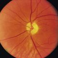
This patient has a glass eye (lesion) that is secondary to trauma in childhood (aetiology).
It is inflammation that attacks the sound eye after injury (usually perforating wound) to the other. It is almost always a plastic iridocyclitis; rarely it manifests as neuroretinitis or choroiditis. It never occurs after excision of an injured eye unless it has already commenced at the time of operation. Steroids have improved the prognosis if such treatment is commenced early.
Rhizopus mucormycosis, originating in the paranasal sinuses and nose, occurs predominantly in patients with poorly controlled diabetes mellitus, malignancy, organ transplantation and those who are receiving long-term desferrioxamine therapy. This fungus can invade the globe or ophthalmic artery to cause blindness.
They arise from a variety of ocular tissues including the conjuctiva, uveal tract (iris, ciliary body and choroid), eyelid, orbit and nasolacrimal ducts. Iris melanomas rarely metastasize, whereas ciliary body and choroidal melanomas readily disseminate.
Unlike cutaneous melanomas, the ocular type consists of two distinct cell types: spindle and epithelioid. Lesions composed of completely or predominantly spindle cells have low aggressiveness, do not tend to metastasize and are associated with a 75% survival rate at 15 years. Consequently, enucleation is usually avoided. The epithelioid type is associated with only a 35% survival rate despite enucleation, because of late metastases.
250 Cases in Clinical Medicine
WhatsApp us