CHAPTER 14 GASTROINTESTINAL PATHOLOGY


ESOPHAGUS
HISTOLOGICAL VARIANTS OF NORMAL ESOPHAGUS


ESOPHAGEAL ATRESIA AND TRACHEO-ESOPHAGEAL FISTULA


Histopathological features (Dutta, Mathur, Bhatnagar 2000)
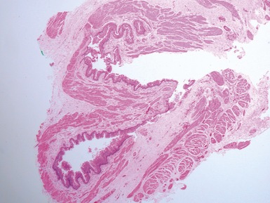
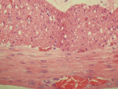
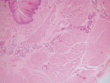
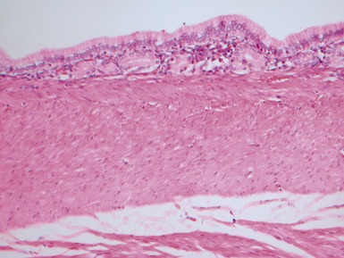
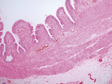
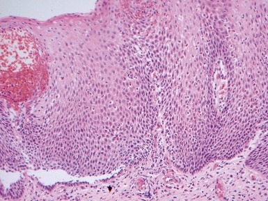
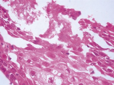
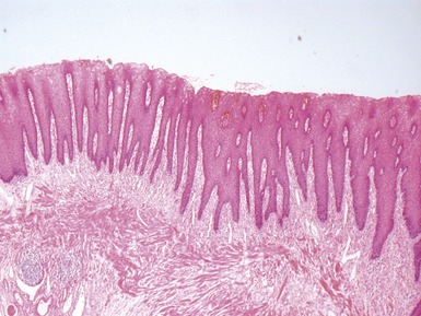
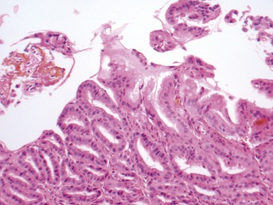
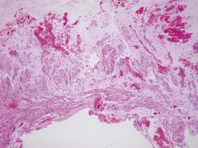
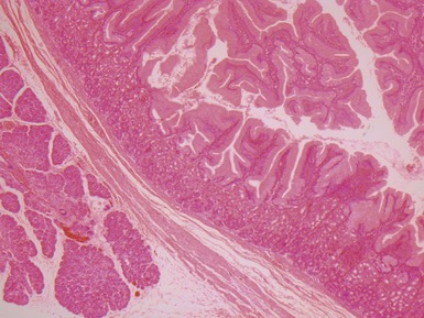
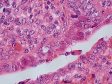
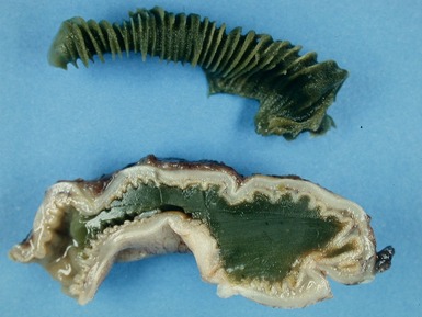
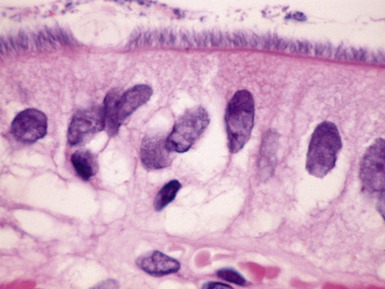
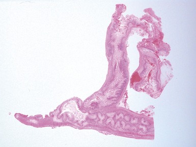
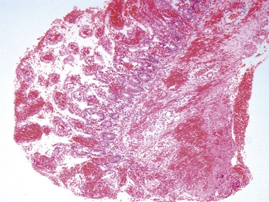
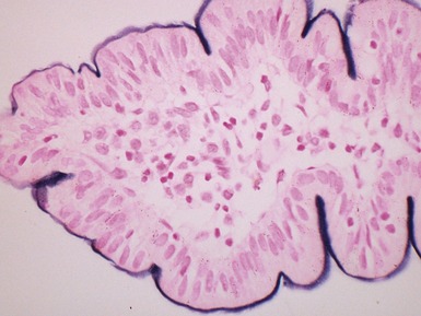
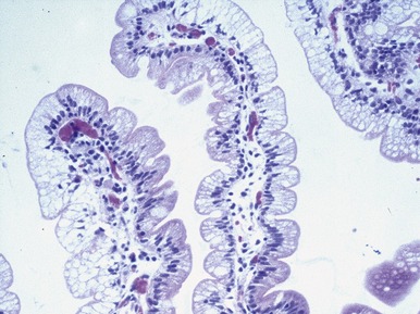
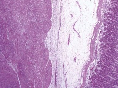
Fig 14.2 One-day-old girl with esophageal atresia and tracheo-esophageal fistula. The specimen is the tip of the proximal esophageal pouch. Photomicrograph of the specimen demonstrating the pouch comprising the usual elements of esophageal wall. The mucosa shows some ciliated epithelium. Unusually, no striated muscle was present in the muscular coat.


GASTRO-ESOPHAGEAL REFLUX DISEASE


COLUMNAR EPITHELIAL LINED ESOPHAGUS (CELE)


INFECTIVE ESOPHAGITIS


CROHN’S DISEASE OF THE ESOPHAGUS


STOMACH

GASTRITIS


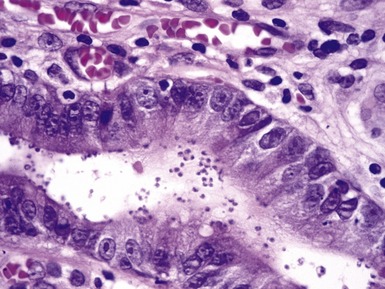
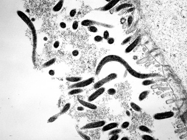
Helicobacter pylori gastritis


Crohn’s disease associated gastritis


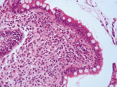
Lymphocytic gastritis


SMALL INTESTINE
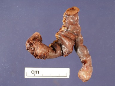
INTESTINAL ATRESIA


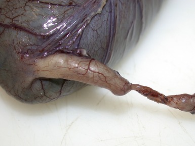


MECKEL’S DIVERTICULUM


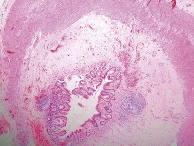
DUPLICATION CYSTS


INTUSSUSCEPTION


VOLVULUS


MECONIUM ILEUS


Histopathological features


CROHN’S DISEASE


INTESTINAL INFECTIONS


Cryptosporidiosis


Astrovirus


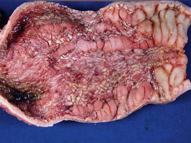
NEONATAL NECROTIZING ENTEROCOLITIS


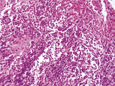
Histopathological features


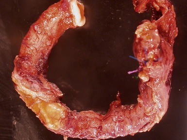
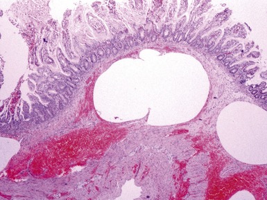
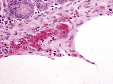
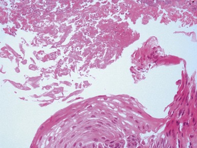
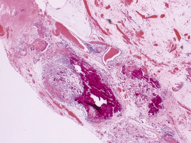
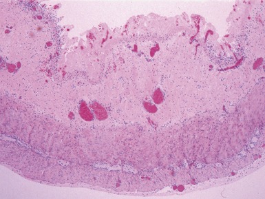
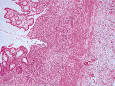
Fig 14.52 Necrotizing enterocolitis. Photomicrograph at a higher power of the same lesion as in Fig 14.51 demonstrating the edge of one of the gas-filled areas. There is hemorrhage and a neutrophil polymorph infiltrate.
FOOD ALLERGY


PRIMARY INTESTINAL FAILURE / NEONATAL-INFANTILE ENTEROPATHIES


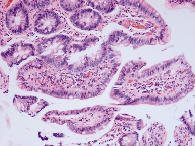
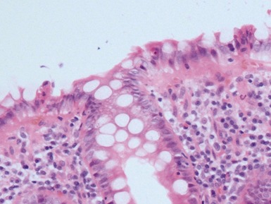
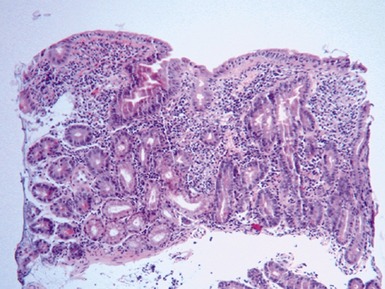
CELIAC DISEASE (GLUTEN SENSITIVE ENTEROPATHY)

Clinical features (Fasano et al 2008)


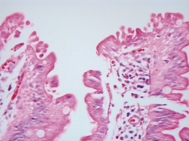
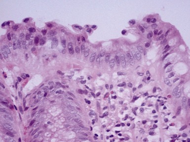
Histopathological features


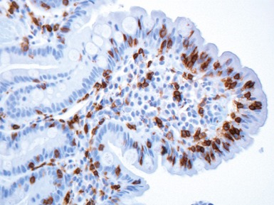
Differential diagnosis and diagnostic pitfalls


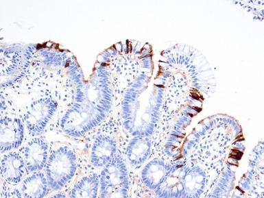
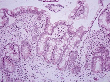
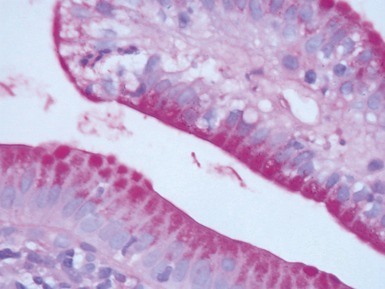
TUFTING ENTEROPATHY


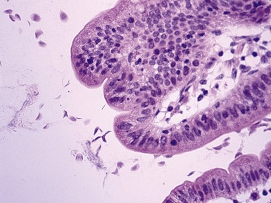
Histopathological features


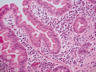
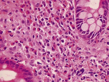
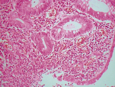
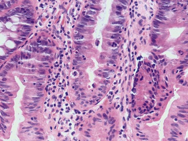
Fig 14.60 Tufting enteropathy inflammation. Photomicrograph demonstrating partial villous atrophy and an acute and chronic inflammatory cell infiltrate in the mucosa. No tufts are present in this field. The degree of inflammation present is very variable, but some degree is usually present.


MICROVILLOUS INCLUSION DISEASE


DISSACHARIDASE ASSESSMENT OF JEJUNAL SPECIMENS


ABETALIPOPROTEINEMIA


PATHOLOGY OF INTESTINAL TRANSPLANTATION


GASTROINTESTINAL NEUROMUSCULAR / MOTILITY DISEASES
Introduction


Investigation of intestinal motility disorders


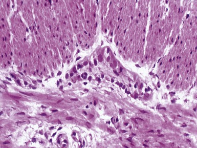
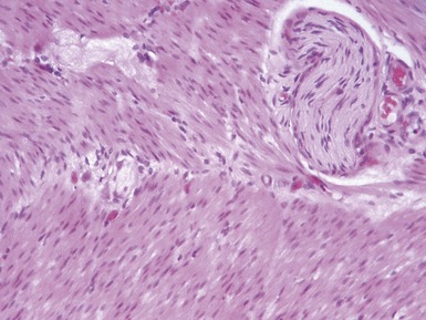
ACHALASIA OF ESOPHAGUS


HYPERTROPHIC PYLORIC STENOSIS


Differential diagnosis of infantile gastric outlet obstruction


HIRSCHSPRUNG’S DISEASE (HSCR)
Genetics


Clinical features


Histopathological features


Primary diagnosis on rectal suction biopsy


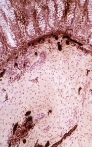
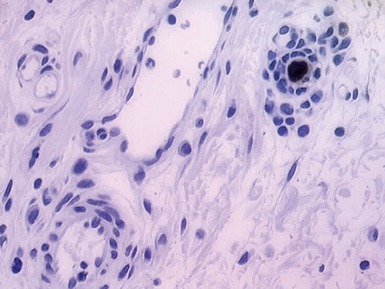
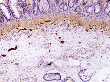
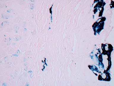
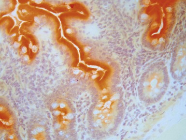
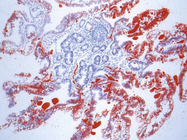
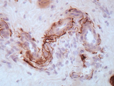
Fig 14.84 Photomicrograph of frozen section of normal rectal suction biopsy stained to display acetylcholinesterase activity demonstrating occasional fine nerves in the muscularis mucosae and lamina propria.
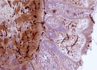
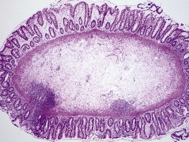
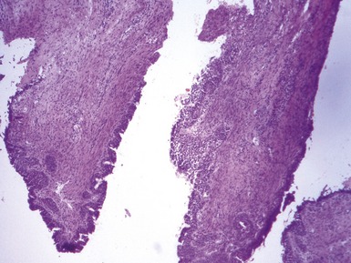
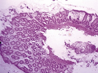
Fig 14.87 Photomicrograph of adequate rectal suction biopsy demonstrating large submucosal surface area.
Urgent


Differential diagnosis and diagnostic pitfalls
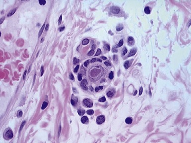
Identification of ganglion cells


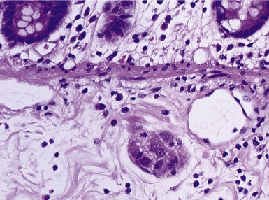
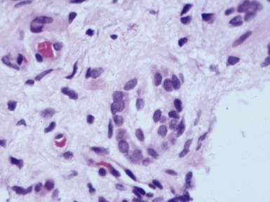
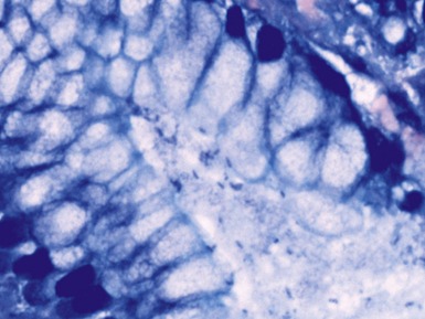
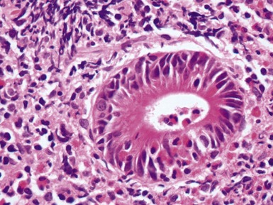
Fig 14.89 Photomicrograph of frozen section of rectal suction biopsy demonstrating normal submucosal ganglion cells. Note cluster of six cells with basophilic cytoplasm and prominent eccentric nucleus. Most do not show a nucleolus, a feature which is typical of neonatal specimens. Note also fine fibrillary neuropil and satellite cell nuclei.