 Evaluate the extent (severity) of the bleeding and assess hemodynamic stability.
 Locate the site of the bleeding:
Hx
 Meds (ASA, steroids, “blood thinners,” NSAIDs)
 Prior GI or vascular surgery
 H/o GI dx or bleeding
 Smoking
 Alcohol intake (gastritis, esophageal varices)
 Sx of PUD
 Associated diseases (CAD, diabetes, HTN, hematologic disorders, renal failure)
 Protracted retching and vomiting (consider gastric or gastroesophageal tear [Mallory-Weiss syndrome])
 Weight loss, anorexia (consider carcinoma)
 Color and character of stool (i.e., hematochezia or melena, constipation or diarrhea)
 Presence or absence of hematemesis
PE
 VS: tachycardia, hypotension, postural changes (orthostatic hypotension). A pulse ↑ >20 bpm or a postural ↑ in systolic BP >10 to 15 mm Hg indicates blood loss >1 L.
 Pts taking β-adrenergic blockers may not demonstrate significant tachycardia w/volume depletion.
 Cardiorespiratory exam: murmurs (↑ incidence of angiodysplasia in pts w/AS), pulmonary rales, JVD to determine rapidity of volume replacement
 Abd exam:
 DRE: Check for masses, strictures, hemorrhoids; test stool for occult blood and inspect it for abnormalities (tarry, blood streaked, bright red, mahogany color).
 Skin: Check for jaundice (liver disease), ecchymoses (coagulation abnormality), cutaneous telangiectasia (Rendu-Osler-Weber disease), buccal pigmentation (Peutz-Jeghers syndrome), and other mucocutaneous changes (Ehlers-Danlos syndrome).
 Look for evidence of metastatic disease (cachexia, firm nodular liver).
 If the pt is not experiencing hematemesis and endoscopy is not immediately available, an NG tube may be placed for gastric lavage while awaiting endoscopy to determine whether the bleeding is emanating from the UGI tract (presence of bright red blood clots or coffee-ground–like guaiac (+) aspirate); however, the sensitivity and specificity of this process are limited. A (−) aspirate does not r/o upper GI bleeding because it could have subsided or the pt could be bleeding from the duodenal bulb w/o reflux into the stomach. Lavage w/500 mL of NS. Failure to clear blood w/gastric lavage indicates persistent bleeding and the need for more urgent endoscopy.
Initial Management
 Stabilize the pt: Insert two large-bore (18-gauge) IV catheters and administer lactated Ringer’s solution or NS; the rate of volume replacement is based on the estimated blood loss, clinical condition, and h/o CVD, including CHF.
 Type and crossmatch for 2 to 8 U of PRBCs, depending on the estimated blood loss, and transfuse as necessary. Aim for Hct >30 for elderly pts w/multiple comorbid conditions and ≥20 for young, healthy individuals.
 Initial labs:
 ECG: R/o myocardial ischemia secondary to severe anemia in patients with risk factors.
Endoscopic EzZvaluation
 EGD is indicated when blood or guaiac (+) coffee ground–like material is obtained from the NG aspirate or if lower endoscopic findings are (−). It should be performed urgently in hemodynamically unstable pts or those found to still be actively bleeding by NG lavage or in those requiring blood transfusion. Otherwise, it should ideally be performed within 24 hr of the hospital admission. In addition, if a therapeutic procedure (e.g., bipolar heater probe, laser cauterization, injection scleroRx, or band ligation) is considered, endoscopy should be done on an emergency basis.
 Colonoscopy should be performed initially if lower GI bleeding is suspected, generally within 24 hr of hospital admission after adequate bowel preparation.
Imaging
 Arteriography can identify briskly bleeding sources. Overall diagnostic sensitivity of arteriography is 41%. Mesenteric arteriography is useful to identify bleeding from AV malformations.
 Radionuclide scans may be used before angiography to determine which pts are bleeding sufficiently to make (+) angiographic result more likely. Bleeding at rates as low as 0.1 mL/min can be detected by radionuclide scans. A (+) “immediate blush” is a good indication for urgent angiography, whereas a (−) “delayed blush” is an indication for observation and elective colonoscopy.
 Technetium-99m (99mTc) pertechnetate scan (Meckel scan) selectively tags acid-secreting cells (gastric mucosa); it is used most often for unexplained bleeding in infants and young adults.
 99mTc-sulfur colloid scan is very sensitive in detecting lesions w/low bleeding rates; its major drawbacks are as follows:
 99mTc-labeled RBC scan: Its major advantage over the sulfur colloid scan is its long duration; it is useful for intermittent bleeding because the pt can be monitored for GI bleeding for 24 to 48 hr. Its disadvantage is that it has a high false-localization rate.
 Selective angiography:
Treatment
 Correct bleeding abnormalities by administering FFP or vitamin K if the pt has a coagulopathy and Plt if the pt is severely thrombocytopenic.
 IV PPIs in cases of probable peptic ulcer or gastritis. After endoscopic Rx of bleeding peptic ulcers, IV PPIs ↓ the risk of recurrent bleeding ↑ pH, ↑ platelet function.
 Octreotide: IV bolus of 50 to 100 μg followed by IV infusion of 25 to 50 μg/hr is useful for acute variceal bleeding. Another useful agent is terlipressin.
 Endoscopy:
 Balloon tamponade is indicated for severe bleeding from esophageal varices if octreotide or other endoscopic Rx modalities are ineffective.
 Radiologic modalities include localized infusion of vasopressin, autologous clots, or foreign coagulating substances (e.g., Gelfoam) in the bleeding vessel during or after arteriography.
 Surgery is indicated at the onset of dx of aortoduodenal fistula, but it is not suggested as the initial Rx in cases of other causes of GI bleeding until a definitive dx is made and other noninvasive modalities are tried. Surgical approach may be necessary in the following situations:
B. Disorders of the Esophagus
1. Dysphagia
a. Oropharyngeal Dysphagia
 Inability to move food from oropharynx via UES → esophagus
 Drooling, postnasal regurgitation, difficulty initiating swallowing, sialorrhea, sensation of food stuck in the neck, coughing/choking during swallowing, dysphonia, and dysarthria
Diagnosis
 First test = modified barium swallow videofluoroscopy, then fiberoptic flexible nasopharyngeal laryngoscopy
b. Esophageal Dysphagia
 Inability to move food from esophagus → stomach
 Dysphagia to solids suggests mechanical obstruction.
 Neuromuscular causes result in dysphagia to both solids and liquids.
 Sx intermittent in pts with esophageal dysphagia from benign causes of structural obstruction or diffuse esophageal spasm; sx progressive in pts with peptic stricture, esophageal carcinoma, scleroderma, and achalasia
 Luminal diameter >18 to 20 mm (rarely sx); diameter <13 mm (sx)
Diagnosis
 Esophageal dysphagia: first test = barium esophagography, then EGD
Treatment
 Goal is airway protection and nutrition maintenance.
 Consider consultation with ENT, head and neck surgeon, radiologist, speech pathologist, physical therapist, dietitian, gastroenterologist, physical medicine and rehabilitation specialist, dentist, neurologist, etc., because nursing home pts with oropharyngeal dysphagia and hx of aspiration have 45% mortality rate over 1 yr
2. Esophageal Motility Disorders
 Table 6-1 compares esophageal motor disorders.
TABLE 6-1
Esophageal Motor Disorders
| Achalasia | Scleroderma | Diffuse Esophageal Spasm | |
| Symptoms | Dysphagia Regurgitation of nonacidic material |
Gastroesophageal reflux disease Dysphagia |
Substernal chest pain (angina-like) Dysphagia with pain |
| Radiographic appearance | Dilated, fluid-filled esophagus Distal bird-beak stricture |
Aperistaltic esophagus Free reflux Peptic stricture |
Simultaneous noncoordinated contractions |
| Manometric Findings | |||
| Lower esophageal sphincter | High resting pressure Incomplete or abnormal relaxation with swallow |
Low resting pressure | Normal pressure |
| Body | Low-amplitude, simultaneous contractions after swallowing | Low-amplitude peristaltic contractions or no peristalsis | Some peristalsis Diffuse and simultaneous nonperistaltic contractions, occasionally high amplitude |

From Andreoli, T E, Benjamin IJ, Griggs RC, Wing EJ: Andreoli and Carpenter’s Cecil Essentials of Medicine, 8th ed. Philadelphia, Saunders, 2010.
3. GERD
 Motility disorder caused by the reflux of gastric contents into the esophagus
Etiology
 Incompetent LES
 Medications ↓ LES pressure (CCBs, β-blockers, theophylline, anti-AChs)
 Foods ↓ LES pressure (chocolate, yellow onions, peppermint)
 Tobacco abuse, alcohol, coffee
 Pregnancy
 Gastric acid hypersecretion
 Hiatal hernia (present in 70% w/GERD ); however, most w/hiatal hernia asx
Diagnosis
H&P
 Heartburn, dysphagia, sour taste, regurgitation of gastric contents into mouth, chronic cough/bronchospasm, chest pain, laryngitis, early satiety, abd fullness and bloating w/belching, dental erosions in children
Additional Testing
 EGD = document type/extent tissue damage; r/o potential malignancy (Barrett’s esophagus)
 24-hr esophageal pH monitoring: generally not done; useful in atypical manifestations of GERD (chest pain and chronic cough)
 Esophageal manometry: useful in refractory reflux pt w/surgical Rx planned
 Upper GI series: identify ulcerations/strictures; may miss mucosal abnormalities. Only one third of pts w/GERD have radiographic signs of esophagitis.
Treatment
 Lifestyle change (wt loss, ↓ fat intake)/avoidance of exacerbating factors: EtOH, tobacco, citrus/tomato–based products, caffeine, β-blockers, CCBs, α-agonists, theophylline
 ↑ Head of bed 4 to 8 in. Avoid lying supine directly after late/large meals.
 Avoid wearing clothing that is tight around the waist.
 PPIs: preferred Rx (H2 blockers less effective)
 Antacids: relief of mild sx; ineffective in severe cases
 Prokinetic agents (metoclopramide): indicated if PPIs not fully effective. May be used in combination Rx; however, side effects limit use.
 Nissen fundoplication (refractory cases)
 Endoscopic radiofrequency heating of GE-jxn (Stretta procedure): pts unresponsive to traditional Rx
4. Barrett’s Esophagus
 Squamous lining of lower esophagus replaced by intestinalized metaplastic columnar epithelium; predisposing to neoplasia
H&P

Diagnosis
 EGD with biopsy
Treatment
 Control GERD sx; maintain healed mucosa via PPI
Monitoring
 Relative risk of adenocarcinoma is 11.3 compared with general population.
 Pts should undergo surveillance EGD and systematic four-quadrant biopsy at intervals determined by the presence and grade of dysplasia.
 Pts who have had two consecutive EGDs showing no dysplasia should have follow-up every 3 to 5 yr.
 Pts with low-grade dysplasia should have extensive mucosal sampling within 6 mo and follow-up every 6 to 12 mo.
 Pts with high-grade dysplasia should have expert confirmation and extensive mucosal sampling. High-grade dysplasia with visible mucosal irregularities should be removed by endoscopic mucosal resection.
 Consider intensive surveillance every 3 mo for patients with focal high-grade dysplasia. Patients with multifocal high-grade dysplasia or carcinoma should be considered for resection or ablation if not an operative candidate.
5. Esophageal Tumors
 15% in proximal third esophagus, 50% in middle third, 35% in lower third
 Risk factors: EtOH, smoking, achalasia (7× > risk), chronic GERD, HPV (types 16, 18), obesity/hiatal hernia/low-vitamin high-fat diet, ingested carcinogens (nitrates, smoked opiates, fungal toxins [pickled vegetables], betel nut chewing), mucosal damage (long-term exposure tea >70° C, lye ingestion), radiation-induced stricture
H&P
 Dysphagia (74%): initially with solid foods; gradually → semisolids/liquids
 Unintentional weight loss; losing >10% of body mass = poor outcome
 Hoarseness (recurrent laryngeal nerve involvement)
 Cervical adenopathy; usually supraclavicular lymph nodes
Diagnosis
 EGD
EGD Endoscopic U/S: locoregional staging (depth of invasion/lymph assessment)
 Chest/abd CT and/or integrated CT-PET scans (tumor spread for preop staging)
 Staging laparoscopy may alter Rx plans (20%-30% cases) by more accurately staging regional lymph nodes/detecting occult peritoneal mets.
Treatment
 Surgical resection of squamous cell carcinoma and adenocarcinoma of lower esophageal third indicated for local, resectable disease in the absence of widespread mets detected by CT-PET. Gastric pull-through/colonic interposition is typically used to provide luminal continuity.
 Squamous cell carcinoma is more radiosensitive than adenocarcinoma; used as palliative monoRx of obstructive sx in unresectable/advanced cases.
 Palliative radiation Rx for bone mets
 Preoperative chemoradioRx + surgery in late stage I (T2,3N0), stage II or III ↑ tumoricidal effects
 5-yr survival 13% (37.3% [local], 18.4% [regional], 3.1% [distant] disease)
C. Disorders of Stomach and Duodenum
1. Peptic Ulcer Disease
Etiology/Epidemiology
 H. pylori infection (70%-90% duodenal ulcers)
 NSAIDs (40%-50% gastric ulcers)
 Cigarette smoking, EtOH
 Neoplasia: gastrinoma (ZE syndrome), carcinoid, mastocytosis
Diagnosis
 EGD (preferred), UGI barium studies
Treatment

 Lifestyle change: d/c smoking/EtOH.
 Add PPI to ↓ acid secretions.
TABLE 6-2
Overview of Antibiotics Used for Helicobacter Pylori Eradication
| Drug Class | Drug | Triple Therapy∗ Dose | Quadruple Therapy† Dose | Sequential Therapy‡ Dose |
| Acid suppression | Proton pump inhibitor | 20-40 mg bid§ | 20-40 mg bid§ | 20-40 mg bid§ |
| Standard antimicrobials | Bismuth compound|| | 2 tablets bid | 2 tablets bid | |
| Amoxicillin | 1 g bid | 1 g bid | ||
| Metronidazole¶ | 500 mg bid | 500 mg tid | 500 mg bid | |
| Clarithromycin | 500 mg bid | 500 mg bid | ||
| Tetracycline | 500 mg qid | |||
| Salvage antimicrobials | Levofloxacin | 300 mg bid | 300 mg bid | |
| Rifabutin | 150 mg bid | |||
| Furazolidone | 100 mg bid | |||
| Doxycycline | 100 mg bid | |||
| Nitazoxanide | 1 g bid |

∗ Triple therapy consists of a proton pump inhibitor or bismuth compound, together with two of the listed antibiotics, usually given for 7-14 days.
† Quadruple therapy consists of a proton pump inhibitor plus either the combination of a bismuth compound, metronidazole, and tetracycline given for 4-10 days, or the combination of levofloxacin, doxycycline, and nitazoxanide for 10 days.
‡ Sequential therapy consists of 10 days of proton pump inhibitor treatment, plus amoxicillin during days 1-5 and a combination of clarithromycin and an imidazole (when available, tinidazole; otherwise, metronidazole) during days 6-10.
§ Proton pump inhibitor dose equivalent to omeprazole 20 mg bid.
|| Bismuth subsalicylate or subcitrate.
¶ An alternative is tinidazole 500 mg bid.
From Goldman L, Schafer AI (eds): Goldman’s Cecil Medicine, 24th ed. Philadelphia, Saunders, Elsevier, 2012.
2. Gastroparesis
 Sx impaired gastric emptying in absence of mechanical obstruction
Differential Diagnosis and Clinical Pearls
 Diabetes mellitus gastroparesis (HBA1c, fasting glucose)
 Gastric surgery
 Pregnancy
 Hypothyroidism (TSH)
 Cannabinoid hyperemesis syndrome (hx of use; hx of relief of N/V with hot baths/showers)
 Rumination syndrome (hx of passive regurgitation of pleasant tasting food w/o nausea)
Management
 Prokinetic agents (e.g., metoclopramide)
 Refractory cases (↑ risk malnutrition), gastroduodenal manometry to distinguish myopathic from neuropathic process
3. Gastric Cancer
 Adenocarcinoma; mostly antral (35%)
 Male-to-female ratio 3:2
 Familiar diffuse gastric cancer (autosomal dominant, mutation E-cadherin gene CDH1 = cancer at young age)
Physical Exam and Labs
 Wt loss (70%-80%), N/V (20%-40%), dysphagia (20%)
 Dyspepsia (unrelieved by antacids, worse w/food), epigastric/abd mass (40%)
 Microcytic anemia, hemoccult (+) stools
 Hypoalbuminemia
Diagnosis
 Upper endoscopy with biopsy (staging via abd CT scan ± lymph node dissection)
D. Disorders of the Pancreas
1. Acute Pancreatitis
 Inflammatory process w/intrapancreatic enzyme activation possibly also involving peripancreatic tissue/remote organ systems
Etiology
 >90% of cases: biliary tract disease (calculi or sludge) or EtOH
 Drugs: thiazides, furosemide, corticosteroids, tetracycline, estrogens, valproic acid, metronidazole, azathioprine, methyldopa, pentamidine, ethacrynic acid, procainamide, sulindac, nitrofurantoin, ACEIs, danazol, cimetidine, piroxicam, gold, ranitidine, sulfasalazine, isoniazid, acetaminophen, cisplatin, opiates, erythromycin
 Abd trauma, surgery, ERCP, viral infections, PUD, pancreas divisum (congenital failure to fuse of dorsal or ventral pancreas), pregnancy, vascular (vasculitis, ischemic), hypolipoproteinemia (types I, IV, and V), hypercalcemia, pancreatic carcinoma (primary/mets), renal failure, hereditary pancreatitis, occupational exposure (methanol, cobalt, zinc, mercuric chloride, creosol, lead, organophosphates, chlorinated naphthalenes)
 Others: scorpion bite, obstruction at ampulla region (neoplasm, duodenal diverticula, Crohn’s disease), hypotensive shock, autoimmune pancreatitis
Scoring System
 Table 6-3 describes various scoring systems to assess severity of acute pancreatitis.
TABLE 6-3
Scoring Systems to Assess Severity of Acute Pancreatitis
| System | Criteria |
| Ranson | At admission Age >55 yr WBC >16,000/μL Glucose >200 mg/dL LDH >350 IU/L AST >250 IU/L Within next 48 hr Decrease in hematocrit by >10% Estimated fluid sequestration of >6 L Serum calcium <8.0 mg/dL Pao2 <60 mm Hg BUN increase >5 mg/dL after hydration Base deficit >4 mmol/L |
| APACHE-II | Multiple clinical and laboratory factors. Calculator available at www.mdcalc.com/apache-ii-score-for-icu-mortality |
| BISAP | BUN >25 mg/dL Impaired mental status Presence of SIRS Age >60 yr Pleural effusion |
| CT | A: Normal pancreas B: Focal or diffuse enlargement of pancreas C: Grade B plus pancreatic and/or peripancreatic inflammation D: Grade C plus a single fluid collection E: Grade C plus two or more fluid collections or gas in pancreas |
| CT severity index | CT grade A = 0 B = 1 C = 2 D = 3 E = 4 Plus necrosis grade No necrosis = 0 <30% necrosis = 2 30-50% necrosis = 4 >50% necrosis = 6 |
APACHE-II, Acute Physiology and Chronic Health Evaluation II; BISAP, bedside index of severity in acute pancreatitis.
From Goldman L, Schafer AI (eds): Goldman’s Cecil Medicine, 24th ed. Philadelphia, Saunders, Elsevier, 2012.
“Severe Acute Pancreatitis”
Diagnosis
H&P
 Fever, epigastric tenderness/guarding; sudden severe pain (peak intensity 10-30 min; lasting several hr w/o relief)
 Hypoactive bowel sounds (secondary to ileus)
 Tachycardia, shock (secondary to ↓ intravascular volume)
 Confusion (secondary to metabolic disturbances)
 ↓ Breath sounds (atelectasis, pleural effusions, ARDS)
 Jaundice (secondary to obstruction or compression of biliary tract)
 Ascites (secondary to tear in pancreatic duct, leaking pseudocyst)
 Palpable abd mass (pseudocyst, phlegmon, abscess, carcinoma)
 Hypocalcemia (Chvostek’s sign, Trousseau’s sign)
 Intra-abd bleeding (hemorrhagic pancreatitis):
 Tender SC nodules (SC fat necrosis)
Labs
 ↑ Serum amylase (initial 3-5 days), ↑ serum lipase, ↑ serum trypsin
 Rapid urinary trypsinogen-2; useful screening test in pts w/abd pain; (−) dipstick r/o acute pancreatitis w/high degree of probability; (+) test result indicates need for further evaluation.
 CBC: leukocytosis; ↑ Hct (secondary to hemoconcentration); ↓ Hct may indicate hemorrhage/hemolysis.
 ↑ BUN (secondary to dehydration)
 ↑ Serum glucose; in previously nl pt correlates w/pancreatic malfunction
 ↑ AST/LDH (tissue necrosis); ↑ bili/alk phos (CBD obstruction); ≥3 ↑ ALT = biliary pancreatitis (95% probability)
 ↓ Serum Ca (saponification, precipitation, and ↓ PTH response)
 ABGs: Pao2 may be ↓ secondary to ARDS, pleural effusions; pH may be ↓ secondary to lactic acidosis, respiratory acidosis, and renal insufficiency.
 Serum electrolytes: K+ may be ↑ secondary to acidosis/renal insufficiency; Na+ may be ↑ secondary to dehydration.
Imaging
 Abd plain film: r/o perforated viscus; may reveal localized ileus (sentinel loop), pancreatic calcifications (chronic pancreatitis), blurring of left psoas shadow, dilation of transverse colon, calcified gallstones
 CXR: elevation of one or both diaphragms, pleural effusions, basilar infiltrates, platelike atelectasis
 Abd U/S: gallstones (sensitivity 60%-70%), pancreatic pseudocysts; limited in presence of distended bowel loops overlying pancreas
 CT abd: superior to U/S in dx extent; also able dx pseudocysts (well-defined area surrounded by high-density capsule); GI fistulation or infection of a pseudocyst (gas within pseudocyst)
 Contrast-enhanced CT (pancreatic necrosis): severity graded by CT scan (see Table 6-3)
 MRCP: useful if surgical procedure not anticipated
 ERCP: avoided during acute stage, unless to remove impacted stone
Treatment
General Measures
 Maintain intravascular volume (vigorous IV hydration).
 NPO until clinically improved, stable, and hungry; enteral feedings preferred to TPN; PN necessary if unable to tolerate enteral/adequate infusion rate cannot be reached within 2 to 4 days.
 NG suction is used to decompress abd in pts w/ileus.
 Control pain with IV morphine or fentanyl.
Specific Measures
 Pancreatic/peripancreatic infection in 40% to 70% pts w/pancreatic necrosis; prophylactic IV abx (5-7 days) justified if septicemia, pancreatic abscess, or pancreatitis secondary to biliary calculi. Cover Bacteroides fragilis/anaerobes (cefotetan, metronidazole, clindamycin, + AG) and enterococcus (ampicillin).
 Surgical Rx indicated: gallstone-induced pancreatitis (cholecystectomy when acute phase subsides), perforated peptic ulcer, excision/drainage necrotic/infected foci w/placement of wide-bore drains for continuous postop irrigation
Complications
 Pseudocyst (dx: CT scan or U/S) Rx: CT scan or U/S-guided percutaneous drainage w/pigtail catheter for continuous drainage (↑ recurrence rate); conservative approach to reevaluate (w/CT scan or U/S) after 6 to 7 wk and surgically drain if no ↓ in size. Pseudocysts <5 cm generally reabsorbed w/o intervention; those >5 cm require surgery after wall maturation.
 Phlegmon (dx: CT scan or U/S) Rx: supportive care
 Pancreatic abscess (dx: CT scan-retroperitoneal bubbles, Gram staining and cultures of fluid from percutaneous biopsy) Rx: surgical/catheter drainage + IV abx (imipenem-cilastatin)
 Pancreatic ascites (dx: paracentesis-amylase/lipase level in fluid, ERCP) Rx: surgery if exudative/does not resolve spontaneously
 GI bleeding: via EtOH gastritis, varices, stress ulcer, or DIC
 Renal failure: via hypovolemia (oliguria or anuria), cortical or tubular necrosis (shock, DIC), or thrombosis of renal artery or vein
 Hypoxia: via ARDS, pleural effusion, or atelectasis
2. Chronic Pancreatitis
 Recurrent/persistent inflammatory process characterized by chronic pain and pancreatic exocrine/endocrine insufficiency
Etiology
 Chronic EtOH, obstruction (ampullary stenosis, tumor, trauma, pancreas divisum, annular pancreas), hereditary pancreatitis, severe malnutrition, untreated hyperparathyroidism (hypercalcemia), mutations of cystic fibrosis transmembrane conductance regulator (CFTR) gene (TF genotype)
 Autoimmune (sclerosing) pancreatitis (5% cases): manifests w/jaundice (63%) + abd pain (35%). CT = diffusely enlarged pancreas, enhanced peripheral rim of hypoattenuation “halo,” and low-attenuation mass in head of pancreas. Labs = ↑ serum IgG4, serum Ig or γ -globulin level, + antilactoferrin Ab, anti–carbonic anhydrase II level, ASMA, or ANA.
Diagnosis
H&P
 Persistent/recurrent epigastric + LUQ pain, may radiate to the back
 Tenderness over the pancreas, muscle guarding
 Significant weight loss, epigastric mass (10%), jaundice (5%-10%)
 Bulky/greasy, foul-smelling stools
Labs
 ↑/Nl serum amylase and lipase
 ↑ Glucose, bili, alk phos, glycosuria
 72-hr fecal fat determination (rarely performed) = excess fecal fat. Fecal elastase test requires 20 g of stool.
 Secretin stimulation test (dx pancreatic exocrine insufficiency)
 Lipid panel: ↑↑ TGs can cause pancreatitis.
 Serum Ca: hyperparathyroidism (rare cause of chronic pancreatitis)
 ↑ Serum IgG4 (sclerosing pancreatitis and autoimmune pancreatitis)
 ↑ Serum Ig or γ-globulin level, antilactoferrin Ab, anti–carbonic anhydrase II level, ASMA, or ANA in autoimmune pancreatitis
Imaging
 Plain abd radiographs: may reveal pancreatic calcifications (95% spec)
 U/S abd: duct dilation, pseudocyst, calcification, presence of ascites
 Contrast-enhanced abd CT scan: calcifications, evaluate ductal dilation, r/o pancreatic cancer
 EUS (97% sens, 60% spec)
 FNAB combined w/EUS = preferred evaluation of modality to r/o malignant cystic/mass lesions
Treatment
 Steatorrhea Rx w/pancreatic supplements (e.g., pancrease, pancrelipase [Creon] PRN on basis of steatorrhea/weight loss)
 Glucocorticoids (autoimmune pancreatitis)
 Surgical intervention if duct obstruction
 Transduodenal sphincteroplasty/pancreaticojejunostomy if intractable pain
Clinical Pearls
 50% of pts die within 10 yr of chronic pancreatitis or malignant neoplasm.
3. Pancreatic Adenocarcinoma
Risk Factors
 Smoking, chronic EtOH, genetics (5%-10% pts have family hx), dipeptidyl peptidase-4 inhibitors, incretin mimetics
Diagnosis
 Labs: ↑ alk phos, bili, amylase
H&P
 Jaundice, abd pain (dull upper abd pain/vague abd discomfort), wt loss
Imaging
 Multidetector helical CT with IV contrast (imaging procedure of choice)
 Endoscopic ultrasonography: useful if no identifiable mass on CT + ↑ clinical suspicion
Treatment
Surgery
 Curative cephalic pancreatoduodenectomy (Whipple’s procedure) in 10% to 20% pts whose lesion <5 cm, solitary, and without metastases. Surgical mortality rate is 5%.
 Palliative surgery (for biliary decompression/diversion)
 Palliative therapeutic ERCP with stents
 Celiac plexus block = pain relief in 80% to 90% of cases
ChemoRx
 Gemcitabine given alone or + platinum agent (erlotinib or fluoropyrimidine)
 Combination consisting oxaliplatin, irinotecan, fluorouracil, and leucovorin (Folfirinox)
Radiation
 External-beam radiation for palliation of pain
4. Neuroendocrine Pancreatic Neoplasms
a. Gastrinoma
 ZE syndrome: hypergastrinemic state via pancreatic/extrapancreatic non–β islet cell tumor (gastrinoma) resulting in peptic ulcer disease
 2/3 gastrinomas (sporadic), 60% assoc (MEN-1; AD including hyperparathyroidism, pituitary tumors)
 60% gastrinomas = malignant (mets to liver, regional lymph nodes)
 Neuroendocrine tumors = 1.3% of all cases of pancreatic cancer
Diagnosis
 Gastric acid secretion: serum gastrin level (fasting) >1000 pg/mL
 Provocative gastrin level tests:
 Gastrinoma localization via arteriography, abd U/S or CT scan or MRI
H&P
 95% sx of peptic ulcer, 60% sx related to GERD, 33% diarrhea, steatorrhea
Treatment
 Surgical resection; total gastrectomy/vagotomy (palliative in some pts)
 Medical Rx: PPIs, somatostatin or octreotide, chemo (mets)
b Insulinoma
Diagnosis
H&P
 Sx typically in AM before meal; fasting hypoglycemia versus reactive hypoglycemia (which is not commonly associated w/insulinoma)
Labs
 Overnight fasting blood sugar level + simultaneous plasma insulin, proinsulin, and/or C peptide level will establish existence of fasting organic hypoglycemia in 60% of pts.
 Plasma proinsulin, C-peptide, antibodies to insulin, and plasma sulfonylurea levels to r/o factitious insulin use/hypoglycemic agents/autoantibodies against the insulin receptor or insulin. Refer to Table 5-5 in Chapter 5.
Imaging
 Abdominal CT scan or MRI
 Octreotide scan
Treatment
 Enucleation of single insulinoma
 Partial pancreatectomy for multiple adenomas
E. Disorders of Small and Large Bowel
1. Diarrhea
 ↑ Frequency (>200 g/24 hr) of stool w/ ↓ consistency compared with baseline; if lasting >3 wk = chronic diarrhea
Diagnosis
Hx
 Travel hx (traveler’s diarrhea)
 Short duration (1-3 days) assoc w/mild sx usually viral (rotavirus, Norwalk virus); >3 wk probably not bacterial or viral
 Nocturnal diarrhea (common w/diabetic neuropathy)
 Onset within minutes: scombroid poisoning (tuna, mahi-mahi, mackerel) (N/V, flushing, diarrhea)
 Onset within hr: toxins (Staphylococcus aureus, toxigenic Escherichia coli, Clostridium perfringens, Bacillus cereus, Vibrio parahaemolyticus, [barracuda, grouper, red snapper: ciguatera toxin, causing paresthesia, weakness])
 Diarrhea secondary to Salmonella, Shigella, Campylobacter, Yersinia = longer incubation period.
 Stress: “functional” diarrhea, IBS
 Diarrhea alternating w/constipation: IBS
 Foods containing sorbitol/mannitol (osmotic diarrhea), fried rice (B. cereus), undercooked hamburger (E. coli 157:H7), poultry, eggs (Campylobacter, Salmonella, S. aureus), diarrhea after dairy ingestion (lactose intolerance)
 Shellfish ingestion (Norwalk virus, Vibrio cholerae, Vibrio mimicus, V. parahaemolyticus, Plesiomonas shigelloides)
 Long-distance runners (bloody diarrhea secondary to bowel ischemia)
 Daycare centers (rotavirus, Giardia, Salmonella, Shigella, Cryptosporidium, Campylobacter)
 Medications: (common agents: Mg-containing antacids, misoprostol, PPIs, methylxanthines [caffeine, theophylline], laxatives, lactulose, colchicine, antiarrhythmic agents (quinidine, digitalis, propranolol), metformin, thyroxine). Abx-induced pseudomembranous colitis should be suspected in any pt receiving abx: (+) Clostridium difficile toxin w/the stool assay, cytotoxin test.
 Sexual habits: male homosexuals ↑ incidence of (Giardia, E. histolytica, Cryptosporidium, Salmonella, Neisseria gonorrhoeae, Campylobacter).
 Relevant medical hx
 Associated sx
 Characteristics of the stool (from pt’s hx)
PE
 Rectal fistulas, RLQ abd mass (Crohn’s disease)
 Arthritis, iritis, uveitis, erythema nodosum (IBD)
 Abd masses (neoplasms of colon, pancreas, or liver; diverticular abscess [LLQ mass], IBD)
 Flushing, bronchospasm (carcinoid syndrome)
 Buccal pigmentation (Peutz-Jeghers syndrome)
 Pigmentation (Addison’s disease)
 Ammoniac/urinary breath odor (renal failure)
 Ecchymosis (vitamin K deficiency secondary to malabsorption, fat-soluble vitamins, celiac)
 Fever (IBD, infectious diarrhea, lymphoma)
 Goiter, tremor, tachycardia (hyperthyroidism)
 Lymphadenopathy (neoplasm, lymphoma, tuberculosis, AIDS, Whipple’s disease)
 Macroglossia (amyloidosis)
 Kaposi’s sarcoma (AIDS)
Initial Evaluation
 Labs (may not be necessary in pts not appearing ill/dehydrated)
 Abd x-rays indicated if abd pain or evidence of obstruction (r/o toxic megacolon, bowel ischemia; pancreatic calcifications = pancreatic insufficiency)
Treatment
 NPO, IV hydration, electrolyte abnl correction, d/c possible causative agents (antacids containing Mg, abx)
 Antiperistaltic agents (diphenoxylate) used w/caution in pts suspected of IBD or infectious diarrhea; loperamide/bismuth subsalicylate helpful in mild cases
 Persistent diarrhea + bacterial/parasitic organism ID, start abx:
 IBS Rx: psyllium (fiber products) + ↓ caffeine, chocolate, EtOH, stress; antispasmodics (dicyclomine, hyoscyamine) if resistant cases
Evaluation of Pt w/Chronic or Recurrent Diarrhea
Etiology
 Drug induced (including laxative abuse), IBS, lactose intolerance, IBD, malabsorption (mucosal/pancreatic insufficiency, bacterial overgrowth), parasitic infections (giardiasis, amebiasis), functional diarrhea, postsurgical (partial gastrectomy, ileal resection, cholecystectomy)
 Endocrine disturbances: DM (↓ sympathetic input to the gut), hyperthyroidism, Addison’s disease, gastrinoma (ZE syndrome), VIPoma (pancreatic cholera), carcinoid tumors (serotonin), medullary thyroid carcinoma (calcitonin)
 Pelvic irradiation, colonic carcinoma (villous adenoma)
 Collagenous colitis (middle-aged woman, nl endoscopy, subepithelial acellular collagen band on sigmoid bx or right colon; sx resolution w/sulfasalazine alone or steroid combination)
 Lymphocytic colitis (lymphocytic infiltration w/o collagen band)
Diagnosis
 H&P/initial labs same as new-onset diarrhea; additional labs:
 Secretory diarrhea from impaired absorption/excessive secretion of electrolytes via enteric infection, neoplasms of exocrine pancreas (VIP, GIP, secretin, glucagon), bile salt enteropathy, villous adenoma, IBD, carcinoid tumor, celiac, cathartic agent ingestion
 Osmotic diarrhea from impaired water absorption secondary to osmotic effect of nonabsorbable intraluminal molecules via lactose and other disaccharide excess, pancreatic insufficiency, lactulose/sorbitol/sodium sulfate/antacid induced, postop (gastrojejunostomy, vagotomy, pyloroplasty, intestinal resection)
 Dx “osmotic gap” in stool analysis = measured osmolality –2([Na+] + [K+])
 Difference between calculated and actual Osm >50 = osmotic diarrhea
2. Constipation
 DDx: metabolic (hypercalcemia, DM, pregnancy, hypothyroidism), structural (colorectal cancer, strictures, hernias, adhesions, endometriosis, rectocele, diverticular disease, volvulus, intussusception, IBD, hematoma of bowel wall secondary to trauma or anticoagulants), drug induced (opiates, anti-Ach, iron, CCB, antipsychotics, antacids w/aluminum, verapamil), neurologic/psychological (IBS, anorexia, depression, MS), neonatal (Hirschsprung’s disease, meconium ileus, atresia), insufficient bulk in diet, travel, old age, spinal cord injury
Treatment
 Fiber (25-30 g/day) and fluid intake essential
 Physiologic testing (DRE, anorectal manometry, rectal balloon expulsion colonic transit) provides insight/possible indication for MR defecography
 Pts >50 yr old or have alarm sx (wt loss, rectal bleeding, change in bowel habits, FHx colon cancer) should undergo colonoscopy
3. Malabsorption

Differential Diagnosis
 Celiac disease (diarrhea, bloating, wt loss)
 Lactose malabsorption (osmotic diarrhea, bloating, excess flatus)
 Short-bowel syndrome (<200 cm of small bowel, nl 600 cm) from Crohn’s disease, ischemia, volvulus, desmoid tumors, trauma
 Small intestinal bacterial overgrowth: bacterial overgrowth from change in colonic flora via dysmotility (scleroderma, amyloidosis), altered secretion/anatomy
4. Celiac Disease
 Chronic disease characterized by malabsorption and diarrhea precipitated by ingestion of food products containing gluten. Celiac sprue is considered an autoimmune-type disease, with TTG suggested as a major autoantigen. It results from an inappropriate T-cell–mediated immune response against ingested gluten in genetically predisposed individuals who carry either HLA-DQ2 or HLA-DQ8 genes. There is sensitivity to gliadin, a protein fraction of gluten found in wheat, rye, and barley. In patients with celiac disease, immune responses to gliadin fractions promote an inflammatory reaction, mainly in the upper small intestine, manifested by infiltration of the lamina propria and the epithelium with chronic inflammatory cells and villous atrophy.
Diagnosis
H&P
 Weight loss, pallor (from anemia), dyspepsia, short stature, and failure to thrive in children and infants
 Weight loss, fatigue, pallor, and diarrhea in adults
 Angular cheilitis, aphthous ulcers, atopic dermatitis, and dermatitis herpetiformis frequently associated w/celiac disease
Labs
 IgA TTG antibody by enzyme-linked immunosorbent assay (TTG test) is the best screening serologic test for celiac disease.
 Iron deficiency anemia (microcytic anemia, ↓ ferritin level)
 Folic acid deficiency
 Serum IfA (small % of patients are IfA deficient)
 Vitamin B12 deficiency, ↓ Mg, ↓ Ca
Treatment
 Gluten-free diet (avoidance of wheat, rye, and barley). Safe grains (gluten free) include rice, corn, oats, buckwheat, millet, amaranth, quinoa.
 Correct nutritional deficiencies w/iron, folic acid, Ca, vitamin B12 as needed.
 Prednisone 20-60 mg qd gradually tapered is useful in refractory cases.
Clinical Pearls
 Celiac disease should be considered in pts w/unexplained metabolic bone disease or hypocalcemia, especially because GI sx may be absent or mild. Clinicians should also consider testing children and young adults for celiac disease if unexplained weight loss, abd pain or distention, and chronic diarrhea are present.
 Pts w/celiac disease have an overall risk of cancer that is almost 2× that in the general population: risk of adenocarcinoma of the small intestine and non-Hodgkin’s lymphoma, especially of T-cell type and primarily localized in the gut.
 Screening for celiac disease is recommended in first-degree relatives. It should also be considered in type 1 DM and autoimmune disorders such as PBC, primary sclerosing cholangitis, autoimmune hepatitis, IBD, thyroid disease (hypothyroidism occurs in up to 15% of patients with celiac disease), SLE, RA, and Sjögren’s syndrome because of the increased risk of celiac disease in these populations. Screening persons with Down’s syndrome or Turner’s syndrome for celiac disease has also been recommended.
5. Inflammatory Bowel Disease
a. Crohn’s Disease
 Inflammatory disease most commonly involving the terminal ileum. Table 6-4 compares Crohn’s disease with ulcerative colitis.
TABLE 6-4
Differentiating Features of Ulcerative Colitis and Crohn’s Disease
| Ulcerative Colitis | Crohn’s Disease | |
| Site of involvement | Involves only colon Rectum almost always involved |
Any area of the gastrointestinal tract Rectum usually spared |
| Pattern of involvement | Continuous | Skip lesions |
| Diarrhea | Bloody | Usually nonbloody |
| Severe abdominal pain | Rare | Frequent |
| Perianal disease | No | In 30% of patients |
| Fistula | No | Yes |
| Endoscopic findings | Erythematous and friable Superficial ulceration | Aphthoid and deep ulcers Cobblestoning |
| Radiologic findings | Tubular appearance resulting from loss of haustral folds | String sign of terminal ileum RLQ mass, fistulas, abscesses |
| Histologic features | Mucosa only Crypt abscesses |
Transmural Crypt abscesses, granulomas (about 30%) |
| Smoking | Protective | Worsens course |
| Serology | p-ANCA more common | ASCA more common |
From Andreoli, T E, Benjamin IJ, Griggs RC, Wing EJ: Andreoli and Carpenter’s Cecil Essentials of Medicine, 8th ed. Philadelphia, Saunders, 2010.
Diagnosis
H&P
 Sx intermittent w/episodic remission:
Labs
 ↓ Hgb/Hct (chronic blood loss, bone marrow inflammation, and vitamin B12 malabsorption)
 ↓ K, Mg, Ca, alb in pts w/chronic diarrhea
 Vitamin B12 and folate deficiency
 ↑ ESR, (+) ASCA, (−) ANCA
Imaging
 Endoscopy: transmural asymmetric/discontinued disease w/deep longitudinal fissures (“cobblestone appearance via submusocal inflammation”), strictures, crypt distortion + inflammation, possible noncaseating granulomas + lymphoid aggregates on bx
 Barium imaging (rarely indicated): deep ulcerations (often longitudinal/transverse) and segmental lesions (skip lesions, strictures, fistulas); “thumbprinting” is common; “string sign” in terminal ileum
 CT abd: helpful in identifying abscesses/complications
 See Table 6-4.
Treatment
 Figure 6-1 describes a treatment algorithm for Crohn’s disease.
b. Ulcerative Colitis
Diagnosis
H&P
 Abd distention and tenderness
 Bloody diarrhea, fever, evidence of dehydration
 Extraintestinal manifestations (25% pts): liver disease, sclerosing cholangitis, iritis, uveitis, episcleritis, arthritis, erythema nodosum, pyoderma gangrenosum, aphthous stomatitis
Labs
 Anemia, ↑ ESR
 ↓ K, Mg, Ca, alb
 If persistent diarrhea: Consider stool exam O&P, stool culture, and testing for C. difficile toxin.
 (−) ASCA (anti-Saccharomyces cerevisiae Ab), (+) p-ANCA (>45% pts; p-ANCA assoc w/relative resistance to medical Rx)
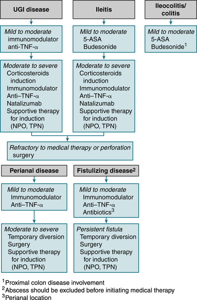
FIGURE 6-1 Crohn’s disease treatment algorithm. (From Goldman L, Schafer AI [eds]: Goldman’s Cecil Medicine, 24th ed. Philadelphia, Saunders, 2012.)
Imaging
 Generally not indicated; double-contrast barium enema w/small bowel follow-through used if colonic strictures prevent evaluation, may reveal continuous involvement (including rectum), pseudopolyps, ↓ mucosal pattern, and fine superficial ulcerations.
Treatment
 Rx is based on disease activity. According to Hanauer and Sanborn, disease activity can be defined as follows:
 TPN in patients with advanced disease
 Oral salicylates, such as mesalamine (Asacol, Rowasa)
 Corticosteroids have been the mainstay for treating moderate to severe active Crohn’s disease. Prednisone 40 to 60 mg/day is useful for acute exacerbation. Steroids are usually tapered over approximately 2 to 3 mo. Some patients require a low dose for a prolonged period of maintenance.
 Steroid analogues are locally active corticosteroids that target specific areas of inflammation in the gastrointestinal tract. Budesonide is available as a controlled-release formulation and is approved for mild to moderate active Crohn’s disease involving the ileum and/or ascending colon.
 Immunosuppressants such as azathioprine 150 mg/day, methotrexate, or cyclosporine can be used for severe, progressive disease. In pts with Crohn’s disease who enter remission after treatment with methotrexate, a low dose of methotrexate maintains remission.
 Metronidazole 500 mg qid may be useful for colonic fistulas and treatment of mild to moderate active Crohn’s disease. Ciprofloxacin 1 g qd has also been found to be effective in decreasing disease activity.
 TNF inhibitors: Infliximab can induce clinical improvement in 80% of pts with Crohn’s disease refractory to other agents. It can be used in combination with other medications such as azathioprine in patients with severe Crohn’s disease. Adalimumab and certolizumab are also effective in inducing remissions and may be useful in adult patients with Crohn’s disease who cannot tolerate infliximab.
 Natalizumab effective in increasing the rate of remission and response in pts with active Crohn’s disease.
 Hydrocortisone enema bid or tid is useful for proctitis.
 Most patients who have anemia associated with Crohn’s disease respond to iron supplementation. ESAs are useful in patients with anemia refractory to treatment with iron and vitamins.
6. Microscopic Colitis
 Painless, watery diarrhea without bleeding via drugs/idiopathic
 High likelihood (acarbose, ASA, PPIs, NSAIDs, ranitidine, sertraline, ticlopidine)
 Intermediate (carbamazepine, lisinopril, simvastatin, paroxetine, flutamide)
7. Irritable Bowel Syndrome
 Recurrent abd pain/discomfort ≥3 days/month in past 3 mo (with onset >6 mo prior) associated w/2/3 of the following:
 Risk factors: physical/sexual abuse, infectious gastroenteritis, somatization/psychological traits
 Alarm sx (onset age >50 yr, anemia, nocturnal sx, wt loss, bleeding, FHx colon cancer) should prompt further testing tailored to the clinical scenario.
 Rx: high-fiber diet and PO linaclotide if constipation IBS, SSRIs, loperamide if diarrheal IBS, behavioral + psychoRx
8. Appendicitis
Etiology
 Obstruction of appendiceal lumen w/subsequent vascular congestion, inflammation, and edema; common causes of obstruction are
 Fecaliths: 30% to 35% of cases (most common in adults)
 Foreign body: 4% (fruit seeds, pinworms, tapeworms, roundworms, calculi)
 Inflammation: 50% to 60% of cases (submucosal lymphoid hyperplasia [most common etiology in children, teens])
 Neoplasms: 1% (carcinoids, metastatic disease, carcinoma)
Diagnosis
H&P
 Abd pain: initially may be epigastric/periumbilical (50%) → localizing to RLQ within 12 to 18 hr. Pain can be found in back/right flank if retrocecal or malrotated appendix.
 (+) psoas sign: pain w/right thigh ext, low-grade fever: Temperature may be >38°C if appendiceal perforation.
 (+) obturator sign: pain w/internal rotation of flexed right thigh
 (+) Rovsing’s sign: RLQ pain on palpation of the LLQ
 PE may reveal right-sided tenderness in pts w/pelvic appendix.
 Point of maximum tenderness = RLQ (McBurney’s point).
 N/V, tachycardia, cutaneous hyperesthesias at the level of T12
Labs

Imaging
 CT abd/pelvis (sens >90%): distended appendix, periappendiceal inflammation, and thickened appendiceal wall
 U/S (sens 75%-90%): useful in younger/pregnant women if dx unclear. Anl U/S should not deter surgery if H&P suggest appendicitis.
Treatment
 Urgent appendectomy (laparoscopic or open), IV hydration/electrolyte replacement
 IV abx prophylaxis to cover gram(−) bacilli and anaerobes (ampicillin-sulbactam 3 g IV q6h or piperacillin-tazobactam 4.5 g IV q8h in adults)
Clinical Pearl
 Perforation is common (20% adult pts): sx perforation = pain >24 hr, leukocytosis >20,000/mm3, temp >102° F, palpable abd mass, and peritoneal findings
9. Diverticular Disease
Definitions
 Colonic diverticula: herniations of mucosa + submucosa through muscularis along the colon’s mesenteric border at anatomic weak point (site where vasa recta penetrates the muscle wall)
 Diverticulosis: asx presence of multiple colonic diverticula
 Diverticulitis: inflammatory process/localized perforation of diverticula
Diagnosis
H&P
 Painful diverticular disease can manifest w/LLQ pain, relieved by defecation; location of pain may be anywhere in lower abd due to redundant sigmoid colon.
 Diverticulitis can cause muscle spasm, guarding, and rebound tenderness predominantly affecting the LLQ.
 Diverticular bleed: painless and stops spontaneously in 60% of pts; 70% occurs in right colon
Labs
 WBC w/left shift: diverticulitis
 Microcytic anemia (chronic bleeding); MCV may be ↑ acute bleeding secondary to reticulocytosis.
Imaging
 CT of abd: (sens of 93%-97%, spec ∼100%) for diverticulitis. Typical findings = bowel wall thickening, fistulas, and abscess formation. May also reveal (appendicitis, tubo-ovarian abscess, Crohn’s disease) accounting for lower abd pain.
 If bleeding suspected:
Treatment
Diverticulosis
 ↑ Fiber intake + regular exercise
Diverticulitis
 Mild: Cipro 500 mg PO bid (aerobic colonic flora) + metronidazole 500 mg q6h (anaerobes) + liquid diet for 7 to 10 days
 Severe: NPO + aggressive IV abx
 Life-threatening: imipenem 500 mg IV q6h or meropenem 1 g IV q8h
 Surgical: resection of involved area + diverting colostomy w/reanastomosis performed when infection controlled
Diverticular Hemorrhage
 Blood replacement/correction of volume and clotting abnl
 Colonoscopic Rx w/epinephrine injection and/or bipolar coagulation, may prevent recurrent bleeding and ↓ need for surgery.
10. Small Bowel Obstruction
Etiology
 Adhesions from prior surgery (60%)
 Hernias (25%)
 Malignant tumors (15%)
Diagnosis
H&P
 Colicky abd pain, N/V, abd distention
 Failure to pass gas/feces
 Tachycardia, hypotension, dehydration, fever (if strangulation present)
 Distended abd, tenderness w/ or w/o palpable mass, hyperactive bowel sounds initially followed by ↓ bowel sounds late in obstruction
Labs
 Electrolytes, BUN, Cr, ALT, amylase (generally not helpful)
Imaging
 Plain abd films: dilated loops of small intestine w/o evidence of colonic distention, air-fluid levels
 CT: sensitive for dx complete or high-grade obstruction (less sensitive for partial obstruction); may also ID cause (abscess, neoplasm).
 Barium studies: enteroclysis (PO insertion of a tube into the duodenum and instillation of air and barium) may be useful for low-grade or intermittent obstruction.
Treatment
 IV fluid resuscitation, NG suction to empty stomach
 Prophylactic broad-spectrum abx
 Operative management
11. Adynamic Ileus
 Abdominal trauma
 Infection (retroperitoneal, pelvic, intrathoracic)
 Laparotomy
 Metabolic disease (hypokalemia)
 Renal colic
 Skeletal injury (rib fracture, vertebral fracture)
 Medications (e.g., narcotics)
12. Ischemic Bowel Disease
a. Mesenteric Ischemia
 Sudden-onset intestinal hypoperfusion via emboli, a/v thrombosis, or vasoconstriction (low-flow states)
Etiology
 Mesenteric arterial embolism: usually LA, LV, or cardiac valves (superior mesenteric artery)
 Mesenteric arterial thrombosis: hx progressive atherosclerotic stenoses, w/superimposed abd trauma or infection
 Mesenteric venous thrombosis: hypercoagulable states, blunt trauma, abd infection, portal HTN, pancreatitis, and portal malignancy
 Nonocclusive mesenteric ischemia: atherosclerotic vascular disease, pt treated w/drugs ↓ intestinal perfusion (recent cardiac surgery/dialysis); cocaine use
Diagnosis
H&P
 Rapid onset of severe periumbilical pain out of proportion to PE findings
 N/V
N/V Initial abd exam may be nl, w/no rebound or guarding, or show minimal distention or occult blood (+) stool.
 Later in course, may present w/gross distention, absent bowel sounds + peritoneal signs (in elderly change in mental status).
Labs
 Nonspecific (early); later may reveal leukocytosis, acidosis, and ↑ Hct (hemoconcentration).
 Protein C/S, AT III, FV Leiden (? hypercoagulable state)
Imaging
 Mesenteric angiography (gold standard)
 Doppler U/S evaluation of intestinal blood flow: often limited by air-filled loops of bowel
 CT scan: nonspecific. Portal venous/intramural gas possible if development of gangrene. CT more useful if mesenteric vein thrombosis causing acute mesenteric ischemia (90% sens).
Treatment
 Rapid blood flow restoration before infarction; correct acidosis, broad-spectrum abx, NG tube decompression
 If peritonitis: laparotomy + infarcted bowel resection
 SMA embolus: embolectomy. Depending on location/degree of occlusion, surgical revascularization, intra-arterial thrombolytic infusion or vasodilators, or systemic anticoagulation may be considered.
 SMA thrombosis: emergent surgical revascularization
 Mesenteric venous thrombosis: if peritoneal sx (laparotomy + infarcted bowel resection), if no peritoneal sx (anticoagulate-heparin)
b. Mesenteric Thrombosis
 Thrombotic occlusion of the mesenteric venous system involving major trunks or smaller branches and leading to intestinal infarction in its acute form
Etiology
 Hypercoagulable states
 Portal HTN
 Inflammation (pancreatitis, peritonitis [appendicitis, diverticulitis, perforated viscus], IBD, pelvic/intra-abd abscess)
 Intra-abd cancer
 Postop state or trauma
 Thrombosis may begin in small mesenteric branches (e.g., in hypercoagulable states) and propagate to the major venous mesenteric trunks or begin in large veins (e.g., in cirrhosis, intra-abd cancer, surgery) and extend distally. If collateral drainage is inadequate, the intestine becomes congested, edematous, cyanotic, and hemorrhagic and eventually may infarct.
Diagnosis
H&P
 Sx: abd pain (90%), typically out of proportion to the physical findings; N/V (50%), GI bleeding (50% occult, 15% gross)
 Early: abd tenderness/distention, ↓ bowel sounds
 Later: guarding and rebound tenderness, fever, and septic shock
Labs
 CBC (leukocytosis), electrolytes (metabolic acidosis [lactic] indicates bowel infarction), amylase
 Hypercoagulable state evaluation: PT, PTT, protein C/S, factor V Leiden, antithrombin III
Imaging
 Abd CT (diagnostic in 90%): bowel wall thickening, venous dilation, venous thrombus
Treatment
 Anticoagulation or thrombolytic Rx
 Laparotomy if intestinal infarction is suspected
13. Colorectal Neoplasia
a. Colorectal Carcinoma
 Descending colon (40%-42%), rectosigmoid and rectum (30%-33%), cecum and ascending colon (25%-30%), transverse colon (10%-13%)
Etiology
 CRC arises via mutations in microsatellite/chromosomal instability, in germline mutations (basis of inherited syndromes), and in accumulation of somatic mutations in cells (basis of sporadic colon cancer).
 Classification and staging
Diagnosis
H&P
 Presentation initially nonspecific (weight loss, anorexia, malaise)
 Right-sided sx:
 Left-sided sx:
Labs
 + Fecal occult blood test
 Microcytic anemia
 ↑ Plasma CEA (poor screening test for CRC as may be ↑ in pts w/ [smoking, IBD, alcoholic liver disease]); normal CEA level does not r/o CRC; main use is to monitor CRC recurrence.
 LFTs
LFTsImaging
 Colonoscopy w/bx (primary assessment tool)
 Virtual colonoscopy (sens/spec detection of polyps >10 mm = 70%-96%, 72%-96%, respectively)
 CT of abd, pelvis, chest (preoperative staging)
 PET scan: accurate in detection of CRC + distant mets; combined PET/CT optimal (colonography) can provide whole-body tumor staging in a single session.
Treatment
 Surgical resection: 70% resectable for cure at presentation; 45% of pts are cured by primary resection.
 Rx mainstay = fluorouracil; addition of leucovorin (folinic acid) enhances fluorouracil effect
 Radiation Rx = useful adjunct to fluorouracil + leucovorin (stage II or III rectal cancers)
 For pts w/standard-risk stage III tumors (e.g., involvement of 1-3 regional lymph nodes), both fluorouracil alone and fluorouracil w/oxaliplatin are reasonable choices. Capecitabine is an alternative to IV fluorouracil as adjuvant Rx for stage III CRC.
 Irinotecan can be used to treat metastatic CRC refractory to other drugs.
 Oxaliplatin, a third-generation platinum derivative, can be used in combination w/fluorouracil and leucovorin for pts w/metastatic CRC whose disease has recurred or progressed despite Rx w/fluorouracil and leucovorin + irinotecan. Fluorouracil + oxaliplatin should be considered for high-risk pts w/stage III cancers (e.g., >3 involved regional nodes [N2] or tumor invasion beyond the serosa [T4 lesion]).
 The monoclonal Abs cetuximab, panitumumab, and bevacizumab can be used for advanced CRC.
 The liver is generally the initial and most common site of CRC mets. Resection of mets limited to the liver is curative in more than 30% of selected pts. In pts who undergo resection of liver mets, postoperative Rx w/a combination of hepatic arterial infusion of floxuridine and IV fluorouracil improves the outcome at 2 yr.

F. Disorders of the Liver
1. Approach to the PT with Abnormal Liver Enzymes
 Table 6-7 describes typical liver test patterns in hepatocellular necrosis and biliary obstruction.
2. Viral Hepatitis
a. Hepatitis A
 Infection by HAV (27-nm, nonenveloped, icosahedral, [+]-stranded RNA virus) transmitted interpersonally via fecal-oral route (contact w/poorly purified food/water); IV transmission rare; vertical transmission also reported.
TABLE 6-5
Colorectal Cancer (CRC) Screening and Surveillance Recommendations∗
| Indication | Recommendations |
| Average risk | Beginning at age 50 yr: colonoscopy every 10 yr, CT colonography every 5 yr Flexible sigmoidoscopy every 5 yr Double-contrast barium enema every 5 yr (stool blood testing annually or stool DNA testing acceptable but not preferred) |
| One or two first-degree relatives with CRC at any age or adenoma at age <60 yr | Colonoscopy every 5 yr beginning at age 40 yr, or 10 yr younger than earliest diagnosis, whichever comes first |
| Hereditary nonpolyposis colorectal cancer | Genetic counseling and screening† Colonoscopy every 1 to 2 years beginning at age 25 yr and then yearly after age 40 yr‡ |
| Familial adenomatous polyposis and variants | Genetic counseling and testing† Flexible sigmoidoscopy yearly beginning at puberty‡ |
| Personal history of CRC | Colonoscopy within 1 yr of curative resection; repeat at 3 yr and then every 5 yr if normal |
| Personal history of colorectal adenoma Inflammatory bowel disease |
Colonoscopy every 3 to 5 yr after removal of all index polyps Colonoscopy every 1 to 2 yr beginning after 8 yr of pancolitis or after 15 yr if only left-sided disease |
∗ Recommendations proposed by the American Cancer Society and U.S. Multi-Society Task Force on Colorectal Cancer; recommendations for average-risk patients also endorsed by the American College of Radiology.
† Whenever possible, affected relatives should be tested first because of potential false-negative results.
‡ Screening recommendation for individuals with positive or indeterminate tests, as well as for those who refuse genetic testing.
From Andreoli, T E, Benjamin IJ, Griggs RC, Wing EJ: Andreoli and Carpenter’s Cecil Essentials of Medicine, 8th ed. Philadelphia, Saunders, 2010.
TABLE 6-6
Hereditary Colorectal Cancer Syndromes
| Type | Trait | Gastric | Small Bowel | Colon | Histology | GI Malignancy | Extraintestinal |
| Familial polyposis | AD | <5% | <5% | 100% | Adenoma | 100% | — |
| Gardner | AD | 5% | 5% | 100% | Adenoma | 100% | Osteoma, others∗ |
| Peutz-Jeghers | AD | 25% | 95% | 30% | Hamartoma | Rare | Perioral pigmentation |
| Juvenile polyposis | AD | — | — | 100% | Inflammatory | ? | — |
| Turcot | AR | — | — | 100% | Adenoma | 100% | Glioma |
| Cronkhite-Canada | Nonhereditary | 100% | 50% | 100% | Inflammatory | None | Ectodermal changes |
| Cowden† | AD | — | — | — | Hamartoma | None | Oral papilloma‡ |
| Ruvalcaba-Myhre† | AD | Yes | Yes | Yes | Hamartoma | None | Macrocephaly, penile macules, mental retardation, SC lipomas |
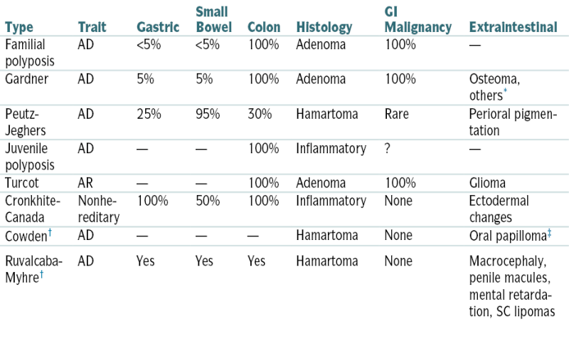
∗ Soft tissue tumors, sarcomas, ampullary carcinoma, ovarian carcinoma.
† Extremely rare.
‡ Gingival hyperplasia, breast cancer, thyroid cancer.
From Weissleder R, Wittenberg J, Harisinghani M, Chen JW: Primer of Diagnostic Imaging, 5th ed. St. Louis, Mosby, 2011.
Diagnosis
H&P
 Infection w/HAV may have acute or subacute presentation, icteric or anicteric. Severity of illness ↑ w/age (90% infections in children <5 yr may be subclinical).
 A preicteric, prodromal phase (1-14 days); 15% no prodrome. Sx onset usually abrupt and may include anorexia, malaise, N/V, fever, headache, abd pain, chills, myalgias, arthralgias, URI sx, constipation, diarrhea, pruritus, urticaria (less common).
 Jaundice (>70%); icteric phase is preceded by dark urine.
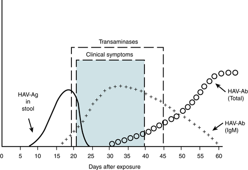
FIGURE 6-2 Serologic test in hepatitis A virus infection. (From Goldberger E: Treatment of Cardiac Emergencies, 5th ed. St. Louis, Mosby, 1990.)
TABLE 6-7
Typical Liver Test Patterns
| Test | Hepatocellular Necrosis | Biliary Obstruction | |||||
| Toxic or Ischemic | Viral | Alcohol | Chronic Complete | Chronic Partial | Acute Complete (first 24 hr) | Infiltration (Chronic) | |
| Aminotransferases | 50-100× | 5-50× | 2-5× | 1-5× | 1-5× | 1-50× | 1-3× |
| Alkaline phosphatase | 1-3× | 1-3× | 1-10× | 2-20× | 2-10× | May be nl | 1-20× |
| Bilirubin | 1-5× | 1-30× | 1-30× | 1-30× | 1-5× | Usually nl | 1-5× |
| PT | Prolonged in severe cases, unresponsive to vitamin K | May be prolonged, responsive to vitamin K | Usually nl | Usually nl | Usually nl | ||
| Albumin | Nl in acute illness, may be ↓ in chronic illness | Usually nl, but may be ↓ in biliary cirrhosis | Usually nl | Usually nl | |||
| Typical disorders | Acetaminophen toxicity, shock liver | Acute hepatitis A or B | Alcoholic hepatitis | Pancreatic carcinoma | Sclerosing cholangitis | Choledocholithiasis | Primary or metastatic carcinoma, Mycobacterium avium-intracellulare infection |
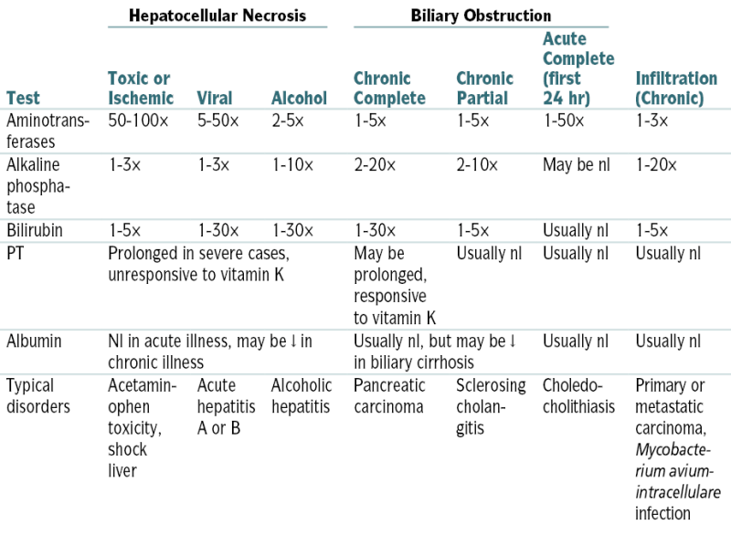
Labs (Fig. 6-2)
 Dx confirmed by IgM anti-HAV; detectable in almost all infected pts at presentation and remains (+) for 3 to 6 mo.
 4× ↑ titer of total Ab (IgM + IgG) to HAV confirms acute infection.
 HAV detection in stool and body fluids by EM
 HAV RNA detection in stool, body fluids, serum, and liver tissue
 ↑ ALT AST (usually >8× nl in acute infection)
 ↑ Bili (usually 5-15× nl)
 Alk phos minimally ↑ but higher level in cholestasis
 Alb and PT generally nl; if ↑ may herald hepatic necrosis
Treatment
 Self-limited (supportive care; activity as tolerated)
 Advise to avoid alcohol and hepatotoxic drugs
 Pts w/fulminant hepatitis may require hospitalization/assessment for liver transplantation.
Clinical Pearls
 Reported to public health authorities
 Common illness in internationally traveled/developing countries; preexposure prophylaxis if travel to endemic area (IG 0.02-0.06 mL/kg IM); Lower dose = effective up to 3 mo; higher dose = effective up to 5 mo.
 Postexposure prophylaxis (IG 0.02 mL/kg given IM) if exposure within 2 wk + no previous vaccination; high-risk pts, may vaccinate with immunoglobulin
 Inactivated vaccines HAVRIX or VAQTA safe/immunogenic in adults/children >12 mo w/protective Ab levels reached 94% to 100% 1 mo combined HepA/HepB vaccine TWINRIX also available
b. Hepatitis B
 Infection by HBV (42-nm hepadnavirus w/an outer surface coat [HBsAg], inner nucleocapsid core [HBcAg; HBeAg], DNA polymerase, and partially double-stranded DNA genome) with transmission via parenteral (needle use, tattooing, piercing, acupuncture, blood transfusion, hemodialysis, sexual contact) and perinatal routes
Diagnosis
H&P
 Often nonspecific sx, malaise
 Prodrome:
Labs (Fig. 6-3)
 Acute HBV infection is best confirmed by IgM HBcAb in acute or early convalescent serum or by HB DNA.
 HBsAg and IgG-HBcAb during acute jaundice are strongly suggestive of remote HBV infection and another etiology for current illness.
 HBsAb alone is suggestive of immunization response.
 W/recovery, HBeAg is rapidly replaced by HBeAb in 2 to 3 mo, and HBsAg is replaced by HBsAb in 5 to 6 mo.
 In chronic HBV, HBsAg and HBeAg are persistent w/o corresponding Ab.
 In chronic carrier state, HBsAg is persistent, but HBeAg is replaced by HBeAb.
 HBcAb develops in all outcomes.
 HBeAg correlation w/highest infectivity; appearance of HBeAb heralds recovery.
 LFTs:
LFTs: Alb and PT: generally nl; if abnl, possible harbinger of impending hepatic necrosis (fulminant hepatitis)
 WBC and ESR: generally nl or mildly ↑
Imaging
 U/S to r/o obstructive jaundice
Treatment
Acute Hepatitis B
 90% of adults spontaneously clear infection; avoid hepatic metabolized drugs; IV hydration if severe vomiting
 Hospitalization if pt risk dehydration via poor PO intake, PT ↑, bili level >15 to 20 μg/dL, or if hepatic failure
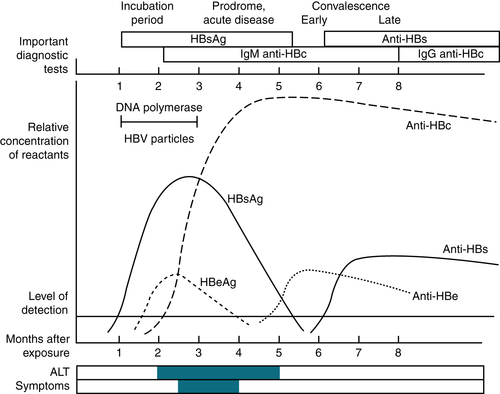
FIGURE 6-3 Serologic and clinical patterns observed during acute hepatitis B virus infection. Patients in whom the hepatitis B infection does not resolve (chronic carrier state) will demonstrate persistence of HBsAg and will not have an elevation of anti-HBs. (From Ravel R: Clinical Laboratory Medicine, 6th ed. St. Louis, Mosby, 1995.)
Chronic Hepatitis B
 Rx: determined by e antigen status and viral qualification
 Documented cirrhosis [(+) or (−) HBeAg] with
Clinical Pearls
 Virus and HBsAg are present in high titers in blood for 1 to 7 wk before jaundice and for a variable time thereafter.
 Transmission is possible during entire period of HBsAg (and especially during HBeAg) in serum.
 Prevention after exposure:
 Preventive Rx w/lamivudine for pts who test (+) for HBsAg and are undergoing chemoRx may ↑ the risk for HBV reactivation and HBV-associated morbidity and mortality.
c. Hepatitis C
 Infection via HCV (single-stranded RNA flavivirus) is mostly via the parenteral route (IV drug use 60% new cases, 20%-50% chronic cases). Occupational needle stick exposure from an HCV(+) source has a seroconversion rate of 1.8% (range, 0%-7%).
Diagnosis
H&P
 Sx usually develop 7 to 8 wk after infection (2-26 wk), but 70% to 80% of cases are subclinical.
 10% to 20% report acute illness w/jaundice and nonspecific sx (abd pain, anorexia, malaise).
 After acute infection, 15% to 25% have complete resolution (absence of HCV RNA in serum, nl ALT).
 Progression to chronic infection is common (50%-84%); 74% to 86% have persistent viremia; spontaneous clearance of viremia in chronic infection is rare. 60% to 70% of pts will have persistent or fluctuating ALT levels; 30% to 40% w/chronic infection have nl ALT levels.
 15% to 20% of those w/chronic HCV will develop cirrhosis during a period of 20 to 30 yr; in most others, chronic infection leads to hepatitis and varying degrees of fibrosis.
 0.4% to 2.5% of pts w/chronic infection develop HCC.
 25% of pts w/chronic infection continue to have an asymptomatic course w/nl LFTs and benign histology.
 In chronic HCV infection, extrahepatic sequelae include a variety of immunologic and lymphoproliferative disorders (e.g., cryoglobulinemia, membranoproliferative GN, and possibly Sjögren’s syndrome, autoimmune thyroiditis, polyarteritis nodosa, aplastic anemia, lichen planus, porphyria cutanea tarda, B-cell lymphoma, others).
Labs (Fig. 6-4)
 Dx is often by exclusion because it takes 6 wk to 12 mo to develop anti-HCV Ab (70% + by 6 wk, 90% + by 6 mo).
 Diagnostic tests include serologic assays for Abs and molecular tests for viral particles.
 Enzyme immunoassay is the test for anti-HCV Ab:
 Recombinant immunoblot is used to confirm + enzyme immunoassays: recommended only in low-risk settings
 Qualitative and quantitative HCV RNA tests using PCR:
 Viral genotyping can distinguish among genotypes 1, 2, 3, and 4, which is helpful in choosing Rx; most of these tests use PCR. (Genotypes 1, 2, 3, and 4 predominate in the United States and Europe [1 is especially common in North America].)
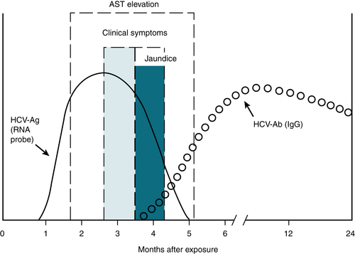
FIGURE 6-4 Hepatitis C virus antigen and antibody. (From Hollinger FB, Dienstag JL. In Murray PR, et al [eds]: Manual of Clinical Microbiology, 6th ed. Washington, DC, American Society for Microbiology, 1995.)
 LFTs:
LFTs:Imaging
 U/S is useful to r/o obstructive jaundice and to evaluate rapid liver size ↓ during fulminant hepatitis or mass in HCC.
Treatment
Acute Hepatitis C
 HCV PCR detected within 13 days (antibody in >36 days)
 If clear HCV viral load via PCR within 3 to 4 mo (no Rx-supportive care)
Chronic Hepatitis C (persistent HCV viral load)
 Rx duration depends on HCV RNA level:
 Null: no ↓ of 2log10 by wk 12
 Partial: 2log10 ↓ by wk12 but detectable by wk 24
 Nonresponsive: HCV RNA not cleared by wk 24
 Early Virologic Response (EVR): >2log10 ↓ by wk 12, undetectable by wk 24
 Complete EVR: undetectable by wk 12 and 24, but not at wk 4
 Rapid Virologic Response (RVR): undetectable by wk 4, no change during Rx
 Extended Virologic Response (eRVR): undetectable by wk 4 (telaprevir) or wk 8 (boceprevir) and wks 12 and 24
 Relapse: undetectable at Rx end, but detectable within 24 wk s/p Rx
 Sustained Virologic Response (SVR): cure; undetectable s/p 24 wk Rx
 Rx options depend on Genotype:
 When Rx HIV-HCV dually infected pt, monitor interactions between direct-acting antivirals (DAAs) and antiretroviral drugs. Rx duration will likely be the full 48 wk.
 Liver transplantation: May be only option if HCV-related deteriorating cirrhosis exists.
d. Hepatitis D
 Incomplete virus requiring presence of HBV for replication; thus, resolution of HBV infection will result in resolution of HDV infection.
e. Hepatitis E
 Similar to HAV (enteric transmission, acute infection only)
 Despite low overall mortality, infection during third trimester mortality rate ≥25%
 Prevention via good hygiene; avoid dirty water/poorly cooked food.
3. Autoimmune Hepatitis
Diagnosis
H&P
 Can manifest asymptomatically → fulminant hepatic failure; most commonly fatigue, pruritus, jaundice, hepatomegaly/splenomegaly
TABLE 6-8
Simplified Diagnostic Criteria for Autoimmune Hepatitis
| Variable | Cutoff | Points | Cutoff | Points |
| ANA or SMA | ≥1:40 | 1 | ≥1:80 | 2 |
| LKM | ≥1:40 | 2 | ||
| SLA | Positive | 2 | ||
| IgG | ≥ULN | 1 | ≥1.1 × ULN | 2 |
| Histology | Compatible with AIH | 1 | Typical of AIH | 2 |
| Absence of viral hepatitis | Yes | 2 | ||
| Maximum number of points for all antibodies = 2, total = 8. | ||||
| Probable AIH ≥6 points, definite AIH ≥7 points. | ||||
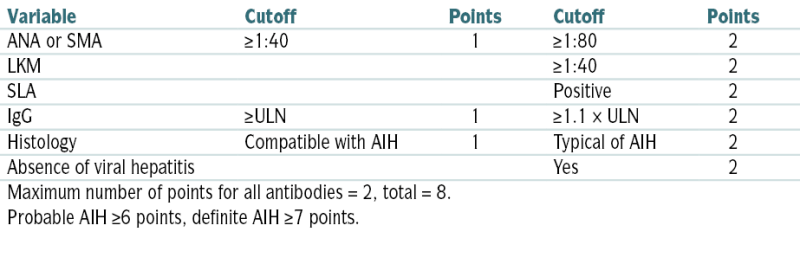
AIH, Autoimmune hepatitis; LKM, liver/kidney microsomes; SLA, soluble liver antigen; SMA, smooth muscle antibody; ULN, upper limit of normal.
From Ferri F: Ferri’s Clinical Advisor: 5 Books in 1. 2013 edition. Philadelphia, Mosby, 2012.
 Autoimmune findings: arthritis, xerostomia, keratoconjunctivitis, cutaneous vasculitis, and erythema nodosum
Labs
 ↑ ALT and AST (both 2-10× nl)
 Women 4× > risk than men
 Bili and alk phos moderately ↑/nl
 ↑ γ-Globulin (>2.0 g/dL)
 Serum ANA + anti–smooth muscle Ab (64% cases); if (−), p-ANCA helpful
 Liver bx = interface hepatitis (lymphoplasmacytic inflammatory infiltrate extending from portal tract → lobule)
 Table 6-8 describes simplified diagnostic criteria for autoimmune hepatitis.
Imaging
 U/S of liver and biliary tree: r/o obstruction or hepatic mass
Indications for Treatment
 AST >10× nl
 AST >5× nl, w/↑ γ-globulin 2× nl
 Fever, N/V, jaundice
 Histologic features of bridging necrosis or multiacinar necrosis
Treatment
 Prednisone 60 mg PO/day or (prednisone 30 mg + azathioprine 50 mg PO/day); both achieve remission in 80% pts w/combo Rx = less steroid SE
 Taper based on lab response striving for minimum dose needed for maintenance Rx (same regimen 12-18 mo) to ↑ remission rate.
 If liver transplant necessary, 10-yr survival rate 75%
4. Cholestatic Liver Disease
a. Primary Biliary Cirrhosis (PBC)
Diagnosis
H&P
 Typical pt middle-aged female (50 yr); most asx but develop fatigue, pruritus, dry eyes/mouth over 10-yr period
Labs
 (+) Antimitochondrial Ab titer >1:40 (if undetectable, need biopsy)
 ↑ alk phos >1.5× nl (with nl total bili); ↑ AST/ALT <5× nl possible
 Histology: florid duct lesion (duct obliteration w/granuloma formation = pathognomic)
Treatment
 Pruritus: cholestyramine (8-24 g qd), sertraline, rifampin
 Hyperlipidemia/malabsorption of fat-soluble vitamins via cholestasis: statin Rx + wt loss and vitamin (especially vitamin D) replacement
 Asymptomatic stage: Follow LFTs q3-4 mo; if alk phos and/or AST >1.5× nl start ursodeoxycholic acid 12 to 15 mg/kg/day (UDCA).
 Liver transplant is only definitive Rx.
b. Primary Sclerosing Cholangitis (PSC)
Diagnosis
H&P
 Mean age 40 yr; 60% men (most asymptomatic, but may have pruritus, abd pain, jaundice)
Labs/Imaging
 ↑ Alk phos (3-10× nl); ↑ ALT/AST (2-3× nl); (+) ANA, smooth muscle abs (20%-50%)
 “Beads on a string” (segmental bile duct fibrosis w/saccular dilatation of nl areas)
 Cholangiography: MRCP (gold standard); if obstruction, ERCP
 Liver bx: periductal fibrosis, fibro-obliterative cholangiopathy
Staging
 See Table 6-9.
TABLE 6-9
Staging of Primary Sclerosing Cholangitis
| Stage | Description |
| I: Portal | Portal edema, inflammation, ductal proliferation; abnormalities do not extend beyond the limiting plate |
| II: Periportal | Periportal fibrosis with or without inflammation extending beyond the limiting plate |
| III: Septal | Septal fibrosis, bridging necrosis, or both |
| IV: Cirrhotic | Biliary cirrhosis |
From: Cameron AM: Current Surgical Therapy, 10th ed. Philadelphia, Saunders, 2011.
Treatment
 Sx relief; liver transplantation
5. Complications of Liver Disease
a. Cirrhosis
 Defined histologically as the presence of fibrosis/regenerative nodules in the liver
Etiology
 EtOH abuse, secondary to biliary cirrhosis/obstruction of the CBD (stone, stricture, pancreatitis, neoplasm, sclerosing cholangitis), drugs (acetaminophen, INH, MTX, methyldopa), hepatic congestion (CHF, constrictive pericarditis, tricuspid insufficiency, hepatic vein thrombosis, vena cava obstruction), PBC, PSC, chronic hepB/C, Wilson’s disease, α1-antitrypsin deficiency, infiltrative diseases (amyloidosis, glycogen storage diseases, hemochromatosis), jejunoileal bypass, parasitic infections (schistosomiasis), idiopathic portal HTN, congenital hepatic fibrosis, systemic mastocytosis, autoimmune hepatitis, hepatic steatosis, IBD
Diagnosis
H&P
 Jaundice; spider angiomas; ecchymosis; gynecomastia in men; small, nodular liver; ascites; hemorrhoids; testicular atrophy
Labs
 Alcoholic hepatitis and cirrhosis: mild ↑ ALT, AST, usually <500 IU; AST > ALT (ratio >2:3)
 Extrahepatic obstruction: moderate ↑ ALT/AST to levels <500 IU
 Viral, toxic, or ischemic hepatitis: ↑↑ (>500 IU) ALT, AST
 ↑ Alk phos: extrahepatic obstruction, PBC, and PSC
 ↑ Serum LDH: liver mets, hepatitis, cirrhosis, extrahepatic obstruction, congestive hepatomegaly
 ↑ Serum GGTP: alcoholic liver disease, PBC, PSC
 ↑ Serum bili + urinary bili: hepatitis, hepatocellular jaundice, biliary obstruction
 ↓ Serum alb: significant liver disease
 ↑ PT: severe liver damage and poor prognosis
 (+) HBsAg: acute or chronic hepatitis B
 (+) Antimitochondrial Abs: PBC, chronic hepatitis
 ↑ Serum copper, ↓ ceruloplasmin: Wilson’s disease
 ↓ α1-Globulins (α1-antitrypsin deficiency), ↑ IgA (alcoholic cirrhosis), ↑ IgM (PBC), ↑ IgG (chronic hepatitis, cryptogenic cirrhosis)
 ↑ Serum ferritin, ↑ transferrin saturation: hemochromatosis
 ↑ Blood ammonia: hepatocellular dysfunction
 (+) ANA: autoimmune hepatitis
Imaging
 U/S: detection of gallstones and dilation of CBDs
 CT abd: detection of mass lesions in liver/pancreas; hepatic fat content; identification of idiopathic hemochromatosis; early dx of Budd-Chiari syndrome, dilation of intrahepatic bile ducts; and detection of varices and splenomegaly
 Percutaneous liver bx: evaluation of hepatic filling defects; dx of hepatocellular disease or hepatomegaly; evaluation of persistently abnl LFTs; and dx of hemochromatosis, PBC, Wilson’s disease, glycogen storage diseases, chronic hepatitis, autoimmune hepatitis, infiltrative diseases, alcoholic liver disease, drug-induced liver disease, and primary or secondary carcinoma
 Scoring system: Table 6-10
Treatment
 Variable w/etiology. Figure 6-5 summarizes the management of compensated and decompensated cirrhosis.
 Liver transplantation: indicated in otherwise healthy pts (age <65 yr) w/sclerosing cholangitis, chronic hepatitis, cirrhosis, or PBC, w/prognostic information suggesting <20% chance of survival w/o transplantation. Contraindications to liver transplantation are AIDS, most metastatic malignant neoplasms, active substance abuse, uncontrolled sepsis, and uncontrolled cardiac or pulmonary disease.
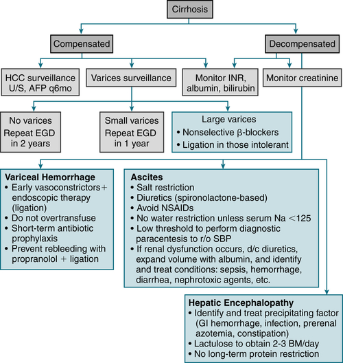
FIGURE 6-5 Summary of the management of compensated and decompensated cirrhosis. (From Goldman L, Schafer AI [eds]: Goldman’s Cecil Medicine, 24th ed. Philadelphia, Saunders, 2012.)
TABLE 6-10
The Two Most Commonly Used Scoring Systems in Cirrhosis
| 1. Child-Turcotte-Pugh (CTP) Score (Range, 5-15) | |||
| Parameters | Points Ascribed | ||
| 1 | 2 | 3 | |
| Ascites | None | Grade 1-2 (or easy to treat) | Grade 3-4 (or refractory) |
| Hepatic encephalopathy | None | Grade 1-2 (or induced by a precipitant) | Grade 3-4 (or spontaneous) |
| Bilirubin (mg/dL) | <2 | 2-3 | >3 |
| Albumin (g/dL) | >3.5 | 2.8-3.5 | <2.8 |
| PT (seconds > control) or INR | <4 | 4-6 | >6 |
| <1.7 | 1.7-2.3 | >2.3 | |
| CTP classification: Child A: score of 5-6; Child B: score of 7-9; Child C: score of 10-15 | |||
| 2. Model of End-Stage Liver Disease (MELD) Score (Range, 6-40) | |||
| [0.957 × LN (creatinine in mg/dL) + 0.378 × LN (bilirubin in mg/dL) + 1.12 × LN (INR) + 0.643] × 10 | |||
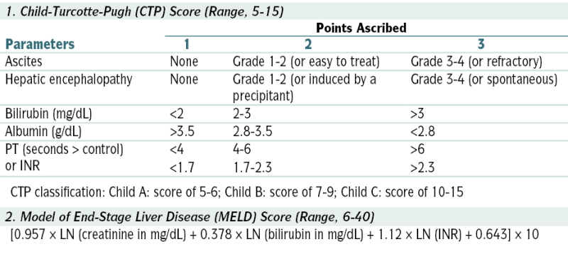
From Goldman L, Schafer AI (eds): Goldman’s Cecil Medicine, 24th ed. Philadelphia, Saunders, 2012.
 Rx of complications of portal HTN (ascites, esophagogastric varices, hepatic encephalopathy, and HRS)
b. Ascites/Paracentesis
Paracentesis
Indications
 Ascites of undetermined etiology
 Evaluation for possible peritonitis
 Relief of abd pain and discomfort caused by tense ascites
 Relief of dyspnea caused by ↑ diaphragm (from ascites)
 Evaluation of possible intra-abd hemorrhage in a pt w/blunt abd trauma
 Institution of peritoneal dialysis
Contraindications
 Bleeding disorders, thrombocytopenia (relative contraindication)
 Bowel distention
 Infection or surgical scars at the site of needle entry
 Acute abdomen
 Distended bladder that cannot be emptied w/Foley catheter
Procedure
Interpretation of Results
 Peritoneal effusion, like pleural effusion, can be subdivided into exudative or transudative on the basis of its characteristics (Fig. 6-6).
 The serum-ascites alb gradient (SAAG) (serum alb level–ascitic fluid alb level) correlates directly w/portal pressure and can also be used to classify ascites (Table 6-11). Pts w/gradients of ≥1.1 g/dL or higher have ascites secondary to portal HTN, and those w/gradients <1.1 g/dL do not; the accuracy of this method is >95%.
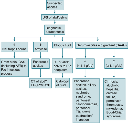
FIGURE 6-6 Diagnostic algorithm for ascites. (From Ferri FF: Ferri’s Best Test: A Practical Guide to Clinical Laboratory Medicine and Diagnostic Imaging, 2nd ed. Philadelphia, Mosby, 2010.)
TABLE 6-11
Causes of Ascites Based on Normal or Diseased Peritoneum and Serum-to-Ascites Albumin Gradient (SAAG)
| Normal Peritoneum | |
| Portal Hypertension (SAAG >1.1 g/dL) | Hypoalbuminemia (SAAG <1.1 g/dL) |
| Hepatic congestion | Nephrotic syndrome |
| Congestive heart failure | Protein-losing enteropathy |
| Constrictive pericarditis | Severe malnutrition with anasarca |
| Tricuspid insufficiency | |
| Budd-Chiari syndrome | |
| Liver disease | Miscellaneous Conditions (SAAG <1.1 g/dL) |
| Cirrhosis | Chylous ascites |
| Alcoholic hepatitis | Pancreatic ascites |
| Fulminant hepatic failure | Bile ascites |
| Massive hepatic metastases | Nephrogenic ascites |
| Urine ascites | |
| Ovarian disease | |
| Diseased Peritoneum (SAAG <1.1 g/dL) | |
| Infections | Other Rare Conditions |
| Bacterial peritonitis | Familial Mediterranean fever |
| Tuberculous peritonitis | Vasculitis |
| Fungal peritonitis | Granulomatous peritonitis |
| HIV-associated peritonitis | Eosinophilic peritonitis |
| Malignant Conditions | |
| Peritoneal carcinomatosis | |
| Primary mesothelioma | |
| Pseudomyxoma peritonei | |
| Hepatocellular carcinoma | |
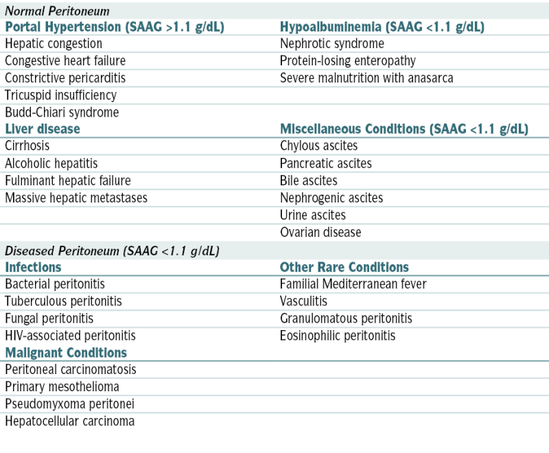
From Vincent JL, Abraham E, Moore FA, et al (eds): Textbook of Critical Care, 6th ed. Philadelphia, Saunders, 2011.
Complications
 Persistent leakage of ascitic fluid
 Hypotension and shock
 Bleeding
 Perforated bowel
 Abscess formation in area of puncture site
c. Varices
 Dilated submucosal veins via portal vein HTN (>10-12 mm Hg), functioning as a shunt between portal and systemic venous circulation; result in severe upper GI hemorrhage (25%-40% cirrhotic pts)
Diagnosis
 EGD (Esophagography with barium noninvasive option)
Labs and W/up
 Anemia (blood loss, nutritional deficiencies, alcohol myelosuppression)
 Thrombocytopenia (hypersplenism, alcohol myelosuppression)
 BUN: often ↑ in setting of upper GI bleeding
 Cr: often ↑ by hypovolemia, monitor for hepatorenal syndrome
 Na+: dilutional hyponatremia
 Heme (+) stools (type/crossmatch: in preparation for blood transfusion)
 INR/PT and PTT: may be ↑ in liver disease or impairment
 LFTs: ALT/AST may be nl in cirrhotic pts because of fibrosis, ↑ alk phos, direct hyperbilirubinemia possible if cholestatic liver disease
 Serum alb: Severe liver disease results in hypoalbuminemia.
Treatment
 Hemorrhage: acute hemodynamic resuscitation (pRBC transfusion, correct coagulopathy/thrombocytopenia), SBP prophylaxis (ceftriaxone or norfloxacin), octreotide 2 to 5 days in conjunction with endoscopic Rx
 Ligation (if ↑ risk hemorrhage: Child Pugh B/C or red wale markings on EGD)
 Follow-up surveillance EGD 1 to 3 mo s/p obliteration, then q6-12mo indefinitely
 Primary prophylaxis: nonselective β-blockers (propranolol 20 mg bid or nadolol 40 mg daily)
d. Portal Hypertension
 Portal vein pressure >10 mm Hg
Etiology
 ↑ Resistance to flow (portal/splenic/hepatic vein thrombosis, cirrhosis, schistosomiasis)
 ↑ Portal blood flow (splanchnic arterial vasodilation, arterial-portal venous fistulae)
Imaging
 Duplex-Doppler U/S (CT/MRI/MRA scanning if results unclear)
Treatment
 ↓ HTN directly, minimize volume overload, correct underlying disorders, and prevent complications (most notably SBP and variceal bleeding).
e. Hepatic Encephalopathy (HE)
 Cognitive disturbance via hepatic insufficiency and portosystemic shunting
Etiology
 Precipitating factors in pts w/underlying cirrhosis (UGI bleeding, hypokalemia, hypomagnesemia, analgesic and sedative drugs, sepsis, alkalosis, dietary protein), acute fulminant viral hepatitis, drugs and toxins (INH, acetaminophen-Reye’s syndrome, diclofenac, statins, methyldopa, loratadine, propylthiouracil, lisinopril, labetalol, halothane, carbon tetrachloride, erythromycin, nitrofurantoin, troglitazone), shock or sepsis, fatty liver of pregnancy, metastatic carcinoma, HCC, other (autoimmune hepatitis, ischemic veno-occlusive disease, sclerosing cholangitis, heat stroke, amebic abscesses)
Diagnosis
 Requires r/o other causes (medications, metabolic disorders, infectious diseases, intracranial lesions/events). Clinical stages of hepatic encephalopathy are described in Table 6-12. The West Haven criteria is noted here.
West Haven Criteria for Classification of Hepatic Encephalopathy
 Stage 0: nl cognitive function/no abnl detected
 Stage 1: lack of awareness, euphoria/anxiety, ↓ attention/concentration
 Stage 2: lethargy/apathy, altered personality, inappropriate behavior, time disorientation
 Stage 3: somnolence to semistupor, confusion, gross disorientation, bizarre behavior
TABLE 6-12
Clinical Stages of Hepatic Encephalopathy
| Stage | Asterixis E | EEG Changes | Clinical Manifestations |
| I (prodrome) | Slight | Minimal | Mild intellectual impairment, disturbed sleep-wake cycle |
| II (impending) | Easily elicited | Usually generalized | Drowsiness, confusion, coma/inappropriate behavior, disorientation, mood swings |
| III (stupor) | Present if patient cooperative | Grossly abnormal slowing of rhythm | Drowsy, unresponsive to verbal commands, markedly confused, delirious, hyperreflexia, positive Babinski’s sign |
| IV (coma) | Usually absent | Appearance of delta waves, decreased amplitudes | Unconscious, decerebrate or decorticate response to pain present (stage IVA) or absent (stage IVB) |
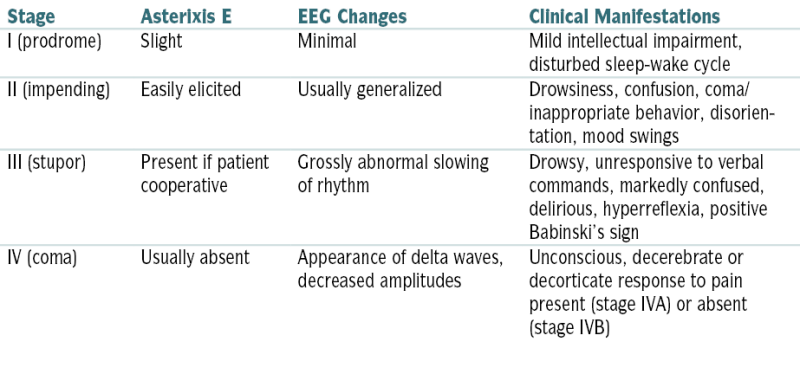
From Fuhrman BP, Zimmerman JJ, Carcillo JA, Clark RS, et al (eds): Pediatric Critical Care, 4th ed. Philadelphia, Saunders, 2011.
 Stage 4: Coma, unable to test mental state
PE
 Variable w/the stage and may reveal the following abnormalities:
Labs
 ALT, AST, bili, alk phos, glucose, Ca, electrolytes, BUN, Cr, alb
 CBC, Plt count, PT, PTT
 Serum/urine toxicology screen (suspected medication/illegal drug use)
 Blood/urine cultures, U/A
 Venous ammonia level (no correlation w/stage HE but helpful in initial evaluation)
 ABGs
ABGsTreatment
 Identification and Rx of precipitating factors
 Restriction of protein intake (30-40 g/day) to ↓ toxic protein metabolites
 ↓ Colonic ammonia production:
 Rx of cerebral edema: Cerebral edema is often present in pts w/acute liver failure, and it accounts for nearly 50% of deaths. Monitoring of ICP by epidural, intraparenchymal, or subdural transducers and Rx of cerebral edema w/mannitol (100-200 mL of 20% solution [0.3-0.4 g/kg of BW]) given by rapid IV infusion is helpful in selected pts (e.g., potential transplantation pts). Dexamethasone and hyperventilation (useful in head injury) are of little value in treating cerebral edema from liver failure.
f. Hepatorenal Syndrome (HRS)
 Type 1: progressive renal function impairment (doubling of initial serum Cr >2.5 mg/dL or 50% reduction initial 24-hr CrCl to <20 mL/min in <2 wk)
 Type 2: stable/slowly progressive renal function impairment (assoc w/refractory ascites)
Etiology
 HRS may occur after significant ↓ effective blood volume (paracentesis, GI bleeding, diuretics) or in absence of any precipitating factors.
Diagnosis
 Serum Cr >1.5 mg/dL; with no improvement (↓ ≤1.5 mg/dL) after 2 days diuretic withdrawal + albumin volume expansion
 Absence of shock, infection, fluid loss, or current Rx w/nephrotoxic drugs
 Absence of proteinuria (<500 mg/day) or hematuria (<50 RBC/hpf)
 Absence of U/S evidence of obstructive uropathy/parenchymal renal disease
 Urinary Na <10 mmol/L
Treatment
 Volume challenge (↑ MAP) then large-volume paracentesis (↑ CO, ↓ renal venous pressure) recommended to distinguish HRS from prerenal azotemia in pts w/FeNa <1% [if prerenal azotemia the ↑ renal perfusion pressure/renal blood flow results in prompt diuresis]; volume challenge can be accomplished by 100 g albumin in 500 mL isotonic saline.
 Vasopressin analogues may ↑ renal perfusion by reversing splanchnic vasodilation (hallmark HRS); IV NE + albumin and furosemide may also be effective.
 Rx w/vasoconstrictors 5 to 15 days in attempt to ↓ serum Cr to <1.5 mg/dL:
 Liver transplantation (most effective Rx) may be indicated pts (<65 yr) w/sclerosing cholangitis, chronic hepatitis w/cirrhosis, or PBC. Contraindications include AIDS, metastatic disease, substance abuse, uncontrolled sepsis, and uncontrolled cardiac/pulmonary disease.
g. Spontaneous Bacterial Peritonitis
Management and Treatment
 Third-generation cephalosporin given until culture report finalized
 If SBP + hepatorenal syndrome, concomitant IV alb ↑ survival rate
 Repeat paracentesis (48 hr) should reveal ↓ PMN count, will be ↑ if secondary peritonitis
 Rx secondary peritonitis with norfloxacin (TMP-SMX if fluoroquinolone allergy)
 Prophylaxis with norfloxacin warranted if low-protein ascitic fluid <1 g/dL (10 g/L)
h. Hepatocellular Carcinoma (HCC)
Diagnosis
H&P
 33% asx at dx; Abd pain may be initial presentation.
 Sx underlying cirrhosis often present; new ascites, encephalopathy, jaundice/bleeding, paraneoplastic syndromes (hypoglycemia, erythrocytosis, hypercalcemia, severe diarrhea) may be present.
Labs
 ↑ LFTs
 ↑ AFP 70% pts (sens 40%-65%; spec 80%-94%)
 Paraneoplastic syndromes associated w/HCC may cause hypercalcemia, hypoglycemia, and polycythemia.
 HBV DNA level (>10,000 copies/mL) is a strong risk predictor of HCC independent of HBeAg, serum aminotransferase level, and liver cirrhosis.
Imaging
 U/S (optimal for screening q6mo if ↑ risk), CT scan, or MRI
Staging and Treatment
 According to the Barcelona Clinic Liver Cancer staging classification, Rx is determined according to stage.
 Early stage: Liver transplantation is the most effective Rx.
 Ablation (via radiofrequency, transarterial hepatic artery chemoembolization, percutaneous ethanol injection) can be used as bridge to transplantation.
 Nonresectable HCC: sorafenib
i. Fulminant Hepatic Failure (FHF)
 Classification:
 Etiologies: acetaminophen overdose (most common cause), infectious (HepA-E, HSV, CMV, EBV, PB19, varicella), drugs (INH, sulfa, NSAIDs, MDMA, cocaine), autoimmune hepatitis
 Rx: Recognition should prompt immediate referral to liver transplantation center (1-yr survival 73%). Table 6-13 summarizes the management of fulminant hepatic failure.
j. Liver Abscess
Etiology
 Pyogenic abscess: polymicrobial (streptococcal species [37%], E. coli [33%], Klebsiella pneumoniae [18%], Pseudomonas, Proteus, Bacteroides)
 Sources of pyogenic abscess: biliary disease w/cholangitis, gallbladder disease, diverticulitis, appendicitis, penetrating wounds, hematogenous
 Amebic abscess: E. histolytica. Transmission is through fecal-oral contamination w/invasion of intestinal mucosa and portal system.
TABLE 6-13
Management of Fulminant Hepatic Failure
From Fuhrman BP, Zimmerman JJ, Carcillo JA, Clark RS, et al (eds): Pediatric Critical Care, 4th ed. Philadelphia, Saunders, 2011.
Diagnosis
H&P
 RUQ abd pain, fever, nausea, cough w/pleuritic chest pain, anorexia, jaundice
Labs
 Leukocytosis, + blood cultures (50%), ↑ INR (70%), ↑ alk phos (>90%), ↑ ALT/AST (50%), + stool samples for E. histolytica (10%-15%). Serologic testing for E. histolytica does not differentiate acute from prior infection.
Imaging
 Abdominal CT (imaging study of choice)
 CXR is abnl in 50% of cases: ↑ right hemidiaphragm, pleural effusion, subdiaphragmatic air fluid levels.
 U/S (80%-95% sensitivity)
 CT or U/S-guided aspiration (50% sterile) in suspected pyogenic abscess.
Treatment
 Medical management (amebic liver abscess) versus percutaneous drainage under CT or U/S guidance + IV abx (pyogenic liver abscess)
 Empiric broad spectrum abx recommended until culture results available:
6. Vascular Disorders of the Liver
a. Budd-Chiari Syndrome
 Primary BCS = endoluminal obstruction (thromboses/webs)
 Secondary BCS = obstruction from nonvascular invasion (malignant/parasitic masses) or extrinsic compression (tumor, abscess, cysts)
Etiology
 Hypercoagulable state and/or underlying risk factor for thromboses (80%), myeloproliferative disease (50%), Behçet’s disease, infection, malignancy (<5%), PNH, abd trauma, UC, celiac disease, dacarbazine
Diagnosis
H&P
 Fulminant/acute (uncommon): severe RUQ abd pain, fever, N/V, jaundice, hepatomegaly, ascites, ↑ serum aminotransferases, ↓ coagulation factors, and encephalopathy. Early recognition and Rx are essential to survival.
 Subacute/chronic (more common): vague abd discomfort, gradual progression to hepatomegaly, portal HTN w/ or w/o cirrhosis; late-onset ascites, LE edema, esophageal varices, splenomegaly, coagulopathy, HRS; asx (15% cases)
 Ascites protein content > 2.5 g/dL and SAAG ≥ 1.1 g/dL = BCS + cardiac disease
Imaging
 Color and pulsed Doppler U/S: (sens >85%) first-line test
 MRI w/gadolinium contrast (second-line test)
 Venography (gold standard); useful if difficult case or precise delineation of venous stenoses required
Treatment
 Lifelong anticoagulation INR 2 to 3 (LMWH → warfarin [Coumadin]) recommended for all
 OCPs contraindicated
 Hepatic vein/IVC angioplasty w/ or w/o stenting if refractory to anticoagulation
 If unresponsive to medical Rx, TIPS insertion recommended
 If unresponsive to TIPS, liver transplant indicated (5-yr survival 70%)
b. Portal Vein Thrombosis
Etiology
 In adults: cirrhosis (common cause), hypercoagulable states, inflammatory diseases, complications of medical intervention: ambulatory dialysis, chemoembolization, liver transplantation, partial hepatectomy, scleroRx, splenectomy, TIPS, infections (appendicitis, diverticulitis, cholecystitis)
 In children: umbilical sepsis
Diagnosis
H&P
 May result in portal HTN → esophageal/GI varices; upper GI hemorrhage (hematemesis or melena) can result from esophageal varices.
 If abd pain is present, mesenteric venous thrombosis should be suspected.
Imaging
 Abd U/S or MRI may show the portal vein thrombosis.
 Esophagogastroscopy (esophageal varices)
Treatment
 Anticoagulation
 Variceal scleroRx or banding
 Surgical mesocaval or splenorenal shunt
7. Hepatic Cysts
 Simple cysts (uniform thin wall, no echoic structures) asx, usually occur more in women
 Cysts <4 cm rarely of clinical significance
 If sx (nausea/abd discomfort), r/o cystadenoma (irregularity/internal echoes) via U/S
 Rx: Laparoscopic fenestration (simple needle aspiration assoc ↑ recurrence) if cystadenoma as malignant transformation to cystadenocarcinoma possible
8. Hepatic Adenoma (HA)
 Sheets of benign hepatocytes without biliary structures/nonparenchymal liver cells
 Assoc with use of OCPs
 Rx: Resection of HA >5 cm or if pregnancy considered, discontinuation OCP with follow-up imaging if <5 cm
G. Metabolic Liver Disease
1. Alcoholic Hepatitis
 Alcohol-induced liver disease: ranges from steatosis (fatty liver) → cirrhosis
Labs
 ↑ AST (2-6× nl); [AST/ALT ratio 2:3 typical, AST >500/ALT >200 rare]
 Alk phos nl to ↑, bili nl to ↑↑↑, ↑ PT, ↓ GGT
 ↑ CRP, hypophosphatemia, hypomagnesia, CBC (possible leukocytosis w/bandemia or anemia)
 Carbohydrate-deficient transferring (CDT) = reliable marker chronic alcoholism
 Screen to r/o: HbsAg, anti-Hep C, ferritin/transferrin sat, AFP, liver biopsy if necessary
Imaging
 U/S (macrovascular steatosis, hepatocyte ballooning/necrosis, Mallory’s bodies, perivenular fibrosis, portal/lobular inflammation)
Prognosis
 Maddrey discriminant function (MDF) score: 4.6× (prothrombin time [s] – control prothrombin time [s]) + total bili (mg/dL)
 MDF ≥32 (severe alcoholic hepatitis): short-term mortality risk 50%
 Model for End-Stage Liver Disease (MELD) score (prognostic index based on serum total bili, Cr, and INR): similar implications as MDF score
Treatment


2. Nonalcoholic Fatty Liver Disease (NAFLD)
Diagnosis
 Abdominal imaging via U/S, CT, MRI (reveal steatosis but not fibrosis)
 Liver biopsy (confirms NASH)
Treatment
 Lifestyle change, wt loss; monitor LFTs q3-6mo; metformin currently investigational Rx
3. Hereditary Hemochromatosis
Diagnosis
H&P
 ↑ Skin pigmentation, hepatomegaly, splenomegaly, hepatic tenderness, testicular atrophy (↓ libido 50% men), amenorrhea (25% women), alopecia, gynecomastia/ascites, fatigue, joint pain, new-onset diabetes, cardiomyopathy, arthropathy
Labs
 Transferrin saturation (iron/TIBC) = best screening test; values >45% indicate need for further testing; values >52% in men and >50% in women suggest hemochromatosis. Serum ferritin is helpful but may be ↑ in inflammatory conditions or malignancy.
 ↑ AST, ALT, alk phos
 Hyperglycemia
 ↓ testosterone, LH, FSH
 Measurement of hepatic iron index (hepatic iron concentration divided by age) in liver bx specimen can confirm dx.
 Genetic testing (HFE genotyping for C282Y/H63D mutations) useful in pts w/liver disease + suspected iron overload (transferrin saturation >40%). Genetic testing should not be performed as part of initial routine evaluation for hereditary hemochromatosis. Once pt confirmed, first-degree relatives should also be screened. Figure 6-7 describes an algorithm for evaluation of possible hereditary hemochromatosis in a person with (−) family hx.
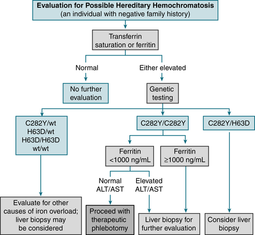
FIGURE 6-7 Algorithm for elevation of possible hereditary hemochromatosis in a person with a negative family history. (From Goldman L, Schafer AI [eds]: Goldman’s Cecil Medicine, 24th ed. Philadelphia, Saunders, 2012.)
 Pts >40 yr old or serum ferritin level >1000 μg/L ↑ risk cirrhosis
Imaging
 CT or MRI of the liver: useful to r/o other causes + may show iron overload in the liver
Treatment
 Weekly phlebotomies of 1 to 2 U of blood (each w/ ≈250 mg iron) should be continued until ferritin level <50 ng/mL and transferrin saturation <30%. Subsequent phlebotomies PRN to maintain a transferrin saturation <50% and a ferritin level <100 ng/mL.
 Deferoxamine 0.5 to 1 g IM qd or 20 mg SC constant infusion (iron chelating agent) is generally reserved for pts w/severe hemochromatosis w/diffuse organ involvement (liver disease, heart disease) and when phlebotomy is not possible.
4. α1-Antitrypsin Deficiency
Diagnosis
 Serum α1-antitrypsin level, genetic analysis for mutation/allele involvement PFTs (obstructive pattern)
 CXR (emphysematous change at lung bases)
 Chest CT (panacinar emphysema)
H&P
 Early-onset, severe, lower-lobe predominant panacinar emphysema (± bronchiectasis)
 Sx similar to “typical” COPD presentation (dyspnea, cough, sputum production)
 Liver involvement (neonatal cholestasis, cirrhosis, primary hepatocarcinoma)
 Dermatologic manifestation (panniculitis) to 85% deficit in plasma α1-antitrypsin
Treatment
 Manage acute exacerbations similar to “typical” COPD
 Avoid smoking/factors that would worsen COPD
5. Wilson’s Disease
 Rare (1/30,000 newborns) autosomal recessive ↑ hepatic uptake and ↓ biliary excretion of copper
Diagnosis
 Usually pts < 45 yr old; ↓ ceruloplasmin, ↑ urinary copper excretion
 ↑ LFTs
 Liver biopsy (excessive intrahepatic copper)
 Kayser-Fleischer rings
 Neuropsychiatric abnormalities (copper deposition in brain)
Treatment
 Chelating agents (trientine, penicillamine) and low-copper diet
H. Disorders of Gallbladder
1. Cholelithiasis
Epidemiology and Demographics
 Incidence ↑ with age (highest in 40s-50s)
 Female sex, pregnancy, FHx, obesity, ileal disease, OCPs, DM
 90% gallstones = cholesterol/mixed type, black-pigmented stones assoc with chronic hemolytic disease/cirrhosis, brown-pigmented stones assoc with biliary tract infection
Physical Exam and Clinical Findings
 (60%-80%) asx
 If biliary colic: episodic, cramping RUQ pain, may radiate to back/right shoulder
Diagnosis
 Labs nl (unless obstruction: ↑ alk phos, ↑ bili)
 U/S; if unclear HIDA scan (90% accuracy)
Treatment


2. Acute Cholecystitis
 Inflammation of the gallbladder
Etiology
 Gallstones (>95% of cases), ischemic damage to the gallbladder, critically ill pt (acalculous cholecystitis), infectious agents (especially in AIDS pts [CMV, Cryptosporidium]), bile duct stricture, neoplasia
Diagnosis
H&P
 Pain/tenderness of right hypochondrium/epigastrium; possibly radiating to infrascapular region. RUQ palpation elicits marked tenderness + stoppage of inspired breath (Murphy’s sign). Guarding, fever (33%), jaundice (25%-50%), palpable gallbladder (20%)
 N/V (>70%), fever and chills (>25%), and ingestion of large, fatty meals before onset of pain in the epigastrium and RUQ
Labs
 Leukocytosis (12,000-20,000) >70% of pts, alk phos, ALT, AST, bili; bili elevation >4 mg/dL is unusual and suggests presence of choledocholithiasis.
Imaging
 U/S of gallbladder: presence of stones, dilated gallbladder w/thickened wall and surrounding edema, fluid stranding
 HIDA scan: sensitivity and specificity >90% for acute cholecystitis. Test is reliable only when bili is <5 mg/dL. (+) Test = absence of gallbladder filling within 60 min after the administration of tracer.
 CT of abd: in cases of suspected abscess, neoplasm, or pancreatitis
Treatment
 Cholecystectomy
 Conservative management w/IV fluids and abx (ampicillin-sulbactam 3 g IV q6h or piperacillin-tazobactam 4.5 g IV q8h) may be justified in some high-risk pts to convert an emergency procedure into an elective one w/lower mortality.
 ERCP w/sphincterectomy and stone extraction can be performed in conjunction w/laparoscopic cholecystectomy for pts w/choledocholithiasis; 7% to 15% of pts w/cholelithiasis also have stones in the CBD.
