190 Fungal nail disease
Salient features
Advanced-level questions
What do you understand by the term onychomycosis?
What are the different causative organisms?
• Dermatophytes (ringworm, tinea unguium): Trichophyton rubrum, T. interdigitale (toenails are more commonly affected than fingernails). T. rubrum infection is associated with distal or lateral involvement of the nail plate that spreads proximally, eventually affecting the whole nail; this may be associated with proximal subungual nail dystrophy associated with separation of the nail from the bed. T. interdigitale infection, characteristically seen in patients with AIDS, produces superficial white onychomycosis with crumbly white areas on the nail surface.
• Yeasts: Candida albicans, C. parapsilosis (fingernails are more commonly affected than toenails).
• Non-dermatophytes (moulds): Fusarium, Scopulariopsis brevicaulis (toenails are more commonly affected than fingernails).
Does the clinical picture vary according to the nature of the infecting organism?
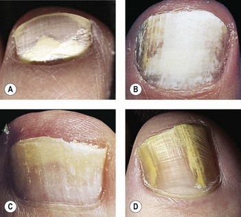
• Dermatophytes: distal or lateral nail involvement spreading proximally, proximal subungual dystrophy, superficial white dystrophy, white or yellow thickened nails, crumbling of nail plate, adjacent web or skin involvement may be present.
• Candida: chronic paronychia, shiny red bolstered nailfold, almost exclusively affects finger nailfolds, loss of cuticle, pus exuding from under nailfold, usually proximal nail involvement, distal nail dystrophy (associated with circulatory disorders), total dystrophic onychomycosis, nails white, green or occasionally black.
• Non-dermatophyte: superficial white onychomycosis, solitary nail involvement, colour of the nail may be influenced by the nature of infecting mould (e.g. the nails are white with Fusarium, white, yellow, brown or green with S. brevicaulis).
What are the patterns of fungal nail involvement?
Distal lateral subungual onychomycosis.
Superficial white onychomycosis.
Proximal white subungual onychomycosis.
Total dystrophic onychomycosis (i.e. all the previous three sites are affected).
What are the poor prognosis factors?
How are specimens obtained for mycological diagnosis?
Scrapings are taken with a blunt scalpel as proximally as possible in the nail:
• In distal and lateral onychomycosis, specimens are taken from the proximal part of the diseased portion of the nail and subungual material
• In superficial white onychomycosis, the scrapings are taken from the diseased nail surface
• In proximal white mycosis, the scrapings are taken from the nail surface.
What is the recommended treatment for onychomycosis?
• Dermatophyte infections (BMJ 1995;311:1277–81):
What is the definition of cure in fungal nail infections?
Persistent onychomycosis is suggested at the end of the observation period by: